

Immunohistochemical detection of Mycoplasma salivarium in oral lichen planus tissue
- PMID: 28295632
- PMCID: PMC5600092
- DOI: 10.1111/jop.12568
Immunohistochemical detection of Mycoplasma salivarium in oral lichen planus tissue
Abstract
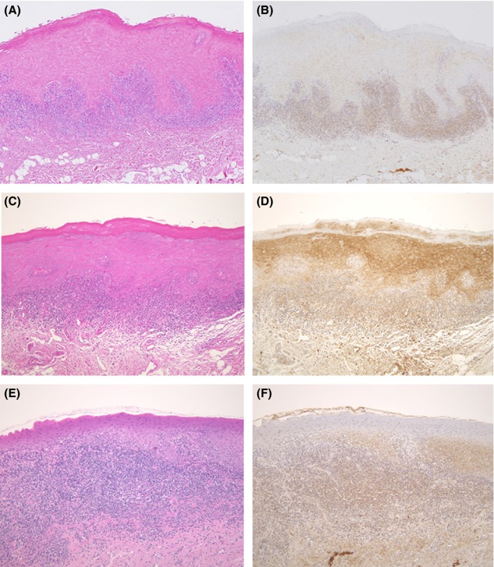
Background: Oral lichen planus (OLP) is a T-cell-mediated inflammatory disease; however, its exact etiology is unknown. Hyperkeratosis is often observed in OLP lesions. Previous studies have revealed the localization of Mycoplasma salivarium in the epithelial cells of oral leukoplakia with hyperkeratosis. Herein, we investigated the presence of M. salivarium in OLP tissue by immunohistochemistry to determine the causative factor of OLP.
Methods: Forty-one formalin-fixed, paraffin-embedded samples obtained from 31 patients with OLP were examined. Ten samples of normal-appearing oral mucosa were used as controls. Immunohistochemistry (IHC) was performed using anti-M. salivarium monoclonal antibodies.
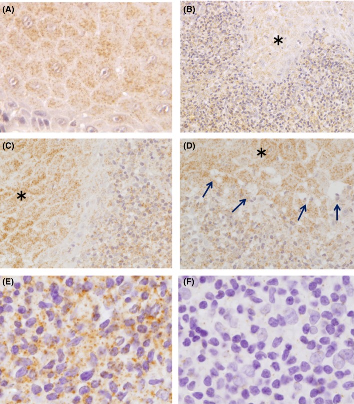
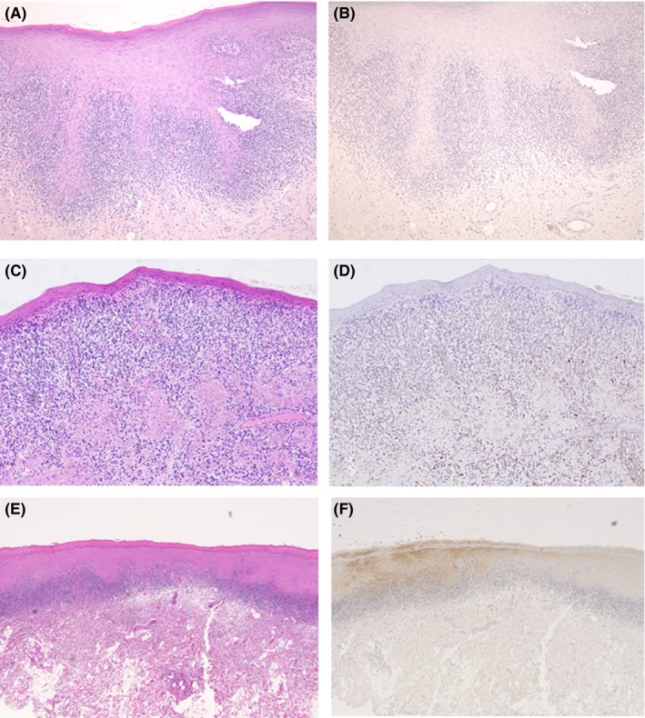
Results and conclusions: Mycoplasma salivarium was detected in the epithelium and lymphocyte infiltrate area in 24 of 41 OLP samples (58.5%). The bacteria were intracellularly localized in epithelial cells, while it was unclear whether they were also localized in lymphocyte cells or in the extracellular spaces among the lymphocytes in the subepithelial lymphocyte infiltrate area. Little or no staining was observed in the epithelium in the normal-appearing mucosa samples. Sawtooth rete ridge formation was observed in 21 OLP samples (51.2%), and a significant positive correlation between sawtooth rete ridge formation and IHC positivity was demonstrated. However, the role of M. salivarium in the epithelium and lamina propria of OLP tissue remains unknown.
Keywords: Mycoplasma salivarium; etiology; immunohistochemistry; oral lichen planus; sawtooth rete ridge.
© 2017 The Authors Journal of Oral Pathology & Medicine Published by John Wiley & Sons Ltd.
Figures
References
-
- Eisen D, Carrozzo M, Bagan Sebastian J‐V, Thongprasom K. Number V Oral lichen planus: clinical features and management. Oral Dis. 2005;11:338‐349. - PubMed
-
- Andreasen JO. Oral lichen planus. 1. A clinical evaluation of 115 cases. Oral Surg Oral Med Oral Pathol. 1968;25:31‐42. - PubMed
-
- Fernandez‐Gonzalez F, Vazouez‐Alvarrez R, Reboiras‐Lopez D, Gandara‐Vila P, Garcia‐Garcia A, Gandara‐Rey JM. Histopathological findings in oral lichen planus and their correlation with the clinical manifestations. Med Oral Pathol Oral Cir Bucal. 2011;16:e641‐e646. - PubMed
-
- Eisenberg E. Clinicopathologic patterns of oral lichenoid lesions. Oral Maxillofac Surg Clin North Am. 1994;6:445‐463.
-
- van der Meij EH, van der Waal I. Lack of clinicopathologic correlation in the diagnosis of oral lichen planus based on the presently available diagnostic criteria and suggestions for modifications. J Oral Pathol Med. 2003;32:507‐512. - PubMed
MeSH terms
LinkOut - more resources
Full Text Sources
Other Literature Sources
