. 2017 Jun;16(3):469-479.
doi: 10.1111/acel.12583. Epub 2017 Mar 14.

Insulin-like growth factor 1 deficiency exacerbates hypertension-induced cerebral microhemorrhages in mice, mimicking the aging phenotype

Affiliations

Insulin-like growth factor 1 deficiency exacerbates hypertension-induced cerebral microhemorrhages in mice, mimicking the aging phenotype

Stefano Tarantini et al. Aging Cell. 2017 Jun.

Abstract

Clinical and experimental studies show that aging exacerbates hypertension-induced cerebral microhemorrhages (CMHs), which progressively impair neuronal function. There is growing evidence that aging promotes insulin-like growth factor 1 (IGF-1) deficiency, which compromises multiple aspects of cerebromicrovascular and brain health. To determine the role of IGF-1 deficiency in the pathogenesis of CMHs, we induced hypertension in mice with liver-specific knockdown of IGF-1 (Igf1f/f + TBG-Cre-AAV8) and control mice by angiotensin II plus l-NAME treatment. In IGF-1-deficient mice, the same level of hypertension led to significantly earlier onset and increased incidence and neurological consequences of CMHs, as compared to control mice, as shown by neurological examination, gait analysis, and histological assessment of CMHs in serial brain sections. Previous studies showed that in aging, increased oxidative stress-mediated matrix metalloprotease (MMP) activation importantly contributes to the pathogenesis of CMHs. Thus, it is significant that hypertension-induced cerebrovascular oxidative stress and MMP activation were increased in IGF-1-deficient mice. We found that IGF-1 deficiency impaired hypertension-induced adaptive media hypertrophy and extracellular matrix remodeling, which together with the increased MMP activation likely also contributes to increased fragility of intracerebral arterioles. Collectively, IGF-1 deficiency promotes the pathogenesis of CMHs, mimicking the aging phenotype, which likely contribute to its deleterious effect on cognitive function. Therapeutic strategies that upregulate IGF-1 signaling in the cerebral vessels and/or reduce microvascular oxidative stress, and MMP activation may be useful for the prevention of CMHs, protecting cognitive function in high-risk elderly patients.

Keywords: arteriole; dementia; gait dysfunction; microbleed; oxidative stress.


Figures

Figure 1
Figure 1
IGF‐1 deficiency exacerbates hypertension (HT)‐induced CMHs in mice. (A) Adeno‐associated viral knockdown of hepatic Ifg1 (Igf1 f/f + TBG‐Cre‐AAV8) significantly decreases the levels of circulating IGF‐1 compared to control animals. Data are mean ± SEM. *P = 0.001 vs. control (t‐test). (B) Treatment with angiotensin II plus lNAME elicited similar increases in systolic blood pressure in control and IGF‐1‐deficient mice. *P < 0.05 vs. control normotensive (one‐way ANOVA, Tukey's post hoc test). (C) Cumulative incidence curves for neurological signs of hypertension‐induced intracerebral hemorrhage in control (n = 24) and IGF‐1‐deficient mice (n = 31). In IGF‐1‐deficient mice, there was a significant increase in CMH incidence compared to control mice (log‐rank test; Mantel‐Cox). (D–G) Representative images of CMHs stained by diaminobenzidine in the cortex, brain stem, white matter, and cerebellum of hypertensive IGF‐1‐deficient mice. (H–K) Black arrows point to cerebral intraparenchymal arterioles in close proximity to the hemorrhages. Hemorrhages were often confined to and spread along the perivascular spaces (arrowheads). Note in (K) the spread of the hemorrhage to the daughter branches of an arteriole along the perivascular spaces. (L): Total number of hypertension‐induced CMHs throughout the entire brain of control and IGF‐1‐deficient mice. Data are mean ± SEM. *P < 0.05 (t‐test). (M) The pie charts illustrate the similar distribution of CMHs by location in both experimental groups. (N) The cumulative frequency distribution of CMHs by volume significantly shifts to the left in IGF‐1‐deficient mice compared to control, indicating that IGF‐1 deficiency specifically increases the number of smaller bleeds. The maximum difference between the cumulative distributions was calculated using the two‐sample Kolmogorov–Smirnov test (D: 0.5202; P < 0.0001).
Figure 2
Figure 2
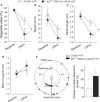
Increased incidence of CMHs is associated with gait dysfunction in hypertensive IGF‐1‐deficient mice. Regularity index (A), body speed (B), stride length (C), and base of support (front paws; D) in control mice and IGF‐1‐deficient mice under baseline conditions and after induction of CMHs. (E) Circular scatter plot showing the distribution of interlimb coupling values (phase dispersion) in control mice and IGF‐1‐deficient mice under baseline conditions and after induction of CMHs (note that the circular plot shows a smaller phase dispersion scatter in the inner circle [before hypertension] as compared to the phase dispersion scatter in the outer circle [assessed after hypertension]). Right panels shows average deviation of phase dispersion (calculated between the right hind paw [RH] and left hind paw [LH]) from the expected value (50%) under baseline conditions and after induction of CMHs. Data are mean ± SEM.*P < 0.05 vs. control baseline, & P < 0.05 vs. IGF‐1‐deficient baseline, # P < 0.05 control vs. IGF‐1 deficient (one‐way ANOVA, Tukey's post hoc test) HT: hypertension.
Figure 3
Figure 3
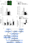
IGF‐1 deficiency exacerbates hypertension‐induced MMP activation. (A) Hypertension‐induced MMP activation, assessed using the MMPsense 645 FAST fluorescent method, in control and IGF‐1‐deficient mice (n = 6 in each group; see Experimental procedures). MMPsense 645 FAST becomes fluorescent upon cleavage by activated MMPs. Data are mean ± SEM. *P < 0.05 vs. control, # P < 0.05 vs. control HT, & P < 0.05 vs. IGF‐1 deficient. (B) Representative confocal image of the longitudinal section of a cerebral intraparenchymal arteriole from a hypertensive IGF‐1‐deficient mouse injected with the MMPsense 645 FAST substrate (scale bar: 25 μm). Note, the strong red fluorescence in the vascular wall indicating increased MMP activity. Intraluminal FITC–dextran is shown for orientation purposes. L (lumen), M (media). (C): Representative compressed Z stacks of confocal images of MCAs showing stronger MMPsense 645 FAST fluorescence (red) in high‐pressure‐exposed MCAs isolated from IGF‐1‐deficient mice as compared to MCAs isolated from control mice, indicating increased MMP activation. MCAs were pressurized at 60 and 160 mmHg for 6 h. (original magnification: 20×, scale bar: 100 μm). Bar graphs (D) are summary data. Data are means ± SEM (n = 6 in each group) *P < 0.05 vs. control (60 mmHg), # P < 0.05 vs. IGF‐1 deficient (160 mmHg); & P < 0.05 vs. IGF‐1 deficient (60 mmHg). (E) IGF‐1 deficiency exacerbates hypertension‐induced oxidative stress. Representative confocal images showing stronger DHE fluorescence (pseudocolored white) indicating increased O2· production in high‐pressure‐exposed MCAs isolated from aged mice as compared to MCAs isolated from young mice. MCAs were pressurized at 60 and 160 mmHg for 6 h. (original magnification: 20×, scale bar: 50 μm). Bar graphs (F) are summary data. Data are means ± SEM (n = 6 in each group). *P < 0.05 vs. control, # P < 0.05 vs. control HT, & P < 0.05 vs. IGF‐1 deficient. G–J show hypertension‐induced changes in mRNA expression of MMP‐2, ‐3, and ‐9 as well as Timp‐1, ‐2, and ‐3 in the cerebral arteries. Data are mean ± SEM (n = 6 in each group). *P < 0.05 vs. control, & P < 0.05 vs. IGF‐1 deficient. Differences between different groups were established using a one‐way ANOVA followed by Tukey's post hoc tests.
Figure 4
Figure 4
IGF‐1 deficiency impairs hypertension‐induced hypertrophy and structural remodeling in cerebral vessels. (A) Representative confocal micrographs from normotensive (NT) and hypertensive (HT) fixed brains. Hypertension induces hypertrophy of penetrating arterioles in control mice, whereas this adaptive response is impaired in IGF‐1‐deficient mice (green fluorescence: immunostaining for alpha smooth muscle actin). Bar graphs are summary data for calculated wall‐to‐lumen ratios. qPCR data showing mRNA expression of alpha smooth muscle actin and collagens in branches of the middle cerebral arteries isolated from normotensive and hypertensive control and IGF‐1‐deficient mice are shown in panels B and C, respectively. Data are mean ± SEM (n = 4–6 in each group), *P < 0.05 vs. control, # P < 0.05 vs. control HT. (D) IGF‐1 deficiency exacerbates hypertension‐induced profragility shift in vascular gene expression signature. Expression of 67 genes related to the pathogenesis of CMHs was determined by qPCR, and vascular fragility signatures (Spearman's ρ) were calculated as described in the Results. A higher ‘fragility signature’ indicates higher expression of profragility genes and lower expression of antifragility regulators. (E) Proposed scheme depicting the mechanisms by which age‐related IGF‐1 deficiency may exacerbate hypertension‐induced microvascular damage, promoting CMHs. Differences between different groups were established using a one‐way ANOVA followed by Tukey's post hoc tests.

Similar articles

Cited by

References

    1. Akoudad S, Wolters FJ, Viswanathan A, de Bruijn RF, van der Lugt A, Hofman A, Koudstaal PJ, Ikram MA, Vernooij MW (2016) Association of cerebral microbleeds with cognitive decline and dementia. JAMA Neurol. 73, 934–943. - PMC - PubMed
    1. Bailey‐Downs LC, Mitschelen M, Sosnowska D, Toth P, Pinto JT, Ballabh P, Valcarcel‐Ares MN, Farley J, Koller A, Henthorn JC, Bass C, Sonntag WE, Ungvari Z, Csiszar A (2012) Liver‐specific knockdown of IGF‐1 decreases vascular oxidative stress resistance by impairing the Nrf2‐dependent antioxidant response: a novel model of vascular aging. J. Gerontol. Biol. Med. Sci. 67, 313–329. - PMC - PubMed
    1. Baumbach GL, Heistad DD (1989) Remodeling of cerebral arterioles in chronic hypertension. Hypertension 13, 968–972. - PubMed
    1. Choi P, Ren M, Phan TG, Callisaya M, Ly JV, Beare R, Chong W, Srikanth V (2012) Silent infarcts and cerebral microbleeds modify the associations of white matter lesions with gait and postural stability: population‐based study. Stroke 43, 1505–1510. - PubMed
    1. Csiszar A, Labinskyy N, Perez V, Recchia FA, Podlutsky A, Mukhopadhyay P, Losonczy G, Pacher P, Austad SN, Bartke A, Ungvari Z (2008) Endothelial function and vascular oxidative stress in long‐lived GH/IGF‐deficient Ames dwarf mice. Am. J. Physiol. Heart Circ. Physiol. 295, H1882–H1894. - PMC - PubMed

Publication types

MeSH terms

Substances

LinkOut - more resources