Case Reports
. 2017 May;173(5):1319-1327.
doi: 10.1002/ajmg.a.38207. Epub 2017 Mar 11.

De novo loss-of-function variants in STAG2 are associated with developmental delay, microcephaly, and congenital anomalies

Affiliations
Case Reports

De novo loss-of-function variants in STAG2 are associated with developmental delay, microcephaly, and congenital anomalies

Sureni V Mullegama et al. Am J Med Genet A. 2017 May.

Abstract

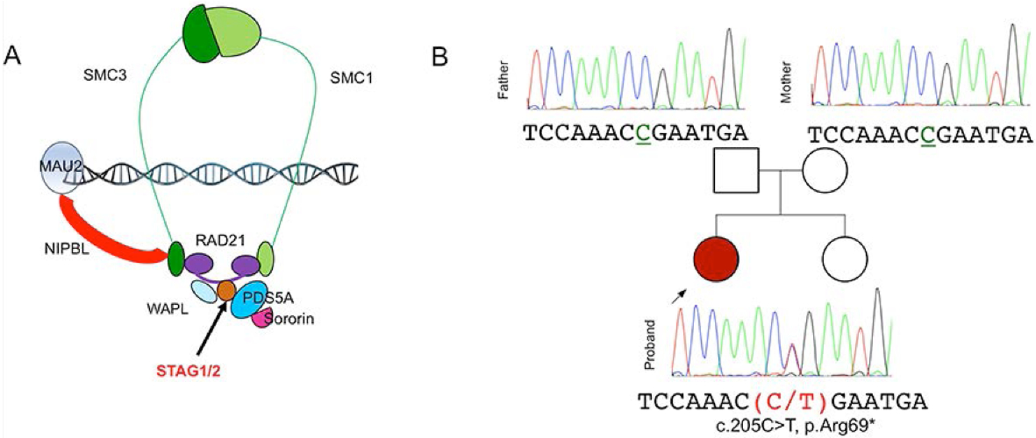
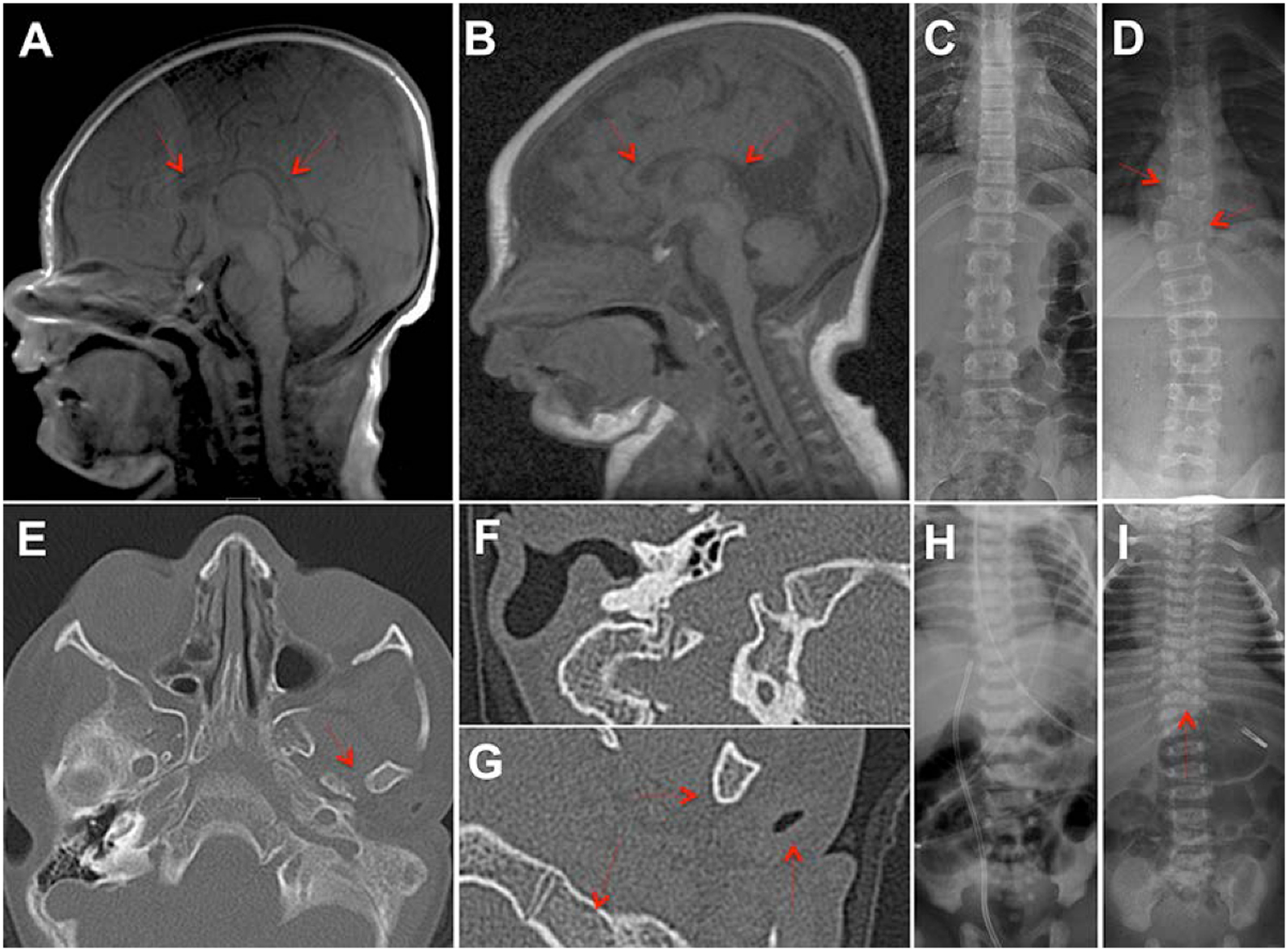
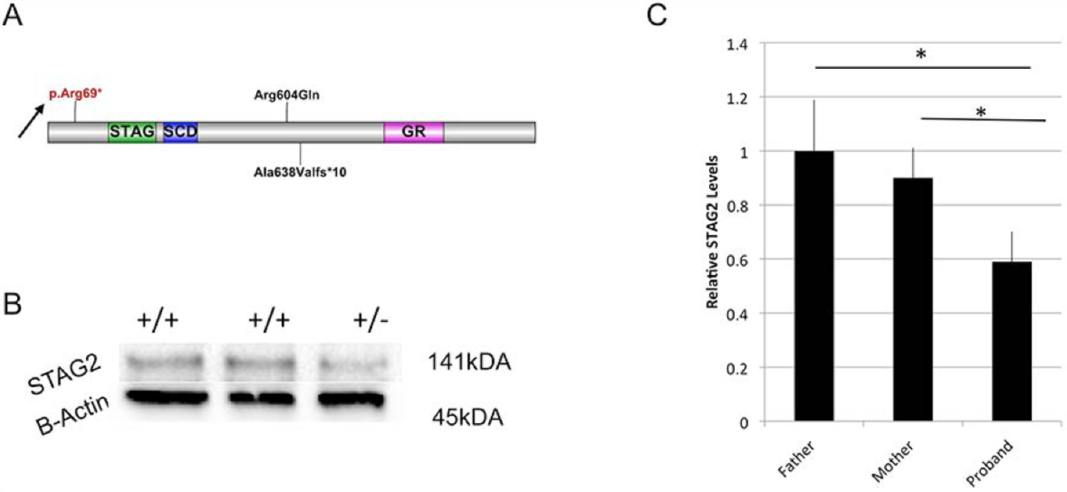
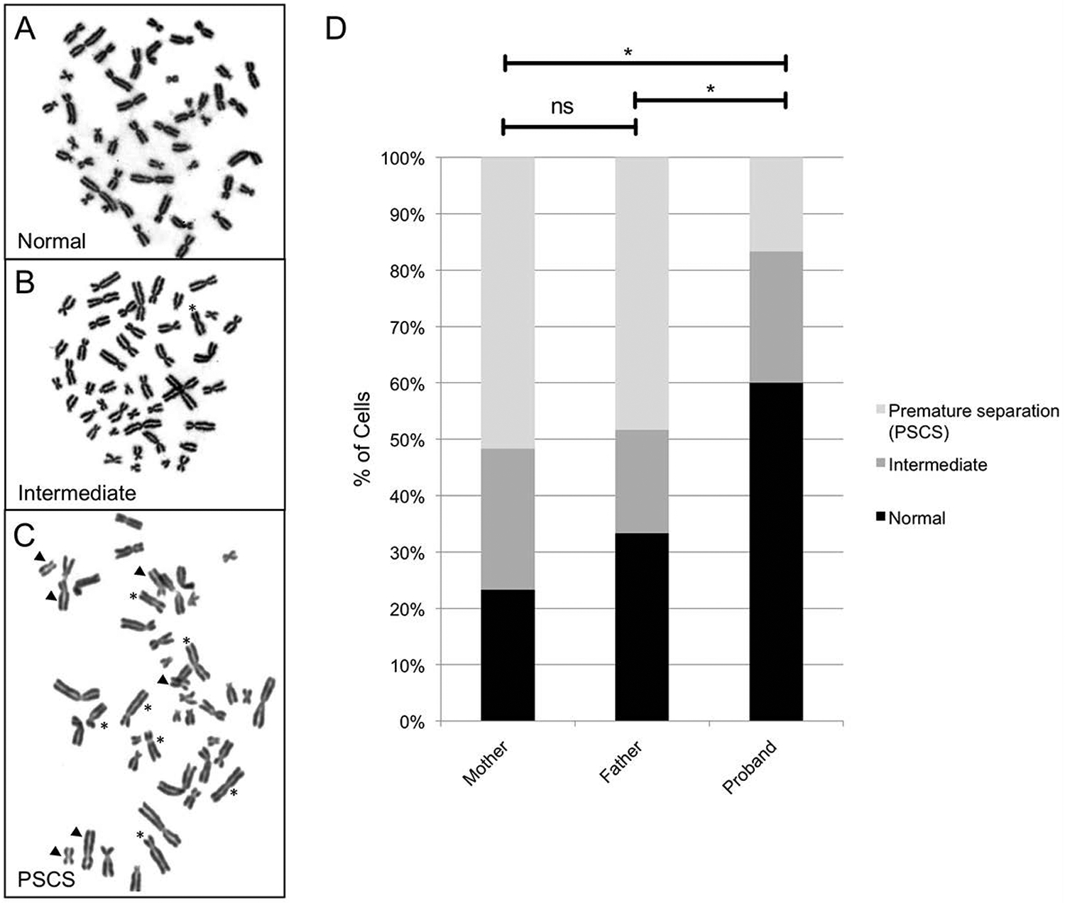
The cohesin complex is an evolutionarily conserved multi-subunit protein complex which regulates sister chromatid cohesion during mitosis and meiosis. Additionally, the cohesin complex regulates DNA replication, DNA repair, and transcription. The core of the complex consists of four subunits: SMC1A, SMC3, RAD21, and STAG1/2. Loss-of-function mutations in many of these proteins have been implicated in human developmental disorders collectively termed "cohesinopathies." Through clinical exome sequencing (CES) of an 8-year-old girl with a clinical history of global developmental delay, microcephaly, microtia with hearing loss, language delay, ADHD, and dysmorphic features, we describe a heterozygous de novo variant (c.205C>T; p.(Arg69*)) in the integral cohesin structural protein, STAG2. This variant is associated with decreased STAG2 protein expression. The analyses of metaphase spreads did not exhibit premature sister chromatid separation; however, delayed sister chromatid cohesion was observed. To further support the pathogenicity of STAG2 variants, we identified two additional female cases from the DECIPHER research database with mutations in STAG2 and phenotypes similar to our patient. Interestingly, the clinical features of these three cases are remarkably similar to those observed in other well-established cohesinopathies. Herein, we suggest that STAG2 is a dosage-sensitive gene and that heterozygous loss-of-function variants lead to a cohesinopathy.

Keywords: cohesin complex; STAG2; X-linked; cohesin-associated genes; cohesinopathy; gene dosage.


Conflict of interest statement

Conflict of interest: No conflicts of interests

Figures

Fig. 1.
Fig. 1.. Characterization of STAG2 variants
(A) Structure of the cohesin ring and its regulatory proteins. (B) Case 1 family pedigree with sequence analysis by Sanger sequencing showed a heterozygous variant, c.205C>T; p.(Arg69*) in Exon 5 of STAG2 in the affected proband (red).
Fig 2.
Fig 2.. MRI and radiological findings in patients with STAG2 mutations.
(A) Healthy three day old female axial T1 weighted MRI view of a normally developed corpus callosum (red arrows) (B) Axial T1 weighted MRI view of patient with STAG2 mutation displaying dysgenesis of the splenium of the corpus callosum (red arrows). (C) Chest x-ray of a healthy one day old female demonstrating normal vertebral development. (D) Chest x-ray of a patient with STAG2 mutation demonstrating scoliosis and vertebral abnormalities including hemi-vertebrae and “butterfly” vertebrae (red arrows). (E) Axial view of bilateral auditory canals in a patient with a STAG2 mutation, showing absence of the left auditory canal (red arrow). (F) Higher magnification of the right auditory canal image shows patent canal and normal bone structures (G) Higher magnification of the left auditory canal demonstrating external auditory canal atresia with a fused ossicular mass, absent stapes, and severely stenotic oval window (red arrows). (H) Chest x-ray of a healthy eight year old female demonstrating normal vertebral development. (I) Chest x-ray of a patient with STAG2 mutation demonstrating scoliosis and vertebral abnormalities including hemi-vertebrae (red arrow).
Fig. 3.
Fig. 3.. Location and consequence of STAG2 Variants
(A) Predicted protein domain structure of STAG2. The 1231 amino acid full-length protein is predicted to contain a STAG domain, a stromalin conservative domain (SCD) and a glutamine-rich region domain (GR). The p.Arg69* mutation is indicated by an arrow. The location of the mutations reported by DECIPHER are depicted (p.Arg604Gln and p.Ala638fs*). (B) Protein expression analysis of STAG2 in patient and her parents reveals decreased levels in proband (+/−) compared to parents (+/+). (C) Western blot analysis was run using two independent antibodies for STAG2. Quantification of band density with normalization to loading controls demonstrates significant reduction in STAG2 levels in the proband when compared to either unaffected parental control. *Denotes significant difference (p<.05, Mann-Whitey U Test (n=6)).
Fig. 4.
Fig. 4.. Sister Chromatid Cohesion Studies on Affected Proband
(A-C) Metaphases that were described as closed, intermediate or having premature sister chromatid separation (PSCS) in our assays. Individual chromatid pairs with abnormal phenotypes are highlighted in (B) and (C), with an arrowhead indicating “open” chromatid pairs, an asterisk indicating pairs with “partial” separation, and all other chromatid pairs being “closed.” In (B) there is one partially separated chromatid pair, while in (C) there are 6 open pairs and 7 partial pairs. By our classification, metaphases with 1–2 open/partial chromatid pairs were described as intermediate, and those with 3 or more open/partial chromatid pairs were described as having PSCS. (D) A graph showing quantification of these results. 60 metaphases were analyzed from the proband and each parent and classified as described above. In the proband having the STAG2 mutation, PSCS was not increased; in contrast, the proportion of nuclei having PSCS showed a significant decrease compared to either parent (significance calculated using a two-tailed Fisher’s Exact test for mother versus proband (P=0.000029), father versus proband (P=0.00075), and mother versus father (P=0.44779)).

Similar articles

Cited by

References

    1. Ball AR Jr., Chen YY, Yokomori K. 2014. Mechanisms of cohesin-mediated gene regulation and lessons learned from cohesinopathies. Biochim Biophys Acta 1839(3):191–202. - PMC - PubMed
    1. Barbero JL. 2013. Genetic basis of cohesinopathies. Appl Clin Genet 6:15–23. - PMC - PubMed
    1. Bonnet C, Leheup B, Beri M, Philippe C, Gregoire MJ, Jonveaux P. 2009. Aberrant GRIA3 transcripts with multi-exon duplications in a family with X-linked mental retardation. Am J Med Genet A 149A(6):1280–1289. - PubMed
    1. Chae JH, Hwang H, Hwang YS, Cheong HJ, Kim KJ. 2004. Influence of MECP2 gene mutation and X-chromosome inactivation on the Rett syndrome phenotype. J Child Neurol 19(7):503–508. - PubMed
    1. Cotton AM, Ge B, Light N, Adoue V, Pastinen T, Brown CJ. 2013. Analysis of expressed SNPs identifies variable extents of expression from the human inactive X chromosome. Genome Biol 14(11):R122. - PMC - PubMed

Publication types