. 2017 Jun;11(6):612-627.
doi: 10.1002/1878-0261.12051. Epub 2017 May 2.

DIMP53-1: a novel small-molecule dual inhibitor of p53-MDM2/X interactions with multifunctional p53-dependent anticancer properties

Affiliations

DIMP53-1: a novel small-molecule dual inhibitor of p53-MDM2/X interactions with multifunctional p53-dependent anticancer properties

Joana Soares et al. Mol Oncol. 2017 Jun.

Abstract

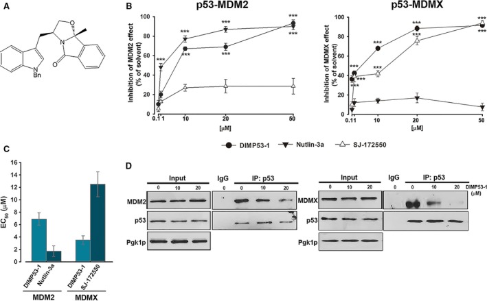
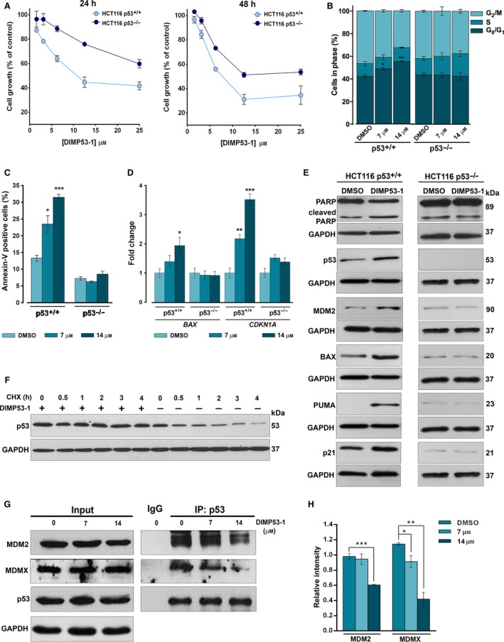
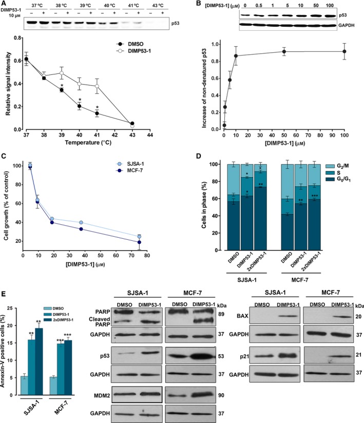
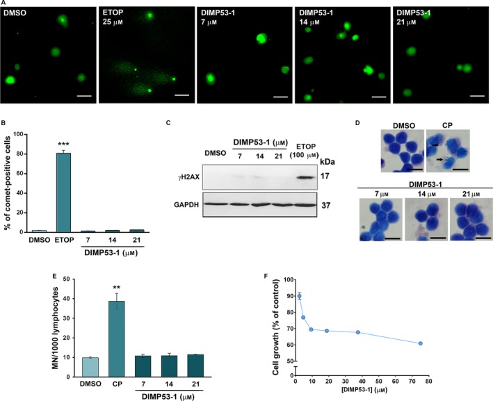
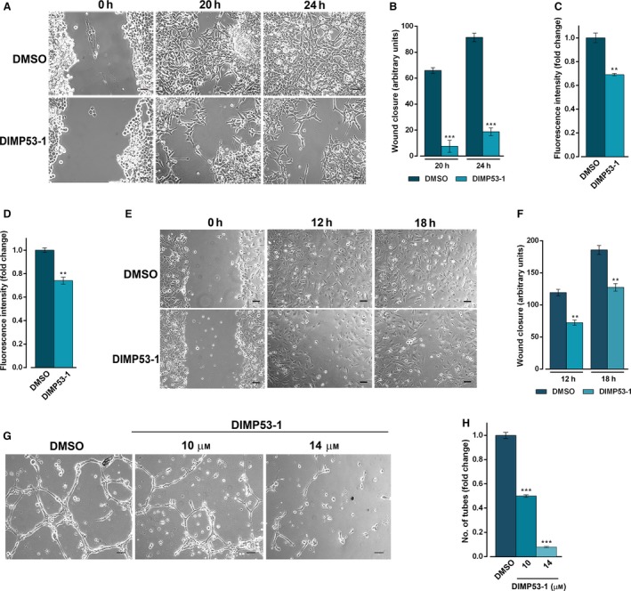
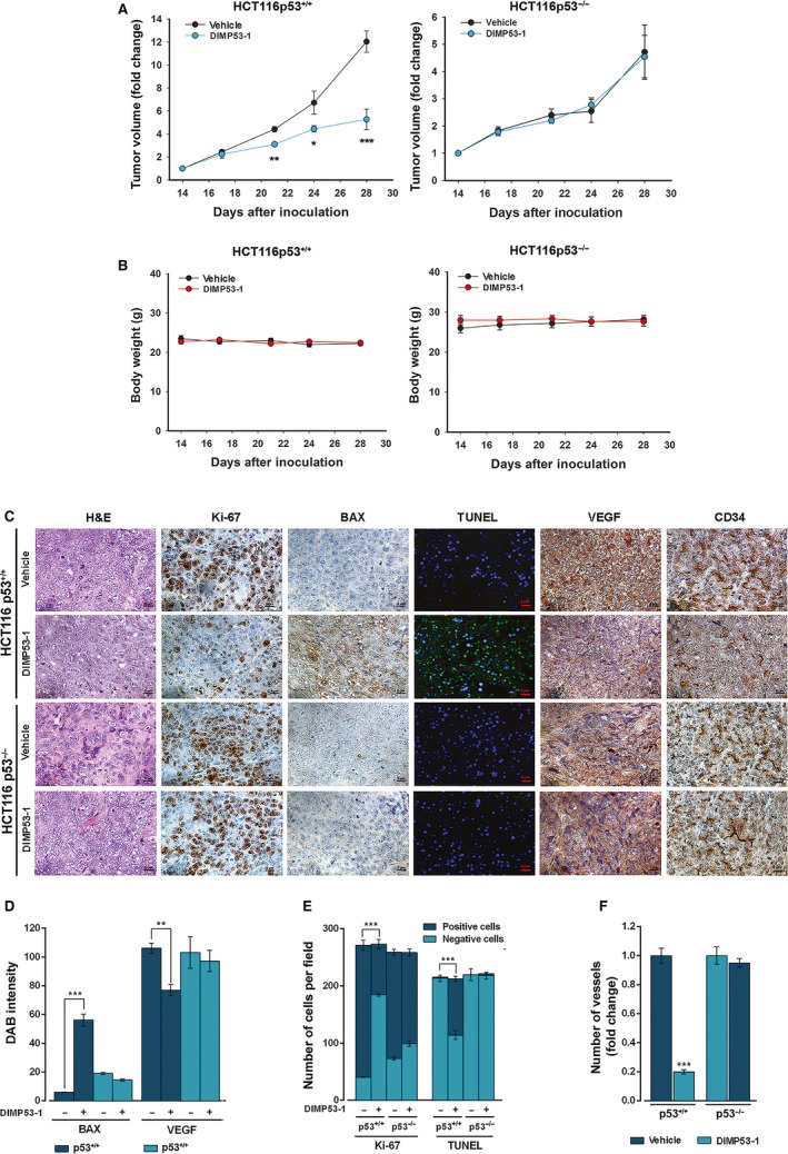
The transcription factor p53 plays a crucial role in cancer development and dissemination, and thus, p53-targeted therapies are among the most encouraging anticancer strategies. In human cancers with wild-type (wt) p53, its inactivation by interaction with murine double minute (MDM)2 and MDMX is a common event. Simultaneous inhibition of the p53 interaction with both MDMs is crucial to restore the tumor suppressor activity of p53. Here, we describe the synthesis of the new tryptophanol-derived oxazoloisoindolinone DIMP53-1 and identify its activity as a dual inhibitor of the p53-MDM2/X interactions using a yeast-based assay. DIMP53-1 caused growth inhibition, mediated by p53 stabilization and upregulation of p53 transcriptional targets involved in cell cycle arrest and apoptosis, in wt p53-expressing tumor cells, including MDM2- or MDMX-overexpressing cells. Importantly, DIMP53-1 inhibits the p53-MDM2/X interactions by potentially binding to p53, in human colon adenocarcinoma HCT116 cells. DIMP53-1 also inhibited the migration and invasion of HCT116 cells, and the migration and tube formation of HMVEC-D endothelial cells. Notably, in human tumor xenograft mice models, DIMP53-1 showed a p53-dependent antitumor activity through induction of apoptosis and inhibition of proliferation and angiogenesis. Finally, no genotoxicity or undesirable toxic effects were observed with DIMP53-1. In conclusion, DIMP53-1 is a novel p53 activator, which potentially binds to p53 inhibiting its interaction with MDM2 and MDMX. Although target-directed, DIMP53-1 has a multifunctional activity, targeting major hallmarks of cancer through its antiproliferative, proapoptotic, antiangiogenic, anti-invasive, and antimigratory properties. DIMP53-1 is a promising anticancer drug candidate and an encouraging starting point to develop improved derivatives for clinical application.

Keywords: MDMX; MDM2; anticancer therapy; p53; small-molecule; tryptophanol-derived oxazoloisoindolinone.


Figures

Figure 1
Figure 1
Identification of DIMP53‐1 as a potential dual inhibitor of the p53–MDM2/X interactions using yeast. (A) DIMP53‐1 chemical structure. (B) Effect of 0.1–50 μm DIMP53‐1, Nutlin‐3a, and SJ‐172550 on the reestablishment of p53‐induced growth inhibition in MDM2/MDMX‐co‐expressing yeast, for 42 h; results were plotted setting the growth of DMSO‐treated cells expressing p53 alone as 100%; data are mean ± SEM of six independent experiments; values were significantly different from DMSO (***< 0.001). (C) EC 50 values of DIMP53‐1, Nutlin‐3a, and SJ‐172550 obtained from concentration–response curves presented in (B). (D) Co‐IP was performed with anti‐p53 (IP:p53) or anti‐immunoglobulin G (IgG) antibodies, followed by immunoblotting with anti‐MDM2, anti‐MDMX, and anti‐p53 antibodies in cells treated with 10 and 20 μm of DIMP53‐1 or DMSO for 42 h; whole‐cell lysate (input).
Figure 2
Figure 2
DIMP53‐1 shows a p53‐dependent growth inhibitory effect in human tumor cells mediated by cell cycle arrest, apoptosis, p53 stabilization, upregulation of p53 target genes, and disruption of the p53–MDM2/X interactions. (A) Concentration–response growth curves for DIMP53‐1 in p53+/+ and p53−/− HCT116, after 24‐ and 48‐h treatments; data are mean ± SEM of four independent experiments; incubation with DMSO, in equivalent % of DIMP53‐1, was used to normalize the results. (B,C) Cell cycle arrest (B) and apoptosis (C) were determined at 7 and 14 μm of DIMP53‐1 for 24 h in p53+/+ and p53−/− HCT116 cells; data are mean ± SEM of three independent experiments; values were significantly different from DMSO (*< 0.05; **< 0.01; ***< 0.001). (D) mRNA levels of BAX and CDKN1A (p21) after 24‐h treatment with 7 and 14 μm of DIMP53‐1 in p53+/+ and p53−/− HCT116 cells; fold expression changes are relative to DMSO and correspond to mean ± SEM of three independent experiments. (E) Western blot analysis was performed after 24‐h (MDM2, p53) and 48‐h (PARP, BAX, PUMA, p21) treatments with 7 μm of DIMP53‐1 or DMSO in p53+/+ and p53−/− HCT116 cells. (F) p53 protein levels in HCT116p53+/+ cells treated for 24 h with DIMP53‐1 or solvent followed with cycloheximide (CHX; 150 μg/mL). (G) Co‐IP was performed with anti‐p53 (IP:p53) or anti‐immunoglobulin G (IgG) antibodies, followed by immunoblotting with anti‐MDM2, anti‐MDMX, and anti‐p53 antibodies in HCT116p53+/+ cells treated with 7 and 14 μm of DIMP53‐1 or DMSO for 8 h; whole‐cell lysate (input); in IP:p53 of MDM2, the cut top band corresponds to the anti‐p53 antibody, and the other two bands correspond to MDM2 isoforms. (H) Quantification of IP:p53 immunoblots; data are mean ± SEM of three independent experiments; values were significantly different from DMSO (*< 0.05; **< 0.01; ***< 0.001). In (E), (F), and (G), immunoblots are representative of three independent experiments; GAPDH was used as loading control.
Figure 3
Figure 3
DIMP53‐1 potentially binds to p53 and inhibits the growth of MDM2‐ and MDMX‐overexpressing tumor cells through induction of cell cycle arrest, apoptosis, and upregulation of p53 target genes. (A and B) CETSA experiments were performed in HCT116p53+/+ cell lysates in the presence or absence of DIMP53‐1. In (A), 10 μm of DIMP53‐1 was used and lysate samples were heated at different temperatures; plot represents the signal intensity normalized to the intensity at 25 °C. In (B), lysate samples were treated with increasing DIMP53‐1 concentrations and heated at 39 °C; plot represents the increase in nondenatured p53 calculated setting the signal obtained with DMSO at 39 °C as 0, and the signal obtained with DMSO at 25 °C (considered the maximum amount of nondenatured p53) as 1. Results are mean ± SEM of three independent experiments. (C) DIMP53‐1 concentration–response growth curves in SJSA‐1 and MCF‐7 cells, after 48‐h treatment; data are mean ± SEM of four independent experiments; incubation with DMSO, in equivalent % of DIMP53‐1, was used to normalize the results. (D,E) Cell cycle arrest (D) and apoptosis (E) were determined in SJSA‐1 and MCF‐7 cells at IC 50 and 2 × IC 50 (2 × DIMP53‐1) concentrations, after 24‐h treatment; data are mean ± SEM of three independent experiments; values were significantly different from DMSO (*< 0.05; **< 0.01; ***< 0.001). (F) Western blot analysis was performed in SJSA‐1 and MCF‐7 cells, after 24‐h (p21) and 48‐h (PARP, p53, MDM2, BAX) treatments with the IC 50 of DIMP53‐1 or DMSO. In (A), (B), and (F), immunoblots are representative of three independent experiments; in (B) and (F), GAPDH was used as loading control.
Figure 4
Figure 4
DIMP53‐1 is nongenotoxic in normal and tumor cells and has low cytotoxicity against normal cells. (A–C) DNA damage was measured in HCT116p53+/+ cells by comet assay (A and B) and by analysis of γH2AX expression levels (C) after treatment with etoposide (ETOP; positive control) or DIMP53‐1. In (A), scale bar = 20 μm; magnification = 200 ×. In (B), quantification of comet‐positive cells (containing more than 5% of DNA in the tail; assessed by open comet/imagej); 100 cells were analyzed in each group. In (C), γH2AX levels were determined by western blot; immunoblots are representative of three independent experiments; GAPDH was used as loading control. (D and E) Genotoxicity of 7, 14, and 21 μm DIMP53‐1 by cytokinesis‐block micronucleus (MN) assay after 72‐h treatment in human lymphocyte cells; 5 μg·mL−1 cyclophosphamide (CP; positive control). In (D), scale bar = 20 μm; magnification = 1000 ×. In (E), the number of MN per 1000 binucleated lymphocytes was recorded. (F) DIMP53‐1 concentration–response growth curve in MCF10A cells, after 48‐h treatment; incubation with DMSO, in equivalent % of DIMP53‐1, was used to normalize the results. In (B), (E), and (F), data are mean ± SEM of three to four independent experiments; in (B) and (E), values were significantly different from DMSO (**< 0.01; ***< 0.001).
Figure 5
Figure 5
DIMP53‐1 prevents in vitro angiogenesis and tumor cell invasion and migration. (A,B) HCT116p53+/+ confluent cells treated with 3 μm DIMP53‐1 or DMSO were observed at different time‐points in the wound healing assay. (B) Quantification of wound closure of HCT116p53+/+ cells in five randomly selected microscopic fields. (C) Effect of 3 μm of DIMP53‐1 on the migration of HCT116p53+/+ cells for 24 h, analyzed by the chemotaxis assay. (D) Effect of 3 μm of DIMP53‐1 on the invasion of HCT116p53+/+ cells for 48 h, analyzed by cell invasion assay. In (C) and (D), migratory cells were quantified by fluorescence signal; fold changes are relative to DMSO and correspond to mean ± SEM of three independent experiments. (E,F) HMVEC‐D endothelial confluent cells treated with 14 μm of DIMP53‐1 or DMSO were observed at different time‐points in the wound healing assay. In (F), quantification of wound closure of HMVEC‐D endothelial in five randomly selected microscopic fields. (G) Antiangiogenic effect of 10 and 14 μm of DIMP53‐1 was evaluated in HMVEC‐D cells for 16 h by the endothelial cell tube formation assay. (H) Quantification of tube‐like structures in five randomly selected microscopic fields; fold changes are relative to DMSO and correspond to mean ± SEM of three independent experiments. In (A), (E), and (G), scale bar = 5 μm and magnification = 100 ×. In (B), (C), (D), (F), and (H), values were significantly different from DMSO (**< 0.01; ***< 0.001).
Figure 6
Figure 6
DIMP53‐1 has in vivo p53‐dependent antitumor activity by inducing apoptosis and inhibiting proliferation and angiogenesis. BALB/c nude mice of about 10 weeks were inoculated subcutaneously, in the dorsal flank, with HCT116p53+/+ and HCT116p53−/− tumor cells; when tumors reached approximately 100 mm3 volume (14 days after the grafts), mice were treated twice a week with 50 mg·kg−1 DIMP53‐1 or vehicle (control) by intraperitoneal injection for two weeks. (A) Tumor volume growth curves of mice carrying p53+/+ or p53−/− HCT116 xenografts treated with DIMP53‐1 or vehicle; data are mean ± SEM of the tumor volume fold change to the start of treatment. (B) Mice body weight during treatment with DIMP53‐1 or vehicle; values were not significantly different from vehicle (> 0.05). (C) Representative images of Ki‐67, BAX, DNA fragmentation (TUNEL), CD34, and VEGF detection in p53+/+ and p53−/− HCT116 xenograft tumor tissues treated with DIMP53‐1 or vehicle at the end of treatment (scale bar = 5 μm; magnification = 400×); H&E (hematoxylin and eosin). (D–F) Quantification of immunohistochemistry of p53+/+ and p53−/− HCT116 xenograft tumor tissues treated with DIMP53‐1 or vehicle. In (D), BAX and VEGF staining quantification by evaluation of DAB (3,3′‐diaminobenzidine) intensity. In (E), quantification of the number of positive and negative Ki‐67 and TUNEL cells. In (F), evaluation of microvessel density by quantification of vessels stained with anti‐CD34; data are mean ± SEM of the number of vessels per mm2 fold change to the vehicle. In (A), (D), (E), and (F), values were significantly different from vehicle (*< 0.05; **< 0.01; ***< 0.001).

Similar articles

Cited by

References

    1. Baeriswyl V and Christofori G (2009) The angiogenic switch in carcinogenesis. Semin Cancer Biol 19, 329–337. - PubMed
    1. Burgess A, Chia KM, Haupt S, Thomas D, Haupt Y and Lim E (2016) Clinical overview of MDM2/X‐targeted therapies. Front Oncol 6, 7. - PMC - PubMed
    1. Cordani M, Pacchiana R, Butera G, D'Orazi G, Scarpa A and Donadelli M (2016) Mutant p53 proteins alter cancer cell secretome and tumour microenvironment: involvement in cancer invasion and metastasis. Cancer Lett 376, 303–309. - PubMed
    1. Gomes S, Leao M, Raimundo L, Ramos H, Soares J and Saraiva L (2016) p53 family interactions and yeast: together in anticancer therapy. Drug Discov Today 21, 616–624. - PubMed
    1. Graves B, Thompson T, Xia M, Janson C, Lukacs C, Deo D, Di Lello P, Fry D, Garvie C, Huang KS et al (2012) Activation of the p53 pathway by small‐molecule‐induced MDM2 and MDMX dimerization. Proc Natl Acad Sci U S A 109, 11788–11793. - PMC - PubMed

MeSH terms

LinkOut - more resources