. 2017 Aug;57(2):162-173.
doi: 10.1165/rcmb.2017-0019OC.

Foxp3+ Regulatory T Cell Expression of Keratinocyte Growth Factor Enhances Lung Epithelial Proliferation

Affiliations

Foxp3+ Regulatory T Cell Expression of Keratinocyte Growth Factor Enhances Lung Epithelial Proliferation

Catherine F Dial et al. Am J Respir Cell Mol Biol. 2017 Aug.

Abstract

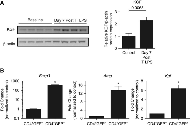
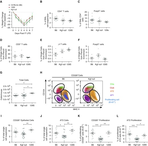
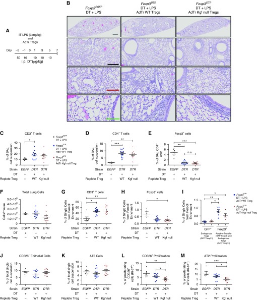
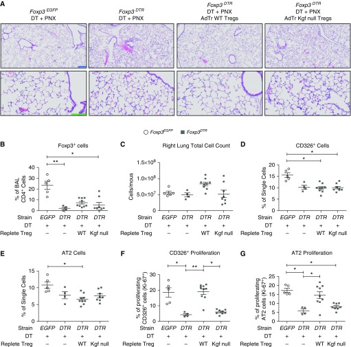
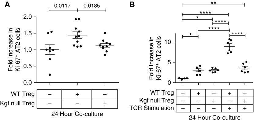
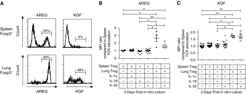
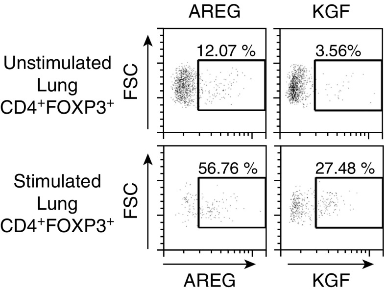
Repair of the lung epithelium after injury is a critical component for resolution; however, the processes necessary to drive epithelial resolution are not clearly defined. Published data demonstrate that Foxp3+ regulatory T cells (Tregs) enhance alveolar epithelial proliferation after injury, and Tregs in vitro directly promote type II alveolar epithelial cell (AT2) proliferation, in part by a contact-independent mechanism. Therefore, we sought to determine the contribution of Treg-specific expression of a growth factor that is known to be important in lung repair, keratinocyte growth factor (kgf). The data demonstrate that Tregs express kgf and that Treg-specific expression of kgf regulates alveolar epithelial proliferation during the resolution phase of acute lung injury and in a model of regenerative alveologenesis in vivo. In vitro experiments demonstrate that AT2 cells cocultured with Tregs lacking kgf have decreased rates of proliferation compared with AT2 cells cocultured with wild-type Tregs. Moreover, Tregs isolated from lung tissue and grown in culture express higher levels of two growth factors that are important for lung repair (kgf and amphiregulin) compared with Tregs isolated from splenic tissue. Lastly, Tregs isolated from human lung tissue can be stimulated ex vivo to induce kgf expression. This study reveals mechanisms by which Tregs direct tissue-reparative effects during resolution after acute lung injury, further supporting the emerging role of Tregs in tissue repair.

Keywords: Foxp3; acute lung injury; alveolar epithelial repair; keratinocyte growth factor; regulatory T cells.


Figures

Figure 1.
Figure 1.
Keratinocyte growth factor (kgf) expression increases in the lung during acute lung injury (ALI) resolution. (A) Wild-type (WT; C57BL/6J) mice were challenged with LPS (3 mg/kg) intratracheally (IT), and whole-lung lysates were measured for kgf and β-actin expression by immunoblot analysis 7 days after injury (n = 4 per group, blots representative of two separate experiments). P value determined by Mann–Whitney. (B) CD4+GFP (CD4+ lymphocyte control) and CD4+GFP+ (Foxp3+ Tregs) cells were sorted from the lungs of Foxp3EGFP mice (cells pooled from > 10 mice) 7 days after LPS administration. Real-time PCR quantification of RNA obtained from both cell populations was used to quantitate forkhead box p3 (Foxp3), Amphiregulin (Areg), and Kgf transcription levels, which were then normalized to 18s ribosomal RNA (n = 3 replicates; data shown are representative of three independent experiments). *P < 0.001 by Student’s t-test. Tregs, regulatory T cells.
Figure 2.
Figure 2.
Alveolar epithelial proliferation is markedly impaired in kgf null mice during ALI resolution. C57BL/6J (B6), 129S, and kgf null mice (n = 6 – 17 per group) were challenged with LPS (3 mg/kg) intratracheally and harvested at Day 7 after LPS treatment. (A) Body weight relative to baseline plotted after injury. (B and C) Percentages of alveolar CD4+ and CD4+Foxp3+ cells 7 days after treatment with LPS. (D and E) Single-cell lung suspensions subjected to Percoll gradients for lymphocyte enrichment were then used with flow cytometry to determine the percentages of (D) CD3+ T, (E) γδ+ T, and (F) Foxp3+ cells in each strain. Data are representative of two separate experiments. No significant difference was determined by Kruskal–Wallis ANOVA in the three populations. (G–L) Single-cell lung suspensions were obtained from harvested lungs at Day 7 after LPS treatment for identification of epithelial populations: (G) total lung cell count; (H) representative dot plot labeling the subgating of CD326+ cells and identifying cilia, club, type II alveolar epithelial cells (AT2), and type I alveolar epithelial cells (AT1) cells; and the percentage of lung digest that consisted of (I) CD326+ epithelial cells or (J) AT2 cells. The percentage of cells that were proliferating was identified by Ki-67 expression for (K) epithelial cells (CD326+Ki-67+) and (L) AT2 cells using flow cytometry. Gating and dot-plot results are representative of at least three independent experiments. P values were determined by Kruskal–Wallis ANOVA followed by post hoc Dunn’s test to determine specific differences between groups. *P ≤ 0.05, **P ≤ 0.01, ***P ≤ 0.001. BAL, bronchoalveolar lavage; MHC, major histocompatibility complex.
Figure 3.
Figure 3.
Adoptive transfer (AdTr) of WT Tregs, but not kgf null Tregs, augments epithelial proliferation after ALI. (A) Foxp3EGFP (EGFP) or Foxp3DTR (DTR) mice (n = 6–11 per group) were challenged with LPS (Day 0) intratracheally and 50 μg/kg diphtheria toxin (DT) administered intraperitoneally (i.p.) at Day −2, and then 10 μg/kg administered on Days −1, 1, 3, and 5. One hour after LPS challenge, the Foxp3DTR mice received 1 × 10⁶ CD4+CD25+ cells from either C57BL/6J (WT) or kgf null mice, and lungs were harvested on Day 7 after LPS administration. (B) Hematoxylin and eosin (H&E) stain of representative lung sections on Day 7 after LPS, intraperitoneal DT, and AdTr of the designated lymphocyte subsets. Gray scale bar, 1 mM; black scale bar, 500 μM; maroon scale bar, 250 μM; green scale bar, 100 μM. (C–E) Identification of BAL immune cells in Foxp3EGFP, Foxp3DTR AdTr WT Treg, and Foxp3DTR AdTr kgf null Treg mice after treatment with LPS and intraperitoneal DT 7 days after LPS. (C) BAL percentage of CD3+ T cells, (D) CD4+ T cells, and (E) CD4+Foxp3+ cells. (F) Total lung cell count from single-cell suspensions. (G–I) Lung single-cell suspensions were subjected to Percoll lymphocyte enrichment and then used with flow cytometry to determine the percentages of (G) CD3+, (H) Foxp3+, and (I) endogenous Treg (GFP+) versus AdTr Treg and endogenous (Foxp3+) cells. (J–M) Single-cell lung suspensions were obtained from harvested lungs at Day 7 after LPS for identification of epithelial populations. (J) Total CD326+ and (K) AT2 cell percentages along with the percentages of (L) proliferating CD326+ cells (CD326+Ki-67+) and (M) AT2 cells as determined by flow cytometry. P values were calculated by Kruskal–Wallis ANOVA followed by post hoc Dunn’s test to determine specific differences between groups. *P ≤ 0.05, **P ≤ 0.01, ***P ≤ 0.001. n.s., not significant.
Figure 4.
Figure 4.
kgf is critical to enhance epithelial proliferation after left-lung pneumonectomy (PNX). Adoptive transfer (AdTr) of C57BL/6J (WT) Tregs augments epithelial proliferation after left-lung PNX compared with AdTr of kgf null Tregs. Foxp3EGFP (EGFP) or Foxp3DTR (DTR) mice underwent left-lung PNX (Day 0) after intraperitoneal administration of DT (50 μg/kg on Day −2 and then 10 μg/kg on Days −1, 1, 3, and 5). One hour after PNX, the Foxp3DTR mice received either 1 × 10⁶ CD4+CD25+ WT or kgf null cells (or no cells), and the right lung was harvested on Day 7 after PNX. (A) H&E stain of representative lung sections after PNX, intraperitoneal DT, and AdTr of the designated lymphocyte subsets to reveal morphologic changes 7 days after the procedure. Blue scale bar, 200 μM; green scale bar, 100 μM. (B) BAL CD4+Foxp3+ cells were identified by flow cytometry for all four conditions after PNX, DT administration, and AdTr of either WT or kgf null Tregs at 7 days after PNX. (C–G) Single-cell lung suspensions were obtained from harvested lungs on Day 7 after LPS treatment for identification of epithelial populations. (C) Total right-lung cell count, (D) percentages of CD326+ and (E) AT2 single cells, and percentages of (F) proliferating CD326+ cells (CD326+Ki-67+) and (G) AT2 cells were determined by flow cytometry 7 days after PNX (n = 4–9 per group). P values were calculated using Kruskal–Wallis ANOVA followed by post hoc Dunn’s test to determine specific differences between groups. *P ≤ 0.05, **P ≤ 0.01.
Figure 5.
Figure 5.
C57BL/6J (WT) Tregs, but not kgf null Tregs, augment epithelial proliferation of AT2 cells in vitro. Primary AT2 cells were isolated by sorting GFP+ cells from SP-CGFP mice and cocultured with either WT or kgf null CD4+CD25+ lymphocytes at a lymphocyte/AT2 ratio of 1:5. The fold increase (compared with media alone) in proliferation (CD326+Ki-67+) was determined after 24 h of coculture of freshly sorted AT2 cells with either purified WT or kgf null CD4+CD25+ lymphocytes isolated from splenocytes. (A) Cocultures of unstimulated lymphocytes and AT2 cells immediately after isolation of both cell types (n ≥ 8, from three independent experiments combined for each condition). P values determined by Mann–Whitney. (B) TCR stimulated (anti-CD3/CD28 beads and IL-2 [500 IU/ml]) or unstimulated WT or kgf null CD4+CD25+ splenic lymphocytes were grown in vitro for 72 h and then cocultured for 24 h with AT2 cells sorted the day of coculture. Data are representative of one of two independent experiments, n ≥ 4 for each condition. P values were determined by Kruskal–Wallis ANOVA with post hoc Dunn’s test used to determine specific differences between groups. *P ≤ 0.05, **P ≤ 0.01, ****P ≤ 0.0001. TCR, T-cell receptor.
Figure 6.
Figure 6.
The Treg expression levels of the growth factors AREG and KGF after 3 days of in vitro expansion are dependent on the tissue from which the lymphocytes were isolated. CD4+CD25+ lymphocytes were isolated from either lung or splenic tissue and grown in TCR-stimulating conditions (anti-CD3/CD28 beads) and IL-2 (500 IU/ml) in the presence or absence of IL-1α, IL-18, or IL-33 (cytokine concentration: 100 ng/ml). (A) Representative histogram of intracellular staining for AREG or KGF expression in Foxp3+ cells. CD4+CD25+ lymphocytes were first isolated from either lung or splenic tissue and then expanded in vitro by TCR stimulation for 3 days. (B and C) Median fluorescent intensity (MFI) of AREG (B) or KGF (C) in Foxp3+ cells after 3 days of in vitro growth with TCR stimulation, IL-2 (500 IU/ml), and in some conditions the cytokine IL-1α, IL-18, or IL-33 at a concentration of 100 ng/ml. The data combine the results of four independent experiments. P values were determined by Kruskal–Wallis ANOVA with post hoc Dunn’s test used to determine specific differences between groups. *P ≤ 0.05, **P ≤ 0.01.
Figure 7.
Figure 7.
Tregs (CD4+FOXP3+) isolated from human lung tissue and stimulated ex vivo express AREG and KGF. Human lymphocytes were isolated from distal human lung tissue from donor lungs that were deemed not acceptable for transplant by enzymatic digestions and Percoll enrichment for lymphocytes. Lymphocytes were unstimulated or stimulated for 3 h with phorbol 12-myristate 13-acetate and ionomycin in the presence of brefeldin A. CD4+FOXP3+ cells expressing AREG or KGF were identified by flow cytometry. Data are representative of one of three independent experiments for KGF and two independent experiments for AREG. FSC, forward scatter.

Similar articles

Cited by

References

    1. Mizgerd JP. Respiratory infection and the impact of pulmonary immunity on lung health and disease. Am J Respir Crit Care Med. 2012;186:824–829. - PMC - PubMed
    1. Huynh ML, Fadok VA, Henson PM. Phosphatidylserine-dependent ingestion of apoptotic cells promotes TGF-β1 secretion and the resolution of inflammation. J Clin Invest. 2002;109:41–50. - PMC - PubMed
    1. Kieran NE, Maderna P, Godson C. Lipoxins: potential anti-inflammatory, proresolution, and antifibrotic mediators in renal disease. Kidney Int. 2004;65:1145–1154. - PubMed
    1. Savill J. Apoptosis in resolution of inflammation. J Leukoc Biol. 1997;61:375–380. - PubMed
    1. Serhan CN, Savill J. Resolution of inflammation: the beginning programs the end. Nat Immunol. 2005;6:1191–1197. - PubMed

Publication types

MeSH terms