Multicenter Study
. 2017 May;41(5):655-662.
doi: 10.1097/PAS.0000000000000835.

RBM10-TFE3 Renal Cell Carcinoma: A Potential Diagnostic Pitfall Due to Cryptic Intrachromosomal Xp11.2 Inversion Resulting in False-negative TFE3 FISH

Affiliations
Multicenter Study

RBM10-TFE3 Renal Cell Carcinoma: A Potential Diagnostic Pitfall Due to Cryptic Intrachromosomal Xp11.2 Inversion Resulting in False-negative TFE3 FISH

Pedram Argani et al. Am J Surg Pathol. 2017 May.

Abstract

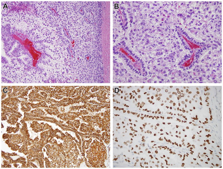
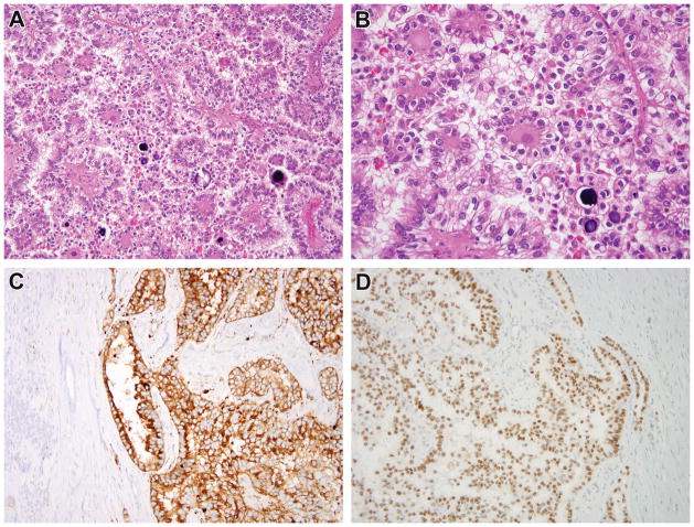
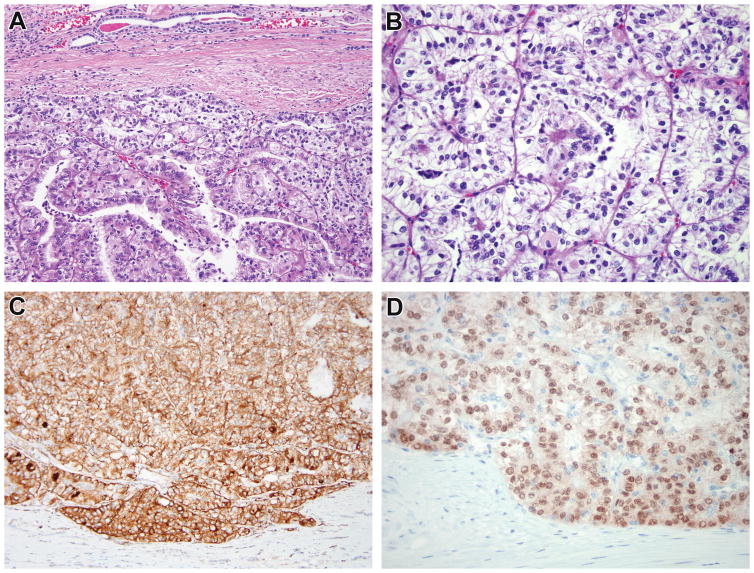
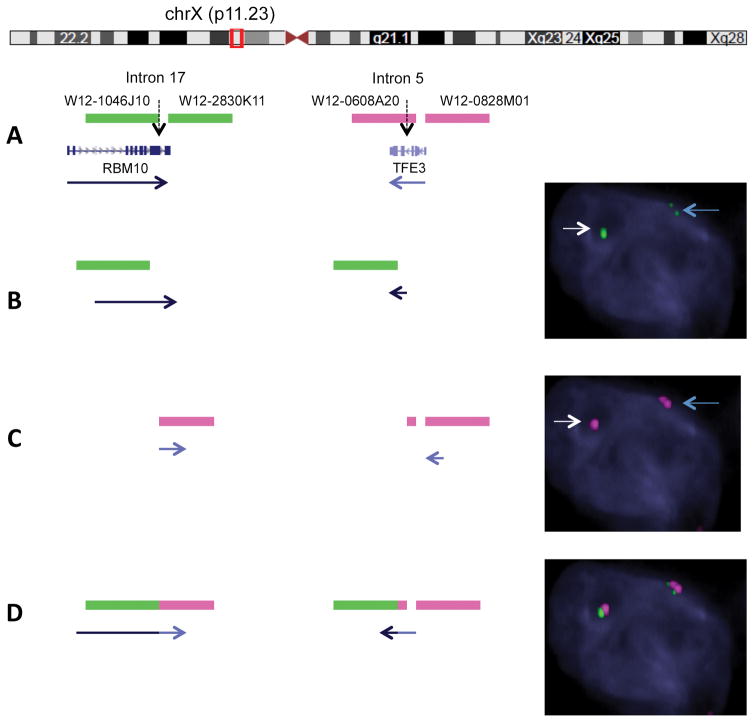
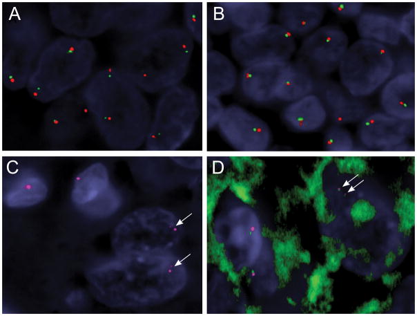
Xp11 translocation renal cell carcinoma (RCC) are defined by chromosome translocations involving the Xp11 breakpoint which results in one of a variety of TFE3 gene fusions. TFE3 break-apart florescence in situ hybridization (FISH) assays are generally preferred to TFE3 immunohistochemistry (IHC) as a means of confirming the diagnosis in archival material, as FISH is less sensitive to the variable fixation which can result in false positive or false negative IHC. Prompted by a case report in the cytogenetics literature, we identify 3 cases of Xp11 translocation RCC characterized by a subtle chromosomal inversion involving the short arm of the X chromosome, resulting in an RBM10-TFE3 gene fusion. TFE3 rearrangement was not detected by conventional TFE3 break-apart FISH, but was suggested by strong diffuse TFE3 immunoreactivity in a clean background. We then developed novel fosmid probes to detect the RBM10-TFE3 gene fusion in archival material. These cases validate RBM10-TFE3 as a recurrent gene fusion in Xp11 translocation RCC, illustrate a source of false-negative TFE3 break-apart FISH, and highlight the complementary role of TFE3 IHC and TFE3 FISH.


Figures

Figure 1
Figure 1
Case 1. This recurrent renal cell carcinoma demonstrates solid papillary architecture and epithelioid cells with clear cytoplasm (A, B). The neoplastic cells are diffusely immunoreactive for cathepsin k (C). While conventional TFE3 FISH did not demonstrate rearrangement, the neoplasm demonstrated diffuse, strong nuclear labeling for TFE3 protein with a clean background (D), prompting additional FISH fusion studies which demonstrated the RBM10-TFE3 gene fusion.
Figure 2
Figure 2
Case 2. The renal cell carcinoma demonstrates papillary architecture, clear cells, and abundant psammoma bodies, highly suggestive of Xp11 translocation RCC (A, B). The neoplasm was diffusely immunoreactive for cathepsin k (C), with surrounding kidney being appropriately negative. While conventional TFE3 FISH was negative for rearrangement, immunostain for TFE3 demonstrated diffuse strong nuclear staining in the neoplastic cells with absence of staining in the normal kidney (D), leading to additional FISH fusion studies which demonstrated the RBM10-TFE3 gene fusion.
Figure 3
Figure 3
Case 3. This renal cell carcinoma demonstrates nested and papillary architecture and epithelioid cells with clear to eosinophilic cytoplasm (A, B). A subpopulation of smaller cells is present within the lumens of acini. The neoplastic cells are diffusely immunoreactive for cathepsin k (C). While conventional TFE3 FISH did not demonstrate rearrangement, the neoplasm demonstrated diffuse, strong nuclear labeling for TFE3 protein with a clean background (D), prompting additional FISH fusion studies which demonstrated the RBM10-TFE3 gene fusion.
Figure 4
Figure 4
Fosmid FISH design and novel application to detect RBM10-TFE3 fusion. A. Diagrammatic representation of custom design of fosmid probes covering the likely breakpoints of RBM10 (2 green probes flanking intron 17) and TFE3 (2 red proves flanking intron 5) genes. Red box illustrates the near proximity (1.8 mb) of the 2 genes on chromosome X (Xp11.23 locus). B. Single color channel assay using the above design for RBM10 in a tumor cell reveals one intact green signal (wild type RBM10 allele, white arrow) and one split green signal into two half-sized fragments, in keeping with a RMB10 break and inversion event (small gap corresponds to the 1.8 mb distance, blue arrow). C. Similar single color assay for TFE3 showing one intact red probe (wild type TFE3 allele, white arrow) and one split signal into two smaller red fragments pieces (TFE3 break/inversion, blue arrow). D. Two-color fusion assay FISH showing one normal sized fused green-red signal due to close proximity of wild type RBM10 and TFE3 genes, and two pairs of split-fused smaller signals RBM10 (green) and TFE3 (red) in keeping with the RBM10-TFE3 fusion/inversion.
Figure 5
Figure 5
Identification of RMB10-TFE3 fusion/inversion by the custom fosmid FISH assay, which is not detected by the standard BAC FISH assay resolution. (Case 2) A,B. Conventional break-apart assay using typical flanking TFE3 probes (red, centromeric; green, telomeric) show small gaps in the tumor cells (A), compared to the normal lymphocytes adjacent to the tumor (B), but interpreted as a negative result in the clinical setting. However the small gaps may suggest the presence of a TFE3 cryptic inversion in the setting of strong TFE3 immunoreactivity and should trigger additional custom FISH assays to investigate a potential RMB10-TFE3 fusion (C, D). C. Single color channel FISH using 2 fosmids flanking the TFE3 breakpoint (red) shows two split smaller red signals in the 2 tumor cells (lower right, arrows), in contrast to 2 normal cells showing one intact red signal (upper left). D. Two-color fusion assay using custom fosmids showing 2 pairs of smaller red-green signals in keeping with a RBM10-TFE3 inversion in a tumor cell (upper right, arrows), compared to two normal cells showing only one pair of intact size red-green signals (lower left).

References

    1. Argani P, Antonescu CR, Illei PB, Lui MY, Timmons CF, Newbury R, Reuter VE, Garvin AJ, Perez-Atayde AR, Fletcher JA, Beckwith JB, Bridge JA, Ladanyi M. Primary renal neoplasms with the ASPL-TFE3 gene fusion of alveolar soft part sarcoma: A distinctive tumor entity previously included among renal cell carcinomas of children and adolescents. Am J Pathol. 2001;159:179–92. - PMC - PubMed
    1. Argani P, Antonescu CR, Couturier J, Fournet J, Sciot R, Debiec-Rychter M, Hutchinson B, Reuter VE, Boccon-Gibod L, Timmons C, Hafez N, Ladanyi M. PRCC-TFE3 renal carcinomas: Morphologic, immunohistochemical, ultrastructural, and molecular analysis of an entity associated with the t(X;1)(p11. 2;q21) Am J Surg Pathol. 2002;26:1553–66. - PubMed
    1. Clark J, Lu YJ, Sidhar SK, Parker C, Gill S, Smedley D, Hamoudi R, Linehan WM, Shipley J, Cooper CS. Fusion of splicing factor genes PSF and NonO (p54nrb) to the TFE3 gene in papillary renal cell carcinoma. Oncogene. 1997;15:2233–9. - PubMed
    1. Argani P, Lui MY, Couturier J, Bouvier R, Fournet J, Ladanyi M. A novel CLTC-TFE3 gene fusion in pediatric renal adenocarcinoma with t(X;17)(p11. 2;q23) Oncogene. 2003;22:5374–8. - PubMed
    1. Huang W, Goldfischer M, Babyeva S, Mao Y, Volyanskyy K, Dimitrova N, Fallon JT, Zhong M. Identification of a novel PARP14-TFE3 gene fusion from 10-year-old FFPE tissue by RNA-seq. Genes Chromosomes Cancer. 2015 May 29; doi: 10.1002/gcc.22261. Epub ahead of print. - DOI - PubMed

Publication types

MeSH terms