. 2017 Mar 15;12(3):e0171836.
doi: 10.1371/journal.pone.0171836. eCollection 2017.

Blue-light induced accumulation of reactive oxygen species is a consequence of the Drosophila cryptochrome photocycle

Affiliations

Blue-light induced accumulation of reactive oxygen species is a consequence of the Drosophila cryptochrome photocycle

Louis-David Arthaut et al. PLoS One. .

Abstract

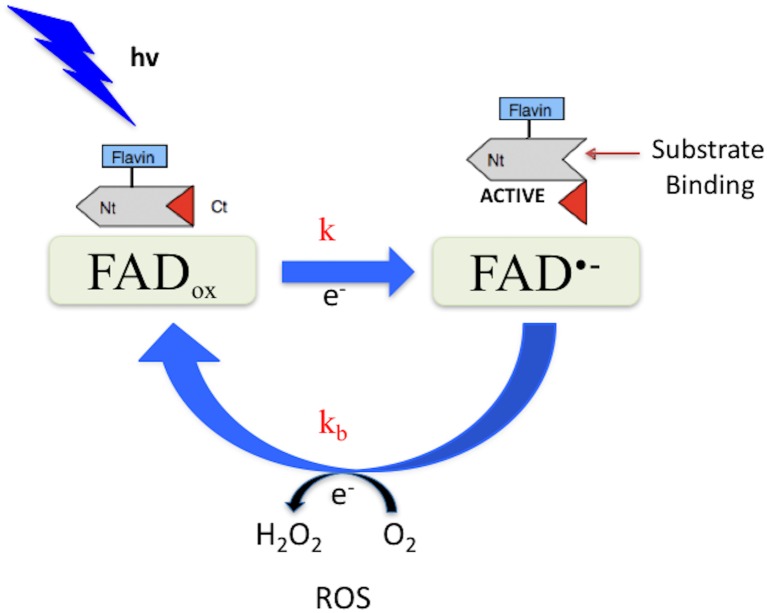
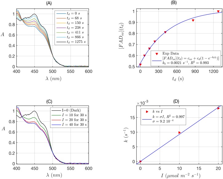
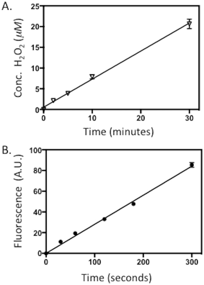
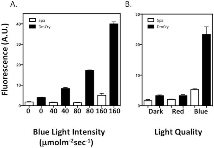
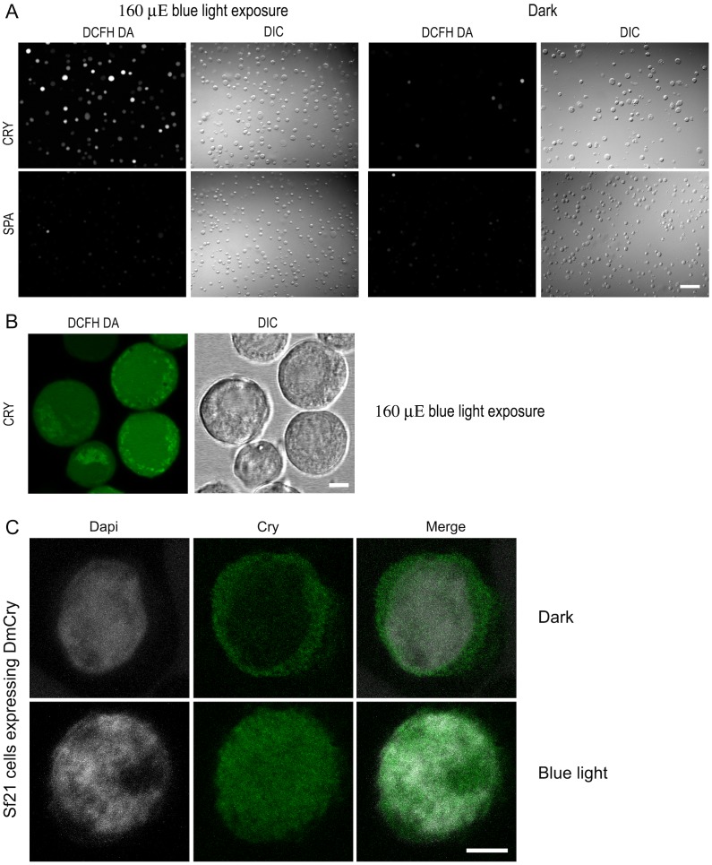
Cryptochromes are evolutionarily conserved blue-light absorbing flavoproteins which participate in many important cellular processes including in entrainment of the circadian clock in plants, Drosophila and humans. Drosophila melanogaster cryptochrome (DmCry) absorbs light through a flavin (FAD) cofactor that undergoes photoreduction to the anionic radical (FAD•-) redox state both in vitro and in vivo. However, recent efforts to link this photoconversion to the initiation of a biological response have remained controversial. Here, we show by kinetic modeling of the DmCry photocycle that the fluence dependence, quantum yield, and half-life of flavin redox state interconversion are consistent with the anionic radical (FAD•-) as the signaling state in vivo. We show by fluorescence detection techniques that illumination of purified DmCry results in enzymatic conversion of molecular oxygen (O2) to reactive oxygen species (ROS). We extend these observations in living cells to demonstrate transient formation of superoxide (O2•-), and accumulation of hydrogen peroxide (H2O2) in the nucleus of insect cell cultures upon DmCry illumination. These results define the kinetic parameters of the Drosophila cryptochrome photocycle and support light-driven electron transfer to the flavin in DmCry signaling. They furthermore raise the intriguing possibility that light-dependent formation of ROS as a byproduct of the cryptochrome photocycle may contribute to its signaling role.


Conflict of interest statement

Competing Interests: The authors have declared that no competing interests exist.

Figures

Fig 1
Fig 1. Possible Drosophila cryptochrome photocycle.
In the dark, the protein-bound cofactor (FADox) is shown in the oxidized redox state. Light absorption triggers flavin photoreduction [ – 14] at a rate constant k. Reoxidation to the FADox state occurs spontaneously in the dark at a rate constant kb. Possible changes in C-terminal conformation linked to redox state interconversion are diagrammed [15].
Fig 2
Fig 2. Rate constants and quantum yield for two-state reduction and reoxidation of DmCry.
(A) Isolated purified DmCry protein was illuminated for 30 s at I = 40 μmol m-2 s-1 blue light and placed in darkness (td = 0 s). Normalized absorption spectra are reported at increasing dark reoxidation times td. (B) Normalized concentration of FADox as a function of the dark reoxidation time td. The FADox concentration was obtained from the absorbance at 450nm from panel A according to Eq. S7 (see S1 Text). The red triangles represent the experimental data, and the blue curve is the fit of the experimental data with the two-states reoxidation model (Eq. S8). From the fit the reoxidation rate resulted kb = 0.0021 s-1, (half-life of τ1/2 = 5.5 min). The goodness of the fit was excellent (R2 ≈ 1). (C) Isolated purified DmCry protein was illuminated for 30 s at the indicated blue light fluence rates I. Normalized absorption spectra are presented. (D) Calculated forward rate constant k versus photon fluence rate I (red triangles). For each I, the rate constant k was calculated by numerically solving the two-states kinetic equations (see S1 Text, Eq. S1), with the concentration of FADox, obtained from panel C and reoxidation kb obtained from panel B. We fit k as function of I by using the linear equation k = σ I. The fit is reported as blue curve in Fig 2. From the fit the photo-conversion cross section was σ = 9.2 x 10⁻⁴ μmol-1 m2. From σ the quantum yield ϕ was calculated according to σ = 2.3 εox(450) ϕ, by using the experimentally calculated extinction coefficient εox(450) = 1130 mol-1 m2 (11300 M-1 cm-1). The quantum yield resulted ϕ = 0.35.
Fig 3
Fig 3. Formation of ROS by purified Drosophila Cryptochrome (DmCry).
30 μM DmCry protein was illuminated at saturating blue light intensity for the indicated times on ice. A. The concentration of H2O2 released in the sample after illumination was determined by the Amplex Red fluorescence detection (see Methods). B. ROS formation in the course of illumination assayed by DCFH-DA fluorescence (ex: 490/em:530) (see Methods). Error is SD of three measurements.
Fig 4
Fig 4. Induction of ROS in insect cell culture expressing DmCry.
A. Living insect cell cultures expressing either DmCry or a SPA1 control construct were treated with the fluorescent substrate DCFH-DA and then exposed to the indicated blue light intensity for 10 min. Subsequently to illumination, DmCry expressing and control cell cultures were harvested, lysed, and evaluated by fluorescence spectroscopy for the formation of ROS (see Methods). B. Cells exposed to dark, red, or blue light. Error bars represent SD of three measurements.
Fig 5
Fig 5. Subcellular localization of ROS and DmCRY in Sf21 insect cells exposed to blue light.
Living Sf21 cells stably expressing DmCRY were treated with DCFH-DA, exposed to dark or blue light and viewed by (A) a Zeiss AxioImager.Z1/ApoTome using a 10x objective (bar 100 μm) (B) an inverted Leica TCS SP5 microscope. Images show single confocal z section that cross the nucleus. Diffused fluorescent ROS staining can be seen in nucleus and cytoplasm. Punctuate and intense fluorescent ROS staining also colocalizes perfectly with ER (endoplasmic reticulum) surrounding the nucleus. Scale bar: 10 μm. (C) Sf21 stably expressing DmCry were fixed with paraformaldehyde, permeabilized with Triton X100, incubated with an anti—DmCry1 rabbit polyclonal antibody and an Alexa 488—conjugated anti—rabbit secondary antibody, DNA were stained with 4′,6′ — diamino—2—phenylindole (DAPI). Cells were observed with a Leica TCS SP5 confocal microscope. Images show projections of optical sections that cross the nucleus. Scale bar, 10 μm.

Similar articles

Cited by

References

    1. Chaves I, Pokorny R, Byrdin M, Hoang N, Ritz T, Brettel K, et al. The cryptochromes: blue light photoreceptors in plants and animals. Annual Review in Plant Biology. 2011; 62: 335–364. - PubMed
    1. Öztürk N, Song SH, Özgür S, Selby CP, Morrison L, Partch C et al. Structure and function of animal cryptochromes. Cold Spring Harbor Symposia on Quantitative Biology. 2007; 72: 119–131. 10.1101/sqb.2007.72.015 - DOI - PubMed
    1. Sancar A. Structure and function of DNA photolyase and cryptochrome blue light photoreceptors. Chemical Reviews. 2003; 103(6): 2203–2237. 10.1021/cr0204348 - DOI - PubMed
    1. Ahmad M. Photocycle and signaling mechanisms of plant cryptochromes. In SI: 33: Cell signalling and gene regulation. Current Opinion in Plant Biology. 2016; 33: 108–115. 10.1016/j.pbi.2016.06.013 - DOI - PubMed
    1. Peschel N, Helfrich-Förster C. Setting the clock—by nature: Circadian rhythm in the fruitfly Drosophila melanogaster. FEBS Letters. 2011; 585(10): 1435–1442. 10.1016/j.febslet.2011.02.028 - DOI - PubMed

LinkOut - more resources