Review
. 2017 Mar;5(2):204-218.
doi: 10.1111/andr.12320.

Molecular changes and signaling events occurring in spermatozoa during epididymal maturation

Affiliations
Review

Molecular changes and signaling events occurring in spermatozoa during epididymal maturation

M G Gervasi et al. Andrology. 2017 Mar.

Abstract

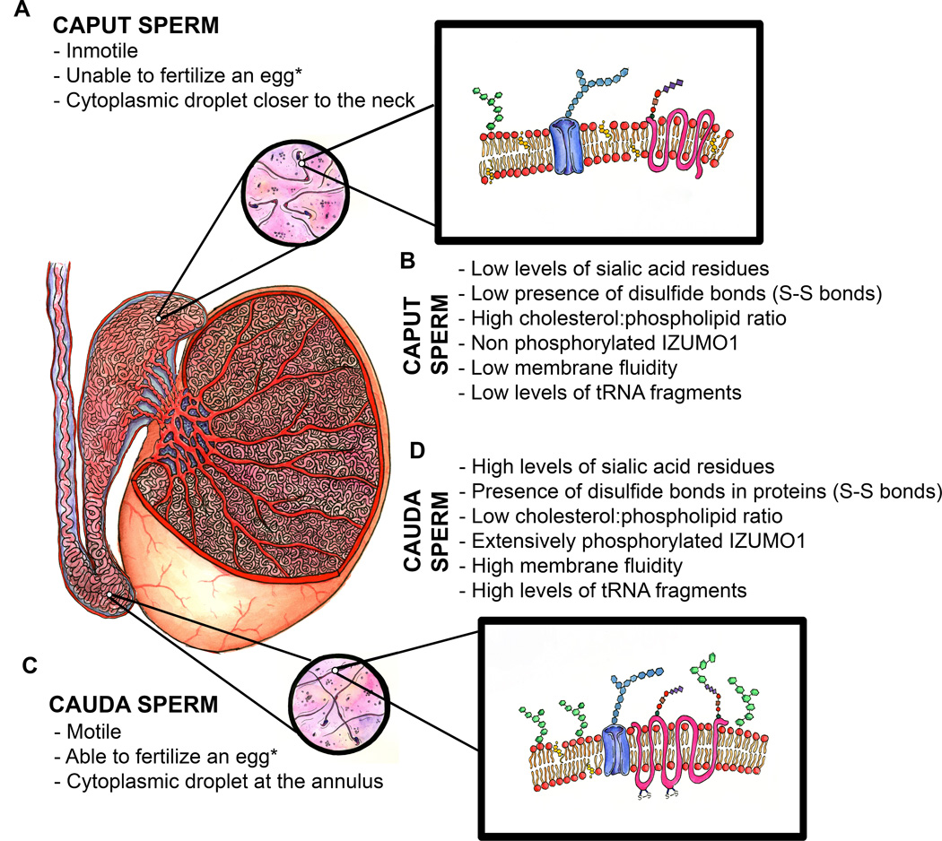
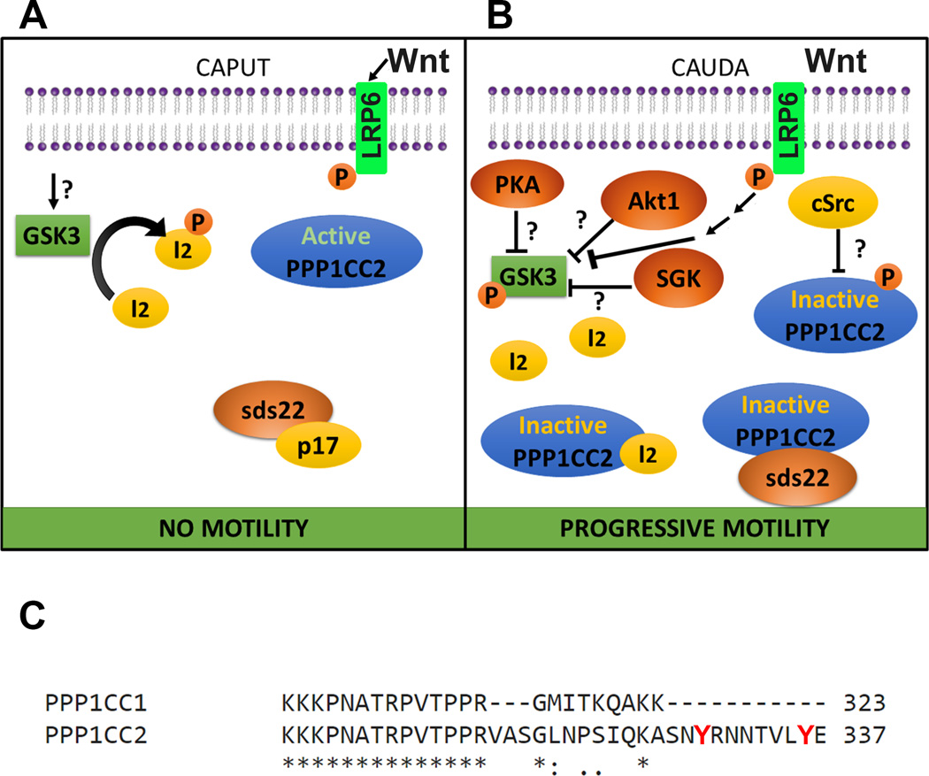
After leaving the testis, spermatozoa have not yet acquired the ability to move progressively and are unable to fertilize oocytes. To become fertilization competent, they must go through an epididymal maturation process in the male, and capacitation in the female tract. Epididymal maturation can be defined as those changes occurring to spermatozoa in the epididymis that render the spermatozoa the ability to capacitate in the female tract. As part of this process, sperm cells undergo a series of biochemical and physiological changes that require incorporation of new molecules derived from the epididymal epithelium, as well as post-translational modifications of endogenous proteins synthesized during spermiogenesis in the testis. This review will focus on epididymal maturation events, with emphasis in recent advances in the understanding of the molecular basis of this process.

Keywords: epididymis; epididymosomes; maturation; signal transduction; spermatozoa.


Conflict of interest statement

COMPETING FINANCIAL INTERESTS

The authors disclose no financial conflicts of interests.

Figures

Figure 1
Figure 1
Schematic representation of a mammalian testis, epididymis and sperm.* after incubation in conditions that support sperm capacitation. A) Principal morphological and functional characteristics of immature caput sperm. B) Molecular characteristics of immature caput sperm. C) Principal morphological and functional characteristics of mature cauda sperm. D) Molecular characteristics of mature cauda sperm.
Figure 2
Figure 2
Model of putative molecular pathways involved in the regulation of PPP1CC2 and progressive sperm motility during epididymal maturation. A) CAPUT: glycogen synthase kinase 3 (GSK3) is active and phosphorylates protein I2. Once phosphorylated, I2 is not able to bind to and inactivate the ser/thr phosphatase PPP1CC2. The PPP1CC2 inhibitory protein sds22 has been shown to be complexed with p17 and cannot bind and inhibit the phosphatase. cSrc kinase is absent from caput sperm, PPP1CC2 is active and caput sperm lack motility. Wnt from epididymosomes activates the primed receptors LRP6. B) CAUDA: GSK3 is phosphorylated on ser residues, and rendered inactive by an unidentified serine/threonine kinase. Among the proposed kinases involved in GSK3 phosphorylation and inactivation are protein kinase A (PKA), RACalpha protein kinase 1 (Akt1) and serum and glucocorticoid induced kinase (SGK). All these kinases have been described in sperm. Active Wnt signaling inhibits GSK3. Due to GSK3 inactivation, protein I2 can no longer undergo phosphorylation and consequently binds to PPP1CC2, inactivating its catalytic activity. On the other hand, sds22 has been found complexed to PPP1CC2 in cauda sperm and can explain PPP1CC2 inhibition. cSrc tyrosine kinase, not present in caput sperm, is incorporated into sperm during epididymal maturation. Because PPP1CC2 is a testis-specific splicing variant containing two unique tyrosine residues in its C-terminal domain, phosphorylation of these residues by cSrc or another tyrosine kinase would also explain PPP1CC2 inactivation. Together, inactivation of PPP1CC2 leads to the ability of the sperm cell to move progressively when exposed to an appropriate medium. C) ClustalW2 sequence alignment of the human protein phosphatases PPP1CC1 and PPP1CC2. These two proteins are products of an alternative splicing and differ only in a small region of the C-terminal, as shown in the Figure. The two unique tyrosine residues (Y) in PPP1CC2 are indicated in red.

References

    1. Aitken RJ, Nixon B. Sperm capacitation: a distant landscape glimpsed but unexplored. Mol Hum Reprod. 2013;19:785–793. - PubMed
    1. Akintayo A, Legare C, Sullivan R. Dicarbonyl L-xylulose reductase (DCXR), a “moonlighting protein” in the bovine epididymis. PLoS One. 2015;10:e0120869. - PMC - PubMed
    1. Alessi DR, James SR, Downes CP, Holmes AB, Gaffney PR, Reese CB, Cohen P. Characterization of a 3-phosphoinositide-dependent protein kinase which phosphorylates and activates protein kinase Balpha. Curr Biol. 1997;7:261–269. - PubMed
    1. Andersen OM, Yeung CH, Vorum H, Wellner M, Andreassen TK, Erdmann B, Mueller EC, Herz J, Otto A, Cooper TG, Willnow TE. Essential role of the apolipoprotein E receptor-2 in sperm development. J Biol Chem. 2003;278:23989–23995. - PubMed
    1. Arya M, Vanha-Perttula T. Lectin-binding pattern of bull testis and epididymis. J Androl. 1985;6:230–242. - PubMed

Publication types

LinkOut - more resources