. 2017 Mar;6(3):799-806.
doi: 10.5966/sctm.2015-0365. Epub 2016 Oct 5.

Surgical Excision of Heterotopic Ossification Leads to Re-Emergence of Mesenchymal Stem Cell Populations Responsible for Recurrence

Affiliations

Surgical Excision of Heterotopic Ossification Leads to Re-Emergence of Mesenchymal Stem Cell Populations Responsible for Recurrence

Shailesh Agarwal et al. Stem Cells Transl Med. 2017 Mar.

Abstract

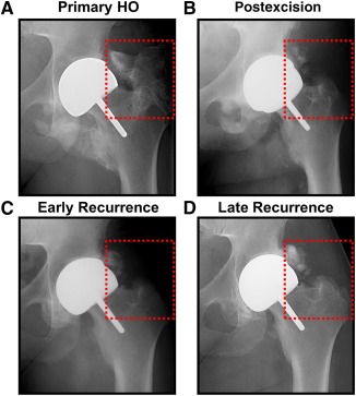
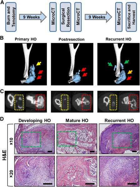
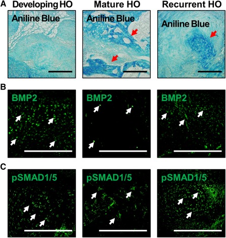
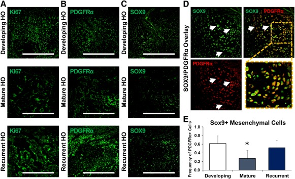
Trauma-induced heterotopic ossification (HO) occurs after severe musculoskeletal injuries and burns, and presents a significant barrier to patient rehabilitation. Interestingly, the incidence of HO significantly increases with repeated operations and after resection of previous HO. Treatment of established heterotopic ossification is challenging because surgical excision is often incomplete, with evidence of persistent heterotopic bone. As a result, patients may continue to report the signs or symptoms of HO, including chronic pain, nonhealing wounds, and joint restriction. In this study, we designed a model of recurrent HO that occurs after surgical excision of mature HO in a mouse model of hind-limb Achilles' tendon transection with dorsal burn injury. We first demonstrated that key signaling mediators of HO, including bone morphogenetic protein signaling, are diminished in mature bone. However, upon surgical excision, we have noted upregulation of downstream mediators of osteogenic differentiation, including pSMAD 1/5. Additionally, surgical excision resulted in re-emergence of a mesenchymal cell population marked by expression of platelet-derived growth factor receptor-α (PDGFRα) and present in the initial developing HO lesion but absent in mature HO. In the recurrent lesion, these PDGFRα+ mesenchymal cells are also highly proliferative, similar to the initial developing HO lesion. These findings indicate that surgical excision of HO results in recurrence through similar mesenchymal cell populations and signaling mechanisms that are present in the initial developing HO lesion. These results are consistent with findings in patients that new foci of ectopic bone can develop in excision sites and are likely related to de novo formation rather than extension of unresected bone. Stem Cells Translational Medicine 2017;6:799-806.

Keywords: Bone; Chondrogenesis; Extremity trauma; Heterotopic ossification; Mesenchymal stem cell; Tissue regeneration.


Figures

Figure 1
Figure 1
Clinical scenario of recurrent HO in a patient with total hip arthroplasty. (A): Primary HO in total hip arthroplasty patient (red square). (B): Postexcision radiograph showing substantial reduction in HO with some residual lesions. (C): Early evidence of recurrence after excision of HO. (D): Recurrent HO lesion 6 months after surgery at excision site. Abbreviation: HO, heterotopic ossification.
Figure 2
Figure 2
Recurrent HO following surgical excision. (A): Experimental design to develop recurrence. (B): Three‐dimensional microCT reconstructions showing images immediately prior to excision, immediately following excision, and recurrence 9 weeks after excision. Green arrows = site of de novo recurrent HO; orange arrow = site of HO developing from remnant HO; red arrows = site of original unresected HO; yellow arrow = region of resected HO. (C): MicroCT cross‐sections of HO lesions. Red box = site of unresected HO; yellow box = site of resected HO. (D): H&E staining showing architecture of early HO 3 weeks after injury, mature HO 9 weeks after injury, and recurrent HO 9 weeks after excision. Green box = site of HO. Abbreviations: H&E, hematoxylin and eosin; HO, heterotopic ossification; microCT, micro‐computed tomography.
Figure 3
Figure 3
Osteoid and BMP signaling characteristics of recurrent HO. (A): Aniline blue staining shows presence of mature osteoid 9 weeks after HO, and emerging aniline blue staining in both early HO and developing recurrent HO lesions. Red arrows = sites of osteoid. (B): BMP2 expression is elevated in the developing HO lesion, reduced in mature osteoid 9 weeks after injury, and recurs in the recurrent HO lesion. (C): pSMAD 1/5 expression is elevated in the developing HO lesion, reduced in mature osteoid 9 weeks after injury, and recurs in the recurrent HO lesion. White arrows = representative sites of positive staining. Scale bar = 200 µm. Abbreviations: BMP2, bone morphogenetic protein‐2; HO, heterotopic ossification.
Figure 4
Figure 4
New cartilage in recurrent lesions undergoes de novo endochondral ossification. (A): Safranin O and COLIIA staining shows cartilaginous extracellular matrix in the developing and recurrent HO lesions, whereas mature HO shows marrow and osteoid with minimal cartilage presence. (B): Hypertrophic chondrocytes (white arrows) labeled by COLX immunostaining are present in developing HO and recurrent HO, but relatively diminished in mature HO. (C): Representative image demonstrating the overlap of COLIIA and COLX in hypertrophic chondrocytes (white arrows) present in areas of cartilage actively undergoing endochondral ossification. (D): Quantification of COLX+ hypertrophic chondrocytes in the cartilage anlagen of developing, mature, and recurrent HO. All values are normalized to developing HO. ∗, p > .05. Error bars represent standard deviation. Scale bars = 200 µm. Abbreviations: COLIIA, collagen II a; COLX, collagen X; HO, heterotopic ossification; HPF, high‐power field.
Figure 5
Figure 5
Contributing mesenchymal cell populations emerge in recurrent HO and undergo de novo chondrogenic differentiation. (A): Mesenchymal cell populations in the developing and recurrent HO are proliferative on the basis of Ki67 immunostaining. (B): Immunostaining for PDGFRα shows the presence of mesenchymal cells in developing HO and recurrent HO, with relative absence in mature HO. (C): Immunostaining for SOX9 shows presence of cells undergoing chondrogenic differentiation in developing HO and recurrent HO, with relative absence in mature HO. (D): Representative image demonstrating the overlap of SOX9 and PDGFRα (white arrows) in areas of mesenchymal cells actively undergoing chondrogenic differentiation. (E): Quantification of SOX9/PDGFRα costaining mesenchymal cells in developing, mature, and recurrent HO. ∗, p > .05. Error bars represent standard deviation. Scale bars = 200 µm. Abbreviations: HO, heterotopic ossification; PDGFRα, platelet‐derived growth factor receptor‐α.

References

    1. Levi B, Jayakumar P, Giladi A et al. Risk factors for the development of heterotopic ossification in seriously burned adults: A National Institute on Disability, Independent Living and Rehabilitation Research burn model system database analysis. J Trauma Acute Care Surg 2015;79:870–876. - PMC - PubMed
    1. Peterson JR, De La Rosa S, Sun H et al. Burn injury enhances bone formation in heterotopic ossification model. Ann Surg 2014;259:993–998. - PMC - PubMed
    1. Peterson JR, Okagbare PI, De La Rosa S et al. Early detection of burn induced heterotopic ossification using transcutaneous Raman spectroscopy. Bone 2013;54:28–34. - PMC - PubMed
    1. Potter BK, Forsberg JA, Davis TA et al. Heterotopic ossification following combat‐related trauma. J Bone Joint Surg Am 2010;92 (suppl): 274–89. - PubMed
    1. Peterson JR, De La Rosa S, Eboda O et al. Treatment of heterotopic ossification through remote ATP hydrolysis. Sci Transl Med 2014;6:255ra132. - PMC - PubMed

Publication types

Substances