. 2017 Mar;6(3):1006-1017.
doi: 10.5966/sctm.2016-0054. Epub 2016 Oct 18.

Oncostatin M-Preconditioned Mesenchymal Stem Cells Alleviate Bleomycin-Induced Pulmonary Fibrosis Through Paracrine Effects of the Hepatocyte Growth Factor

Affiliations

Oncostatin M-Preconditioned Mesenchymal Stem Cells Alleviate Bleomycin-Induced Pulmonary Fibrosis Through Paracrine Effects of the Hepatocyte Growth Factor

Ying-Wei Lan et al. Stem Cells Transl Med. 2017 Mar.

Abstract

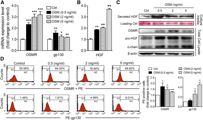
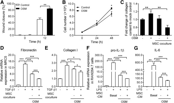
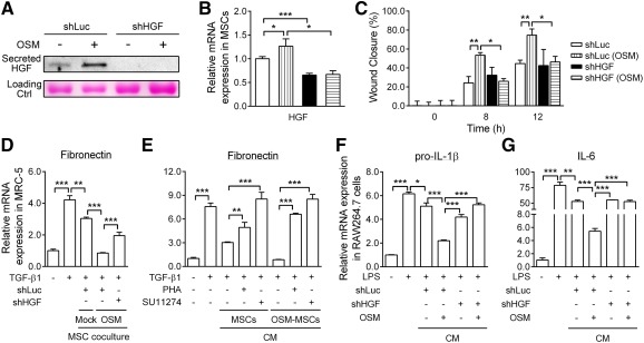
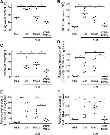
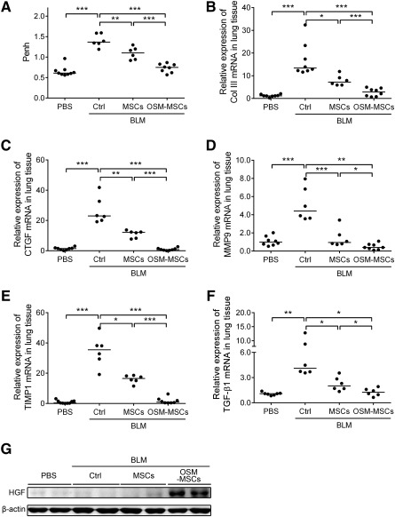
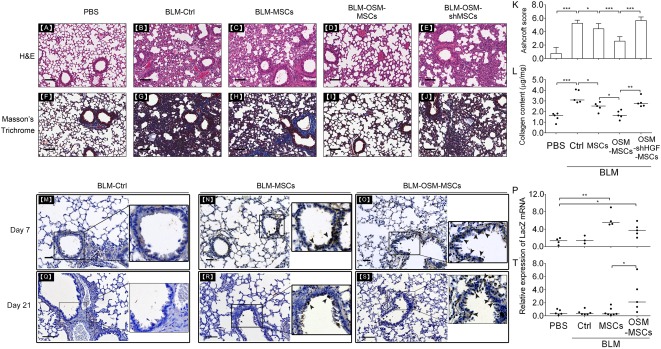
Mesenchymal stem cells (MSCs) are widely considered for treatment of pulmonary fibrosis based on the anti-inflammatory, antifibrotic, antiapoptotic, and regenerative properties of the cells. Recently, elevated levels of oncostatin M (OSM) have been reported in the bronchoalveolar lavage fluid of a pulmonary fibrosis animal model and in patients. In this work, we aimed to prolong engrafted MSC survival and to enhance the effectiveness of pulmonary fibrosis transplantation therapy by using OSM-preconditioned MSCs. OSM-preconditioned MSCs were shown to overexpress type 2 OSM receptor (gp130/OSMRβ) and exhibited high susceptibility to OSM, resulting in upregulation of the paracrine factor, hepatocyte growth factor (HGF). Moreover, OSM-preconditioned MSCs enhanced cell proliferation and migration, attenuated transforming growth factor-β1- or OSM-induced extracellular matrix production in MRC-5 fibroblasts through paracrine effects. In bleomycin-induced lung fibrotic mice, transplantation of OSM-preconditioned MSCs significantly improved pulmonary respiratory functions and downregulated expression of inflammatory factors and fibrotic factors in the lung tissues. Histopathologic examination indicated remarkable amelioration of the lung fibrosis. LacZ-tagged MSCs were detected in the lung tissues of the OSM-preconditioned MSC-treated mice 18 days after post-transplantation. Taken together, our data further demonstrated that HGF upregulation played an important role in mediating the therapeutic effects of transplanted OSM-preconditioned MSCs in alleviating lung fibrosis in the mice. Stem Cells Translational Medicine 2017;6:1006-1017.

Keywords: Bleomycin-induced pulmonary fibrosis; Hepatocyte growth factor; Oncostatin M; Preconditioning; Stem cells.


Figures

Figure 1
Figure 1
Oncostatin M preconditioning upregulates expression of OSM receptor complex and hepatocyte growth factor in mesenchymal stem cells. Real‐time polymerase chain reaction quantification is shown of the relative expression levels of mRNAs of OSM receptor and gp130 (A) and HGF (B) in MSCs treated with different dosages of OSM for 24 hours. Values were normalized to glyceraldehyde‐3‐phosphate dehydrogenase and are expressed in relation to the respective control group. (C): Western blot analysis of OSMR and HGF in total cell lysates and secreted HGF in culture media. Ponceau S staining of the membrane was presented (row 2) as the loading control. (D): Flow cytometry analysis of the percentage of MSCs positive for OSMRβ and gp130. The quantitative histogram on the right shows the data of three independent MSC samples of phycoerythrin‐positive cells. ∗, p < .05; ∗∗, p < .01; ∗∗∗, p < .001. Abbreviations: Ctrl, control; HGF, hepatocyte growth factor; OSM, oncostatin M; OSMR, OSM receptor; PE, phycoerythrin.
Figure 2
Figure 2
Oncostatin M preconditioning enhances mesenchymal stem cell proliferation, increases wound healing, attenuates inflammatory effects, and decreases extracellular matrix production in fibroblast cells. (A): Scratch wound assay of the paracrine effect of OSM‐MSCs on MRC‐5 fibroblast migration. Quantification of wound closure was assessed by TScratch software after 12 hours. (B): Proliferation of MSCs under serum‐starved condition and treated with 2 ng/ml of OSM for 24 and 48 hours; cell number was counted by a hemocytometer. (C): Inhibitory effects of MSCs on collagen production. Cocultured MSCs (upper chamber) and MRC‐5 (lower chamber) were simultaneously incubated with OSM for 24 hours, followed by measurement of collagen production with Sirius Red/Fast Green Collagen detection kit. (D, E): To assess the antifibrotic effects of MSCs, we seeded untreated MSCs or OSM‐MSCs in the upper chamber, and transforming growth factor‐β1‐treated MRC‐5 cells were seeded in the lower chamber in the coculture experiments. After 24‐hour incubation, quantitative real‐time reverse transcription‐polymerase chain reaction (RT‐qPCR) was performed to determine the mRNA levels of fibronectin and collagen type I in the cells. (F, G): To assess the anti‐inflammatory effects, we incubated lipopolysaccharides (10 ng/ml)‐treated RAW264.7 cells simultaneously with a mixture of RAW264.7 basal media and OSM‐MSC‐conditioned medium or MSC basal media (Dulbecco's modified Eagle's medium/Ham's nutrient mixture F‐12; control) (1:1) for 7 hours. RT‐qPCR was performed to examine changes in the pro‐interleukin (IL)‐1β and IL‐6 mRNA levels in the cells. Values were normalized to the glyceraldehyde‐3‐phosphate dehydrogenase gene and are expressed in relation to the control group. ∗, p < .05; ∗∗, p < .01; ∗∗∗, p < .001. Abbreviations: CM, conditioned medium; h, hour; IL, interleukin; LPS, lipopolysaccharides; MSC, mesenchymal stem cell; OSM, oncostatin M; TGF, transforming growth factor.
Figure 3
Figure 3
Hepatocyte growth factor secreted in oncostatin M‐preconditioned mesenchymal stem cells attenuates wound healing, extracellular matrix (ECM) production, and inflammatory effects. The HGF‐specific gene is silenced by lentivirus‐mediated shRNA. (A): Western blot analysis of the levels of secreted HGF in conditioned medium and (B) quantitative real‐time reverse transcription‐polymerase chain reaction (RT‐qPCR) quantification of HGF mRNA levels in cells of the specific HGF knockdown (shHGF‐MSC) under OSM treatments. shLuc‐MSCs are transduced with a scrambled control. Ponceau S staining of the membrane was used as loading control. (C): Scratch wound assay of the effect of HGF on MRC‐5 migration. Quantification of wound closure was assessed by TScratch software after 8 and 12 hours. (D): Effects of HGF silencing in MSCs on the collagen production. Cocultured OSM‐pretreated shLuc‐ or shHGF‐MSCs (upper chamber) and transforming growth factor‐β1‐treated MRC‐5 (lower chamber) were performed. After 24 hours of incubation, fibronectin mRNA levels were determined. (E): Effects of inhibiting HGF signaling with a c‐Met inhibitor on collagen production. MRC‐5 cells were preincubated with PHA‐665752 or SU11274. The cells were then treated with MSCs or OSM‐MSC‐conditioned medium for 24 hours, followed by determination of fibronectin mRNA levels. (F, G): Effects of HGF silencing in MSCs on proinflammatory cytokine production, lipopolysaccharides‐treated RAW264.7 cells simultaneously incubated with a mixture of RAW264.7 basal media, and OSM‐pretreated shLuc‐ or shHGF‐MSCs CM or MSCs basal media (1:1). After 7 hours of incubation, RT‐qPCR was performed to examine the pro‐interleukin‐1β and IL‐6 mRNA levels in the cells. Values were normalized to the glyceraldehyde‐3‐phosphate dehydrogenase gene and are expressed in relation to the control group. ∗, p < .05; ∗∗, p < .01; ∗∗∗, p < .001. Abbreviations: CM, conditioned medium; Ctrl, control; h, hour; HGF, hepatocyte growth factor; IL, interleukin; LPS, lipopolysaccharides; MSC, mesenchymal stem cell; OSM, oncostatin M; PHA, PHA‐665752; shHGF, HGF knockdown; TGF, transforming growth factor.
Figure 4
Figure 4
Transplantation of oncostatin M‐preconditioned mesenchymal stem cells attenuates lung edema and bronchoalveolar lavages fluid differential cell counts and downregulates the expression of inflammatory mediators in the early‐stage of a bleomycin‐induced pulmonary fibrosis mouse model. (A): Effects of OSM‐MSC treatment on pulmonary edema. The lung water content (in percent) was determined at day 7 post‐BLM treatment. After collecting BALF, the total cells in BALF were counted by a hemocytometer, and cell viability was measured by trypan blue exclusion (B) and differential cells in BALF were counted by flow‐cytometric hematology system (XT‐2000iV) (C). Then the lung tissues were collected for inflammatory‐related genes, pro‐interleukin‐1β (D), IL‐6 (E), and OSM mRNA (F) expression level detection by quantitative real‐time polymerase chain reaction. Values were normalized to the glyceraldehyde‐3‐phosphate dehydrogenase gene and expressed in relation to the phosphate‐buffered saline group. Each dot represents an individual mouse with the mean shown for n ≥ 5 per group. ∗, p < .05; ∗∗, p < .01; ∗∗∗, p < .001. Abbreviations: BALF, bronchoalveolar lavages fluid; BLM, bleomycin; Ctrl, control; IL, interleukin; MSC, mesenchymal stem cell; OSM, oncostatin M; PBS, phosphate‐buffered saline.
Figure 5
Figure 5
Transplantation of oncostatin M‐preconditioned mesenchymal stem cells improves lung function and downregulates the expression of fibrosis‐related genes in the late‐stage of bleomycin‐induced pulmonary fibrosis mouse model. (A): Effects of OSM‐MSCs on pulmonary function. Lung functions were detected as Penh values in animals that received phosphate‐buffered saline (PBS), BLM‐control, BLM‐MSCs, or BLM‐OSM‐MSCs for 18 days following BLM administration. Whole‐body plethysmograph was used, and baseline‐enhanced respiratory pause was used as a noninvasive index of airway dysfunction. Then the lung tissues were collected for fibrotic indicators, Col III (B), connective tissue growth factor (C), matrix metalloproteinase‐9 (D), TIMP1 (E), and transforming growth factor‐β1 (F) mRNA expression level detection by quantitative real‐time reverse transcription‐polymerase chain reaction. Values are normalized to the glyceraldehyde‐3‐phosphate dehydrogenase gene and are expressed in relation to the PBS group. Each dot represents an individual mouse with the mean shown for n > 5 per group. (G): Western blot analysis of HGF in whole lung tissues from each group of mice, treated as described above. ∗, p < .05; ∗∗, p < .01; ∗∗∗, p < .001. Abbreviations: BLM, bleomycin; Col III, collagen III; CTGF, connective tissue growth factor; Ctrl, control; HGF, hepatocyte growth factor; MMP9, matrix metalloproteinase‐9; MSC, mesenchymal stem cell; OSM, oncostatin M; PBS, phosphate‐buffered saline; Penh, baseline‐enhanced respiratory pause; TGF, transforming growth factor; TIMP1, tissue inhibitor of metalloproteinase 1.
Figure 6
Figure 6
Transplantation of oncostatin M (OSM)‐preconditioned mesenchymal stem cells (MSCs) reduces fibrotic histopathologic changes through hepatocyte growth factor (HGF) signaling in the bleomycin (BLM)‐induced pulmonary fibrosis mouse model. Representative images at day 21 after BLM administration of hematoxylin and eosin (A‐E) and Masson’s trichrome‐stained (F‐J) histological sections from each group. (K): The fibrotic changes in the lung were quantified by using Ashcroft scores, ranging from 0 (normal lung) to 8 (complete fibrosis). (L): Total collagen content of whole lung tissues from each group was determined by Sircol Collagen Assay. Immunohistochemistry staining was performed to observe the distribution of LacZ in lung tissues at day 7 (M‐O) and day 21 (Q‐S) post‐BLM treatments. Quantitative real‐time polymerase chain reaction was performed to detect LacZ expression in the lung tissues of mice that received phosphate‐buffered saline (PBS), BLM‐control, BLM‐MSCs, and BLM‐OSM‐MSCs on day 7 (P) and day 21 (T) after BLM administration. (A, F): PBS control. (B, G, M, Q): BLM + PBS. (C, H, N, R): BLM + MSCs. (D, I, O, S): BLM + OSM‐MSCs. (E, J): BLM + OSM‐preconditioned HGF knockdown MSCs. Scale bar, 100 μm. Solid arrows around the bronchi indicate LacZ‐positive cells (brown spots). Values are normalized to the glyceraldehyde‐3‐phosphate dehydrogenase values and expressed in relation to the PBS group. Each dot represents an individual mouse with the mean shown for n ≥ 3 per group. ∗, p < .05; ∗∗, p < .01; ∗∗∗, p < .001. Abbreviations: BLM, bleomycin; Ctrl, control; H&E, hematoxylin and eosin; MSC, mesenchymal stem cell; OSM, oncostatin M; PBS, phosphate‐buffered saline; shHGF, HGF knockdown.

Similar articles

Cited by

References

    1. Ji X, Wu B, Fan J et al. The anti‐fibrotic effects and mechanisms of microRNA‐486‐5p in pulmonary fibrosis. Sci Rep 2015;5:14131–14142. - PMC - PubMed
    1. Spagnolo P. Novel treatments for idiopathic pulmonary fibrosis. Am J Med 2015;128:447–449. - PubMed
    1. Antunes MA, Laffey JG, Pelosi P et al. Mesenchymal stem cell trials for pulmonary diseases. J Cell Biochem 2014;115:1023–1032. - PubMed
    1. Glassberg MK, Toonkel RL. Moving stem cell therapy to patients with idiopathic pulmonary fibrosis. Respirology 2014;19:950–951. - PubMed
    1. Yang J, Jia Z. Cell‐based therapy in lung regenerative medicine. Regen Med Res 2014;2:7–13. - PMC - PubMed

Publication types