. 2017 Mar 23;543(7646):573-576.
doi: 10.1038/nature21671. Epub 2017 Mar 15.

RNA m6A methylation regulates the ultraviolet-induced DNA damage response

Affiliations

RNA m6A methylation regulates the ultraviolet-induced DNA damage response

Yang Xiang et al. Nature. .

Erratum in

Abstract

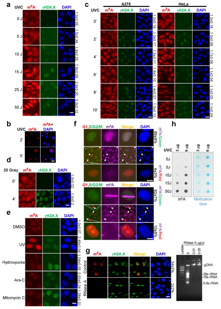
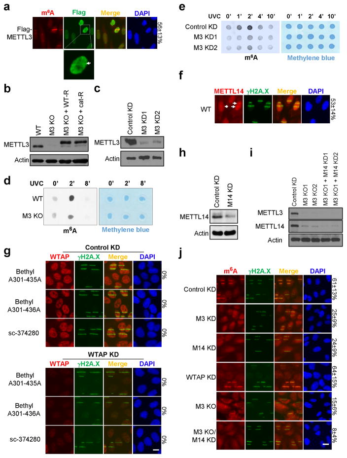
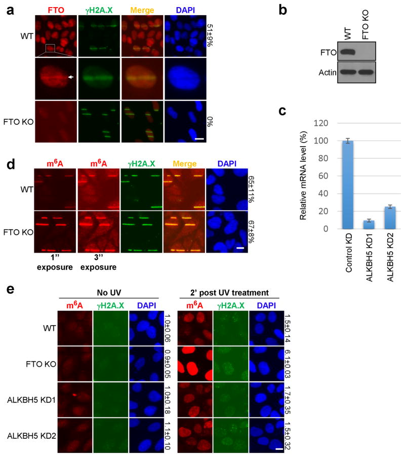
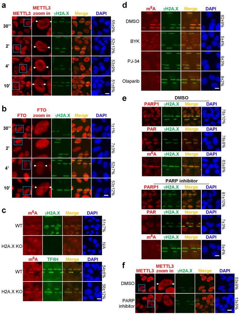
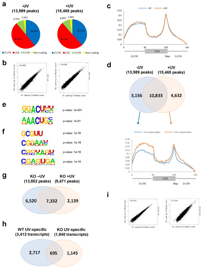
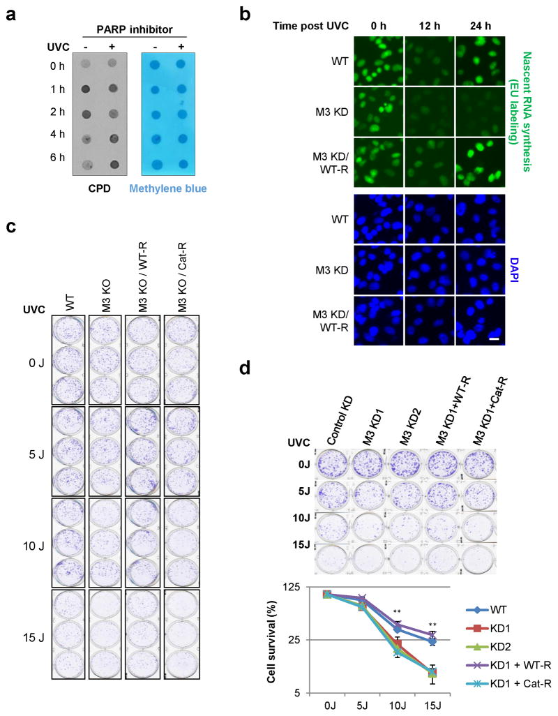
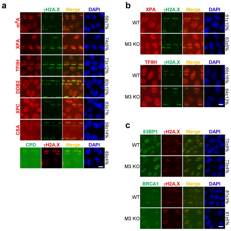
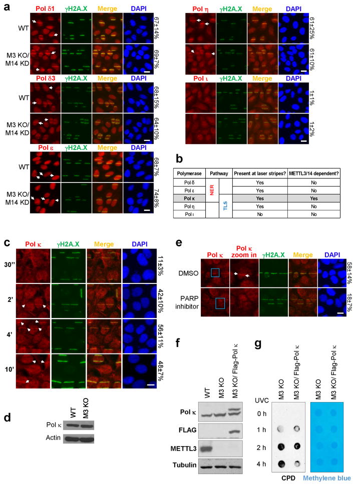
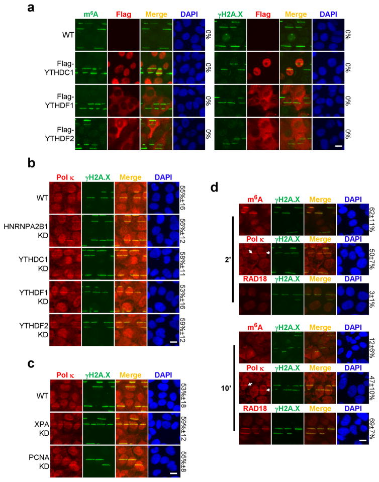
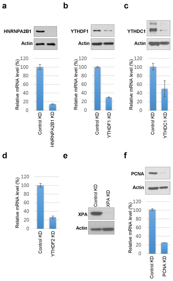
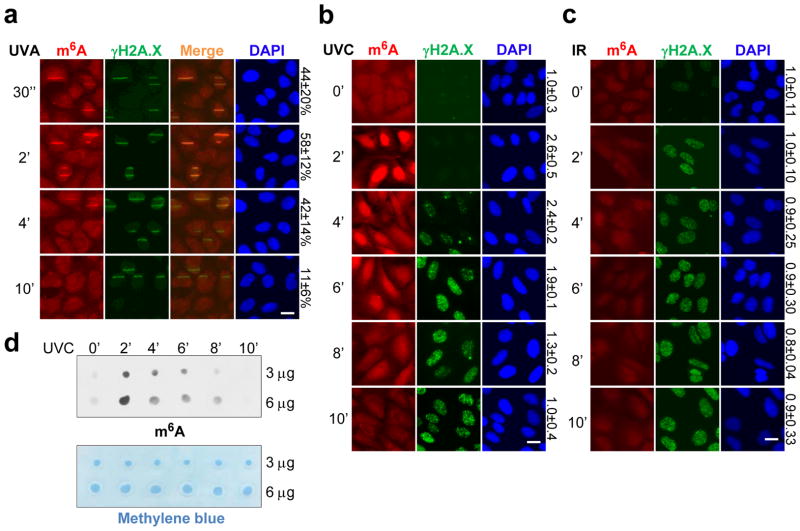
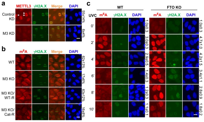
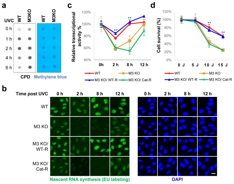
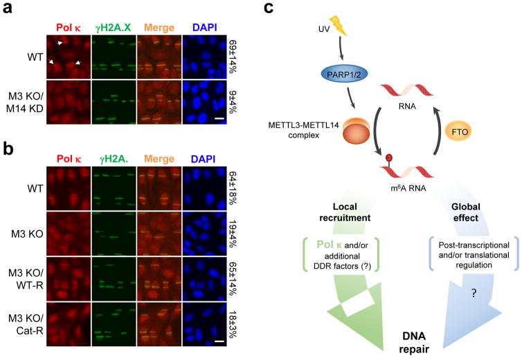
Cell proliferation and survival require the faithful maintenance and propagation of genetic information, which are threatened by the ubiquitous sources of DNA damage present intracellularly and in the external environment. A system of DNA repair, called the DNA damage response, detects and repairs damaged DNA and prevents cell division until the repair is complete. Here we report that methylation at the 6 position of adenosine (m6A) in RNA is rapidly (within 2 min) and transiently induced at DNA damage sites in response to ultraviolet irradiation. This modification occurs on numerous poly(A)+ transcripts and is regulated by the methyltransferase METTL3 (methyltransferase-like 3) and the demethylase FTO (fat mass and obesity-associated protein). In the absence of METTL3 catalytic activity, cells showed delayed repair of ultraviolet-induced cyclobutane pyrimidine adducts and elevated sensitivity to ultraviolet, demonstrating the importance of m6A in the ultraviolet-responsive DNA damage response. Multiple DNA polymerases are involved in the ultraviolet response, some of which resynthesize DNA after the lesion has been excised by the nucleotide excision repair pathway, while others participate in trans-lesion synthesis to allow replication past damaged lesions in S phase. DNA polymerase κ (Pol κ), which has been implicated in both nucleotide excision repair and trans-lesion synthesis, required the catalytic activity of METTL3 for immediate localization to ultraviolet-induced DNA damage sites. Importantly, Pol κ overexpression qualitatively suppressed the cyclobutane pyrimidine removal defect associated with METTL3 loss. Thus, we have uncovered a novel function for RNA m6A modification in the ultraviolet-induced DNA damage response, and our findings collectively support a model in which m6A RNA serves as a beacon for the selective, rapid recruitment of Pol κ to damage sites to facilitate repair and cell survival.


Conflict of interest statement

Competing financial interests

Y.S. is a cofounder of Constellation Pharmaceuticals and a member of its scientific advisory board, and a consultant for Active Motif, Inc.

Figures

Extended Data Figure 1
Extended Data Figure 1. 6A-modified RNA accumulates at damage sites in response to UV irradiation
a, U2OS cells were subjected to the indicated doses of UVC irradiation, incubated at 37°C for 2 min, and costained for m6A and γH2A.X. Relative m6A intensity is indicated on the right. b, U2OS cells were subjected to 50 J UVC irradiation through a micropore filter, incubated at 37°C for 2 or 5 min, and costained for m6A and DNA (DAPI). c, A375 (melanoma) or HeLa cells were subjected or not (0′) to 25 J UVC irradiation, incubated at 37°C for the indicated times, and then costained for m6A and γH2A.X. d, U2OS cells were subjected or not (0′) to 20 Gray of γ-irradiation, incubated at 37°C for 4 min, then costained for m6A and γH2A.X. e, U2OS cells were either irradiated with 25 J UVC or treated with DMSO, mitomycin C, hydroxyurea, or arabinoside-C, then costained for m6A and γH2A.X. c–e, relative m6A intensity is indicated on the right. f, Fluorescent Ubiquitination-based Cell Cycle Indicator (FUCCI) cells were subjected to 25 J UVC irradiation (top panel) or microirradiation by UVA laser (bottom panel), incubated at 37°C for 2 min, and stained for m6A. Top and bottom panels show m6A signal in representative S/G2/M or G1 phase cells, respectively. Arrows in middle panels denote representative G1-phase cells negative for m6A signal. The percentage of G1 (red) or S/G2/M (green) cells positive for m6A signal is indicated on the right. g, U2OS cells were microirradiated by UVA laser, permeabilized and treated with or without RNase A, and costained for m6A and γH2A.X (left panel). The percentage of γH2A.X-positive cells displaying colocalizing m6A signal is indicated. Nucleic acids (DNA and RNA) from cells treated with or without RNase A were isolated and analyzed on an agarose gel (right panel). Ladder (1kb DNA ladder). h, Poly(A)+ RNA was extracted from the samples in (a), and subjected to dot-blot analysis with an antibody recognizing m6A. Methylene blue staining was used as a loading control.
Extended Data Figure 2
Extended Data Figure 2. METTL3/14, but not WTAP, regulate m6A RNA at damage sites
a, U2OS cells stably expressing Flag-tagged METTL3 were microirradiated by UVA laser, incubated at 37°C for 2 min, and costained for m6A and Flag. The percentage of m6A-positive cells with colocalizing Flag signal is indicated on the right. Lower panel shows a representative cell with METTL3 localized at a damage site (arrow). b, Western blot for METTL3 in WT, METTL3 KO (M3 KO), or METTL3 KO U2OS cells stably expressing Flag-tagged METTL3 (M3 KO/WT-R) or its catalytic mutant (M3 KO/Cat-R). Actin is shown as a loading control. c, Western blot for METTL3 in U2OS cells expressing control (Control KD) or 2 independent shRNAs targeting METTL3 (M3 KD1, M3 KD2). Actin is shown as a loading control. d, WT or METTL3 KO (M3 KO) U2OS cells were irradiated or not (0′) with 15 J UVC, incubated at 37°C for 2 or 8 min, then subjected to poly(A)+ RNA extraction. Isolated poly(A)+ RNA was analyzed by dot-blot with an antibody recognizing m6A. Methylene blue staining was used as a loading control. e, Cells shown in (c) were subjected or not (0′) to 15 J UVC irradiation, incubated at 37°C for 1, 2, 4 or 10 min, and then subjected to poly(A)+ RNA extraction and dot-blot analysis as described in (d). f, WT cells were microirradiated by UVA laser, incubated at 37°C for 2 min, and stained for γH2A.X and METTL14. Arrows indicate representative γH2A.X-positive cells with colocalizing METTL14. The percentage of γH2A.X-positive cells showing colocalization with METTL14 is indicated on the right. g, WT U2OS cells expressing control (Control KD) or an shRNA targeting WTAP (WTAP KD) were subjected to UVA laser microirradiation, incubated at 37°C for 2 min, then costained for γH2A.X and WTAP, using three independent WTAP antibodies as indicated. The percentage of γH2A.X-positive cells displaying colocalizing WTAP signal is indicated on the right. h, Western blot for METTL14 in U2OS cells expressing control shRNA (Control KD) or shRNA targeting METTL14 (M14 KD). Actin is shown as a loading control. i, Western blot of METTL3 and METTL14 in WT, 2 independent METTL3 KO (M3 KO), or 2 independent METTL3 KO/METTL14 KD (M3 KO + M14 KD) U2OS cell lines. Actin is shown as a loading control. M3 KO1 and M3 KO1 + M14 KD1 were used for all subsequent experiments. j, WT, METTL3 KD (M3 KD), METTL14 KD (M14 KD), WTAP KD, METTL3 KO (M3 KO), or METTL3 KO/METTL14 KD (M3 KO/M14 KD) U2OS cells were microirradiated by UVA laser, incubated at 37°C for 2 min, and costained for m6A and γH2A.X. The percentage of γH2A.X-positive cells displaying colocalizing m6A signal is indicated on the right. All images are representative of at least 50 cells in triplicate. Scale bar, 20 μm.
Extended Data Figure 3
Extended Data Figure 3. FTO, but not ALKBH5, modulates m6A RNA levels and duration at damage sites
a, WT or FTO KO U2OS cells were subjected to UVA laser microirradiation, incubated at 37°C for 4 min, then stained for FTO and γH2A.X as indicated. Middle panel shows a representative γH2A.X-positive cell with FTO colocalizing at the damage site (arrow). The percentage of cells showing colocalization of signals is indicated on the right. b, Western blot of FTO in WT or FTO KO U2OS cells. Actin is shown as a loading control. c, U2OS cells expressing control (Control KD) or one of two independent shRNAs targeting ALKBH5 (ALKBH5 KD1, KD2) were subject to qPCR analysis of ALKBH5 mRNA levels and normalized to GAPDH. d, WT or FTO KO U2OS cells were microirradiated by UVA laser, incubated at 37°C for 4 min, and costained for m6A and γH2A.X. Two different exposures for m6A are shown. The percentage of γH2A.X-positive cells showing colocalizing m6A signal is indicated on the right. e, WT, FTO KO, and two ALKBH5 KD U2OS cell lines were irradiated with 50 J UVC, incubated at 37°C for 2 min, and costained for m6A and γH2A.X. Relative m6A intensity is indicated on the right. All images are representative of at least 50 cells in triplicate. Scale bar, 20 μm.
Extended Data Figure 4
Extended Data Figure 4. METTL3, FTO, and PARP1 regulate m6A RNA at damage sites
a–b, U2OS cells were subjected to UVA laser microirradiation, incubated at 37°C for the indicated time, then stained for γH2A.X and METTL3 (a) or FTO (b) as indicated. Cells in blue boxes are shown at higher zoom level in the second column. Arrows denote γH2A.X-positive damage sites with colocalizing METTL3 (a) or FTO (b). The percentage of γH2A.X-positive cells displaying colocalizing METTL3 or FTO signal is indicated on the right. c, WT or H2A.X KO MEF cells were microirradiated by UVA laser, incubated at 37°C for 2 min, and co-stained for m6A and TFIIH or γH2A.X as indicated. Where possible, the percentage of γH2A.X-positive cells showing colocalizing m6A signal is indicated on the right. d, U2OS cells pre-treated with DMSO or PARP inhibitors BYK, PJ-34, or Olaparib were microirradiated by UVA laser, incubated at 37°C for 2 min, and then costained for m6A and γH2A.X. e, U2OS cells pre-treated with DMSO or PARP inhibitor (Olaparib, 10 μM) were microirradiated by UVA laser, incubated at 37°C for 2 min, and then stained for PARP1, Poly(ADP-ribos)ylation (PAR), m6A, and γH2A.X as indicated. The percentage of γH2A.X-positive cells showing colocalizing signal is indicated on the right. f, U2OS cells pre-treated with DMSO or PARP inhibitor (Olaparib, 10 μM) were microirradiated by UVA laser, incubated at 37°C for 2 min, and then costained for γH2A.X and METTL3. Arrows denote representative γH2A.X-positive cells displaying colocalizing METTL3 signal. The percentage of γH2A.X-positive cells showing colocalizing METTL3 is indicated on the right. All images are representative of at least 50 cells in triplicate. Scale bar, 20 μm.
Extended Data Figure 5
Extended Data Figure 5. Sequencing analysis of m6A-methylated RNAs responding to UV
a, Pie-chart representation of the distribution of m6A peaks in different transcript segments in U2OS cells before (left panel) or 2 minutes after 50 J UVC irradiation (right panel). CDS, coding sequence region. b, Correlation of m6A peaks between two independent samples from U2OS cells before (top panel) or 2 minutes after (bottom panel) 50 J UVC irradiation. A total of 4 replicates was performed for unirradiated cells, and 3 for irradiated cells. c, Metagene profiles of m6A distribution across the transcriptome of U2OS cells before (−UV) or 2 minutes after 50 J UVC irradiation (+UV). d, Overlap of m6A peaks identified in mRNAs isolated from U2OS cells before (13,989 peaks) or 2 minutes after 50 J UVC irradiation (15,465 peaks) (top panel), and Metagene profiles of m6A peaks uniquely present in unirradiated (3,156 peaks) or irradiated cells (4,632 peaks) (bottom panel). e, Consensus motifs enriched in m6A peaks from transcripts identified in UV-irradiated cells (15,465 peaks). f, Consensus motifs enriched in m6A peaks from transcripts uniquely methylated in irradiated cells (4,632 peaks). g, Overlap of m6A peaks identified in mRNAs isolated from METTL3 KO cells before (13,852 peaks) or 2 minutes after 50J UVC irradiation (9,471 peaks). h, Overlap of transcripts that are uniquely methylated after 50 J UV irradiation in WT and METTL3 KO cells (3,412 and 1,840 transcripts respectively). i, Correlation of m6A peaks between two independent samples from METTL3 KO cells before (left panel) or 2 minutes after 50J UVC irradiation (right panel). A total of 3 replicates was performed for each condition.
Extended Data Figure 6
Extended Data Figure 6. m6A RNA is required for efficient DNA repair and cell survival after UV exposure
a, U2OS cells pre-treated with DMSO or PARP inhibitor (Olaparib, 10 μM) were subjected or not to 15 J UVC irradiation and incubated at 37°C for 1, 2, 4 or 6 hours. Genomic DNA was purified from unirradiated or irradiated cells, and then subjected to dot blot analysis with an antibody recognizing CPDs. Methylene blue staining was used as a loading control. b, WT A375 cells, A375 cells expressing METTL3 shRNA (METTL3 KD), or METTL3 KD cells stably expressing shRNA-resistant METTL3 (M3 KD/WT-R) were subjected or not to 25 J UVC irradiation and incubated at 37°C for 12 or 24 hours. Nascent RNA was labeled in unirradiated or irradiated cells at each time-point by incubating for an additional 3 hours with 1 mM 5-ethynyl uridine (EU) (green). Images are representative of at least 50 cells in triplicate. Bar, 20 μm. c, WT, METTL3 KO (M3 KO), or METTL3 KO U2OS cells stably expressing Flag-tagged METTL3 (M3 KO/WT-R) or its catalytic mutant (M3 KO/Cat-R) were subjected to different dosages of UVC irradiation (0, 5, 10 and 15 J) and used to perform a colony formation assay. Colonies were stained by crystal violet solution 10–14 days after seeding, and the quantification is shown in Fig. 3d. d, U2OS cells infected with control (Control KD), two independent METTL3 shRNAs (M3 KD1 and M3 KD2), or M3 KD1 cells stably expressing shRNA-resistant WT METTL3 (KD1/WT-R) or its catalytic mutant (KD1/Cat-R), were subjected to different dosages of UVC irradiation (0, 5, 10 and 15 J) and used to perform a colony formation assay. Colonies were stained by crystal violet solution 10–14 days after seeding (top panel), and the number of colonies was determined (lower panel). Y-axis represents colony survival normalized to unirradiated control. Results are shown as mean ± SEM from at least three independent experiments. (**) P ≤ 0.001.
Extended Data Figure 7
Extended Data Figure 7. m6A RNA does not interact with canonical NER or DSB repair pathways
a, U2OS cells were subjected to UVA laser microirradiation, incubated at 37°C for 2 min and costained for γH2A.X and m6A, XPA, TFIIH, DDB2, XPC, CSA, or CPD as indicated. b–c, WT or METTL3 KO (M3 KO) U2OS cells were subjected to UVA laser, incubated at 37°C for 2 min, then costained for γH2A.X and XPA or TFIIH (b), and 53BP1 or BRCA1 (c) as indicated. a–c, The percentage of γH2A.X-positive cells displaying the indicated signal colocalizing with γH2A.X is indicated on the right.
Extended Data Figure 8
Extended Data Figure 8. m6A RNA recruits Pol κ to damage sites
a, WT or METTL3 KO/METTL14 KD (M3 KO/M14 KD) U2OS cells were microirradiated by UVA laser, incubated at 37°C for 30 seconds to 10 min, and then stained for the indicated polymerases and γH2A.X. Staining for each polymerase was conducted at the peak of its localization, which occurred at 4 min post-UV irradiation for Pol κ, and 8 min post-UV irradiation for Pol δ, Pol ε, and Pol η. Pol ι was not detected during the first 10 min following irradiation, using either of the two antibodies listed in Methods section (ABclonal A1942 shown). Arrows denote representative γH2A.X-positive damage sites with colocalizing polymerase. The percentage of γH2A.X-positive cells displaying the indicated signal colocalizing with γH2A.X is indicated on the right. b, Table summarizing the observed localization patterns of the tested polymerases (from (a) and Fig. 4a). c, U2OS cells were subjected to UVA laser microirradiation, incubated at 37°C for indicated time, then costained for Pol κ and γH2A.X as indicated. Arrows denote representative γH2A.X-positive damage sites with colocalizing Pol κ. The percentage of γH2A.X-positive cells displaying colocalizing Pol κ is indicated on the right. The 10 min timepoint displayed greater cell-to-cell heterogeneity in the intensity of Pol κ staining, as compared to the other timepoints. d, Western blot of Pol κ in WT or METTL3 KO (M3 KO) U2OS cells. Actin is shown as a loading control. e, U2OS cells pre-treated with DMSO or PARP inhibitor (Olaparib, 10 μM) were microirradiated by UVA laser, incubated at 37°C for 2 min, and then costained for γH2A.X and Pol κ, as indicated. Arrows denote representative γH2A.X-positive damage sites with colocalizing Pol κ, and the percent of cells showing colocalization is indicated on the right. All images are representative of at least 50 cells in triplicate. Scale bar, 20 μm. f, Western blot of Pol κ, Flag, and METTL3 in WT, METTL3 KO, and METTL3 KO U2OS cells stably expressing Flag-tagged Pol κ (M3 KO/Flag- Pol κ). g, METTL3 KO (M3 KO) or METTL3 KO U2OS cells stably expressing Flag-tagged Pol κ (M3 KO/Flag- Pol κ) were irradiated or not (0 h) with UVC, and incubated at 37 °C, at which point genomic DNA was extracted. DNA was subjected to dot-blot analysis with an antibody recognizing CPDs. Methylene blue staining was used as a loading control.
Extended Data Figure 9
Extended Data Figure 9. m6A readers and known Pol κ interactors are not responsible for early Pol κ recruitment to UV damage sites
a, WT U2OS cells, stably expressing Flag-tagged YTHDC1, YTHDF1, or YTHDF2, were subjected to UVA laser microirradiation, incubated at 37°C for 2 min and costained for Flag and m6A or γH2A.X as indicated. b, WT, HNRNPA2B1 KD, YTHDC1 KD, YTHDF1 KD, or YTHDF2 KD U2OS cells were subjected to UVA laser microirradiation, incubated at 37°C for 2 min and costained for Pol κ and γH2A.X. c, XPA KD or PCNA KD U2OS cells were subjected to UVA laser microirradiation, incubated at 37°C for 2 min and costained for Pol κ and γH2A.X. a–c, data from one representative experiment shown. d, U2OS cells were subjected to UVA laser microirradiation and incubated at 37°C for 2 or 10 min, then costained for γH2A.X and m6A, Pol κ, or RAD18 as indicated. Arrows denote representative γH2A.X-positive damage sites with colocalizing Pol κ. n=3, 50 cells per replicate. a–d, The percentage of cells displaying the indicated signal colocalizing with γH2A.X (or with m6A, panel a, left) is indicated on the right. Scale bar, 20 μm.
Extended Data Figure 10
Extended Data Figure 10. Knockdown efficiencies of candidates tested for their effects on Pol κ recruitment
a–f, U2OS cells were infected with control (Control KD) or shRNA targeting (a) HNRNPA2B1 (HNRNPA2B1 KD), (b) YTHDF1 (YTHDF1 KD), (c) YTHDC1 (YTHDC1 KD), (d) YTHDF2 (YTHDF2 KD), (e) XPA (XPA KD), or (f) PCNA (PCNA KD). mRNA levels were measured by qPCR and normalized to that of GAPDH. Results are shown as mean ± SEM from at least 3 independent experiments. Protein expression levels were examined in control and KD cells by qPCR and western blot; actin is shown as a western blot loading control.
Figure 1
Figure 1. m6A modification on RNA accumulates at sites of DNA damage after UV exposure
a–c, U2OS cells were subjected or not (0′) to UVA laser (a), 15 J UVC irradiation (b), or 10 Gray γ-irradiation, incubated at 37 °C for the indicated time, and costained for m6A and γH2A.X. The percentage of γH2A.X-positive cells displaying colocalizing m6A signal (a) and relative m6A intensity (b–c) are indicated. d, Poly(A)+ RNA from samples in (b) was subjected to dot-blot analysis with an antibody recognizing m6A. Loading control: methylene blue.
Figure 2
Figure 2. METTL3/14 and FTO oppositely regulate m6A RNA at DNA damage sites
a, Control or METTL3 (M3 KD) U2OS cells were microirradiated, incubated at 37°C for 2 min, and stained as indicated. Arrows: representative γH2A.X-positive damage sites with METTL3 colocalization. b, WT or METTL3 KO U2OS cells expressing or not WT (WT-R) or catalytically inactive (Cat-R) METTL3, treated as in (a). c, WT or FTO KO U2OS cells subjected or not (0′) to 50 J UVC, treated as in (a). The percentage of γH2A.X-positive damage sites displaying colocalizing METTL3 (a), m6A (b), or relative m6A intensity (c), are indicated. n=3, 50 cells per replicate. Scale bar, 20 μm.
Figure 3
Figure 3. METTL3 is important for UV-induced DNA damage repair and cell survival
a, Dot-blot detection of CPDs in genomic DNA of WT or METTL3 KO (M3 KO) U2OS cells, subjected or not (0 h) to 15 J UVC. Loading control: Methylene blue. b, Nascent RNA in cells from (a) expressing or not WT (WT-R) or catalytically inactive (Cat-R) METTL3, subjected or not (0 h) to 25 J UVC and incubated at 37°C as indicated. (a–b) n=3, 50 cells per replicate. Scale bar, 20 μm. c, Quantification of (b) with each time-point normalized to its unirradiated control. d, Normalized colony formation of UVC-irradiated cell lines from (b). n≥3, results shown as mean ± SEM. (**) P ≤ 0.01.
Figure 4
Figure 4. Pol κ localization to damage sites is METTL3-dependent
a, WT or METTL3 KO/METTL14 KD (M3 KO/M14 KD) U2OS cells were microirradiated, incubated at 37°C for 4 min, and stained as indicated. Arrows: representative γH2A.X-positive damage sites with colocalizing Pol κ. b, WT or METTL3 KO (M3 KO) U2OS cells expressing or not WT (WT-R) or catalytically inactive (Cat-R) METTL3 treated as in (a). (a–b) The percentage of cells showing Pol κ and γH2A.X colocalization is indicated. n=3, 50 cells per replicate. Scale bar, 20 μm. c, Proposed model of m6A RNA regulation and role in DNA repair.

Similar articles

Cited by

References

    1. Liu J, et al. A METTL3-METTL14 complex mediates mammalian nuclear RNA N6-adenosine methylation. Nat Chem Biol. 2014;10:93–95. doi: 10.1038/nchembio.1432. - DOI - PMC - PubMed
    1. Jia G, et al. N6-methyladenosine in nuclear RNA is a major substrate of the obesity-associated FTO. Nat Chem Biol. 2011;7:885–887. doi: 10.1038/nchembio.687. - DOI - PMC - PubMed
    1. Hanawalt PC, Spivak G. Transcription-coupled DNA repair: two decades of progress and surprises. Nat Rev Mol Cell Biol. 2008;9:958–970. doi: 10.1038/nrm2549. - DOI - PubMed
    1. Waters LS, et al. Eukaryotic translesion polymerases and their roles and regulation in DNA damage tolerance. Microbiol Mol Biol Rev. 2009;73:134–154. doi: 10.1128/MMBR.00034-08. - DOI - PMC - PubMed
    1. Ogi T, Lehmann AR. The Y-family DNA polymerase kappa (pol kappa) functions in mammalian nucleotide-excision repair. Nat Cell Biol. 2006;8:640–642. doi: 10.1038/ncb1417. - DOI - PubMed

Publication types

MeSH terms