. 2017 Apr 28;120(9):1466-1476.
doi: 10.1161/CIRCRESAHA.116.310557. Epub 2017 Mar 15.

Angiogenic Mechanisms of Human CD34+ Stem Cell Exosomes in the Repair of Ischemic Hindlimb

Affiliations

Angiogenic Mechanisms of Human CD34+ Stem Cell Exosomes in the Repair of Ischemic Hindlimb

Prabhu Mathiyalagan et al. Circ Res. .

Abstract

Rationale: Paracrine secretions seem to mediate therapeutic effects of human CD34+ stem cells locally transplanted in patients with myocardial and critical limb ischemia and in animal models. Earlier, we had discovered that paracrine secretion from human CD34+ cells contains proangiogenic, membrane-bound nanovesicles called exosomes (CD34Exo).

Objective: Here, we investigated the mechanisms of CD34Exo-mediated ischemic tissue repair and therapeutic angiogenesis by studying their miRNA content and uptake.

Methods and results: When injected into mouse ischemic hindlimb tissue, CD34Exo, but not the CD34Exo-depleted conditioned media, mimicked the beneficial activity of their parent cells by improving ischemic limb perfusion, capillary density, motor function, and their amputation. CD34Exo were found to be enriched with proangiogenic miRNAs such as miR-126-3p. Knocking down miR-126-3p from CD34Exo abolished their angiogenic activity and beneficial function both in vitro and in vivo. Interestingly, injection of CD34Exo increased miR-126-3p levels in mouse ischemic limb but did not affect the endogenous synthesis of miR-126-3p, suggesting a direct transfer of stable and functional exosomal miR-126-3p. miR-126-3p enhanced angiogenesis by suppressing the expression of its known target, SPRED1, simultaneously modulating the expression of genes involved in angiogenic pathways such as VEGF (vascular endothelial growth factor), ANG1 (angiopoietin 1), ANG2 (angiopoietin 2), MMP9 (matrix metallopeptidase 9), TSP1 (thrombospondin 1), etc. Interestingly, CD34Exo, when treated to ischemic hindlimbs, were most efficiently internalized by endothelial cells relative to smooth muscle cells and fibroblasts, demonstrating a direct role of stem cell-derived exosomes on mouse endothelium at the cellular level.

Conclusions: Collectively, our results have demonstrated a novel mechanism by which cell-free CD34Exo mediates ischemic tissue repair via beneficial angiogenesis. Exosome-shuttled proangiogenic miRNAs may signify amplification of stem cell function and may explain the angiogenic and therapeutic benefits associated with CD34+ stem cell therapy.

Keywords: cardiovascular disease; cell transplantation; exosomes; hindlimb; ischemia; microRNA; stem cells.


Figures

Figure 1
Figure 1
CD34Exo improve revascularization and functional benefits in mouse HLI. Day28 of mouse ischemic (L) hind limb treated as indicated (A); day28 LDPI images, severely restricted limb perfusion in blue and normal in red (B); quantification of LDPI perfusion (C); limb motor function (D) and salvage (E) scores of the ischemic limb (in a scale of 1–5; 1-poor and 5-strong); n=10–21; *P< 0.05.
Figure 2
Figure 2
CD34Exo improve revascularization in mouse HLI. Capillaries shown by lectin staining from day28 ischemic tissues in CD34Exo-, CD34+ cells-, CD34-CM, CD34Exo-depleted CM-, MNCExo- or PBS-treated ischemic limbs (A); Quantification of ratio of capillary density between ischemic and non-ischemic limbs in each treatment groups. *p< 0.05 vs PBS, MNCExo and CD34Exo-depleted CM (B); n=4–5.
Figure 3
Figure 3
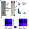
CD34Exo carry pro-angiogenic miRNA. Human angiogenesis protein array comparing CD34Exo and MNCExo (A); quantification of proteins present in CD34Exo and MNCExo using human angiogenesis protein array (B); miRNA expression profiles comparing CD34+ cells with MNCs and CD34Exo with MNCExo, shown using heat map expression analysis (C).
Figure 4
Figure 4
Loss of miiR-126 in CD34Exo results in loss of its angiogenic function in vitro and in vivo. MiR-126 expression in CD34+ cells or exosomes isolated from CD34+ cells, treated as indicated, n=3–6 (A); tube formation of HUVECs on Matrigel, treated as indicated; *P<0.05, n=3 (B); experimental scheme illustrating CD34Exo injection to ischemic (I) hindlimb (C); Day28 of mouse ischemic hind limb treated as indicated and LDPI images, severely restricted limb perfusion in blue and normal in red (D); quantification of LDPI perfusion (E); limb motor function (F) and salvage (G) scores of the ischemic limb (in a scale of 1–5; 1-poor and 5-strong); Quantification of ratio of capillary density between ischemic (I) and non-ischemic (NI) limb (H, I); *p< 0.001, vs. scrambled-control-cells or scrambled-control-Exo, n=6–8.
Figure 5
Figure 5
Loss of miiR-126 in CD34Exo results in loss of angiogenic gene activation function. Expression of miR-126 (A) and mouse pri-miRNA (B) normalized to U6; mSPRED1 (C); mVEGF (D); ANG1 (E) and MMP9 (F) mRNAs normalized to 18s RNA, in tissue homogenates of non-ischemic (NI) or ischemic (I) limbs treated as indicated, at 4h; n=4–9; P<0.05, *vs. PBS or, PBS I; †vs. scrambled-control-Exo.
Figure 6
Figure 6
Uptake and transfer of CD34Exo and exosomal miRNAs by endothelial cells in vitro and in vivo. Flow cytometry analysis of CD34+ cells (A) or exosomes isolated from CD34+ cells (B), transfected as indicated; confocal image of HUVECs treated with Cy3miRNA-CD34Exo (C); flow cytometry analysis of single cell suspensions from post-ischemic hindlimb tissue injected with R-PE-CD34Exo at 2h (D); % of cells uptaking exosomes (PE) from total number of each cell types quantified from Figure 5D (E), (n=3–5, *p<0.05).
Figure 7
Figure 7
CD34Exo induce proliferation of endothelial cells. Confocal image of post-ischemic hindlimb tissue injected with R-PE-CD34Exo at 12h (A); flow cytometry analysis of back-tracked CD31-positive endothelial cells (in blue) from single cell suspensions of ischemic hindlimbs treated as indicated (B); expression of Cyclin A (C) and Cyclin B (D) mRNA normalized to 18S rRNA in tissue homogenates of ischemic limbs treated as indicated, at 28d; n=5–6; P<0.05.
Figure 8
Figure 8
Summary of the new knowledge based on our hypothesis. Exosomes secreted by CD34+ cells induce tissue repair in mouse ischemic hind limb by delivering exosomal angiomiR-126 to endothelial cells inducing cell cycle changes and angiogenesis.

References

    1. Losordo DW, Henry TD, Davidson C, Lee JS, Costa MA, Bass T, Mendelsohn F, Fortuin FD, Pepine CJ, Traverse JH, Amrani D, Ewenstein BM, Riedel N, Story K, Barker K, Povsic TJ, Harrington RA, Schatz RA. Intramyocardial, Autologous CD34+ Cell Therapy for Refractory Angina. Circ Res. 2011 - PMC - PubMed
    1. Losordo DW, Kibbe MR, Mendelsohn F, Marston W, Driver VR, Sharafuddin M, Teodorescu V, Wiechmann BN, Thompson C, Kraiss L, Carman T, Dohad S, Huang P, Junge CE, Story K, Weistroffer T, Thorne TM, Millay M, Runyon JP, Schainfeld R. A randomized, controlled pilot study of autologous CD34+ cell therapy for critical limb ischemia. Circ Cardiovasc Interv. 2012;5:821–30. - PMC - PubMed
    1. Kawamoto A, Iwasaki H, Kusano K, Murayama T, Oyamada A, Silver M, Hulbert C, Gavin M, Hanley A, Ma H, Kearney M, Zak V, Asahara T, Losordo DW. CD34-positive cells exhibit increased potency and safety for therapeutic neovascularization after myocardial infarction compared with total mononuclear cells. Circulation. 2006;114:2163–9. - PubMed
    1. Kumar AH, Caplice NM. Clinical potential of adult vascular progenitor cells. Arteriosclerosis, thrombosis, and vascular biology. 2010;30:1080–7. - PubMed
    1. Sahoo S, Klychko E, Thorne T, Misener S, Schultz KM, Millay M, Ito A, Liu T, Kamide C, Agrawal H, Perlman H, Qin G, Kishore R, Losordo DW. Exosomes From Human CD34+ Stem Cells Mediate Their Proangiogenic Paracrine Activity. Circ Res. 2011;109:724–8. - PMC - PubMed

MeSH terms