. 2017 Jun 1;117(6):2125-2136.
doi: 10.1152/jn.00002.2017. Epub 2017 Mar 15.

Nonreciprocal homeostatic compensation in Drosophila potassium channel mutants

Affiliations

Nonreciprocal homeostatic compensation in Drosophila potassium channel mutants

Eugene Z Kim et al. J Neurophysiol. .

Abstract

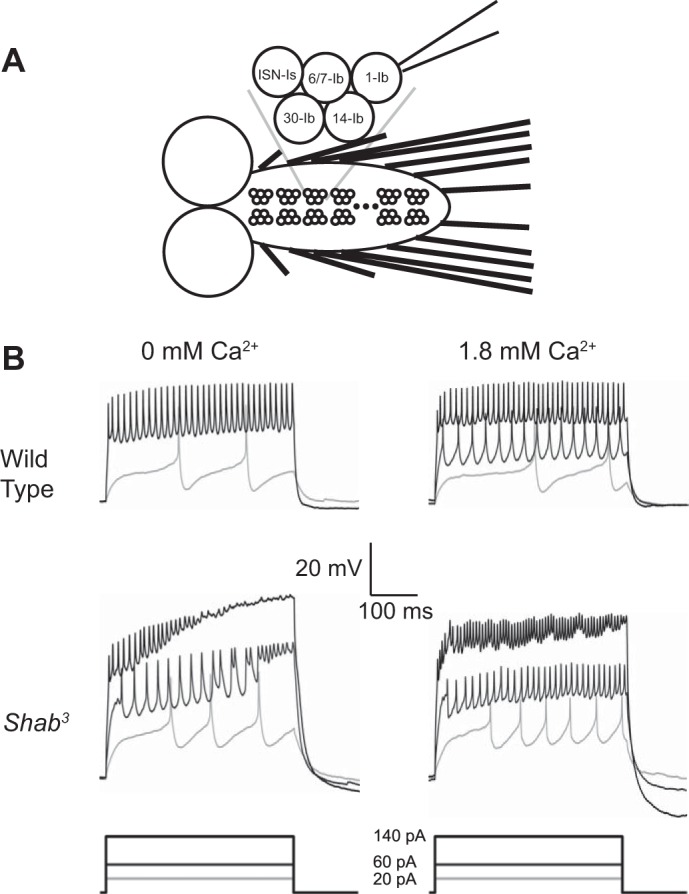
Homeostatic control of intrinsic excitability is important for long-term regulation of neuronal activity. In conjunction with many other forms of plasticity, intrinsic homeostasis helps neurons maintain stable activity regimes in the face of external input variability and destabilizing genetic mutations. In this study, we report a mechanism by which Drosophila melanogaster larval motor neurons stabilize hyperactivity induced by the loss of the delayed rectifying K+ channel Shaker cognate B (Shab), by upregulating the Ca2+-dependent K+ channel encoded by the slowpoke (slo) gene. We also show that loss of SLO does not trigger a reciprocal compensatory upregulation of SHAB, implying that homeostatic signaling pathways utilize compensatory pathways unique to the channel that was mutated. SLO upregulation due to loss of SHAB involves nuclear Ca2+ signaling and dCREB, suggesting that the slo homeostatic response is transcriptionally mediated. Examination of the changes in gene expression induced by these mutations suggests that there is not a generic transcriptional response to increased excitability in motor neurons, but that homeostatic compensations are influenced by the identity of the lost conductance.NEW & NOTEWORTHY The idea that activity-dependent homeostatic plasticity is driven solely by firing has wide credence. In this report we show that homeostatic compensation after loss of an ion channel conductance is tailored to identity of the channel lost, not its properties.

Keywords: Drosophila; excitability; homeostasis; ion channel; motor neuron.


Figures

Fig. 1.
Fig. 1.
Shab3 mutants exhibit Ca2+-suppressed firing instability. A: schematic of the third instar larval brain. Ventral ganglion is denoted by bracket. Individual motor neuron clusters are arranged in a stereotyped repeating pattern. The MN1-Ib neuron, shown attached to a patch electrode, is distinguishable from other identified neurons in each cluster by position. B: representative voltage traces from current-clamp experiments. Wild type (Canton S) is represented in top row. Traces from Shab3 recordings are shown in bottom row. Recordings taken in 0 mM external Ca2+ are shown at left, with 1.8 mM Ca2+ recordings shown at right. Removal of SHAB results in a destabilization of firing behavior at higher current injection levels in 0 mM Ca2+. This instability was seen in 12/13 cells recorded and is suppressed by addition of external Ca2+.
Fig. 2.
Fig. 2.
Mutation of Shab reduces total outward current and alters the balance of Ca2+-dependent and Ca2+-independent steady-state currents. A: current traces from voltage-clamp experiments on cells shown in Fig. 1B. Wild type (Canton S) is represented in the top row. Traces from Shab3 recordings are shown in bottom row. Recordings taken in 0 mM external Ca2+ are shown at left, with 1.8 mM Ca2+ recordings shown at right. B: total current density from voltage-clamp experiments done in 0 or 1.8 mM Ca2+ are shown for both genotypes. No significant difference in the outward currents can be seen in wild type when Ca2+ is added. In contrast, outward currents in Shab3 are substantially increased in Ca2+ (P < 0.0005). C: gene dosage effect on Ca2+-dependent currents. The percentage of currents that are Ca2+ dependent is significantly higher in Shab3 motor neurons than in wild type (P < 0.05), whereas the Shab3/+ heterozygote falls midway between the homozygote and wild type and is not significantly different from either wild type or Shab3. A,BDifferent letters indicate groups with a significant difference. D: IKd and IKCa currents in wild type and Shab3 mutants. IKd and IKCa for wild type and Shab3 were significantly different (P < 0.0005). E: difference plot for Shab3 mutant and wild type. The steady-state current at each holding potential for wild type was subtracted from currents produced in Shab3 neurons at the same potential. The downward deflection of the IKd curve demonstrates the loss of SHAB-mediated currents, whereas the upward deflection of the IKCa line indicates that there is an increase in this current in Shab3. The shape of the curve shows the voltage-dependence profile of the difference current. All error bars are SE; n = 5 for both wild type and Shab3. For data in B and D, 2-way ANOVAs with holding potential and genotype were performed and the Tukey-Kramer post hoc test was used to determine significant differences between genotypes. For data in C, significance was determined by 1-way ANOVA with the Tukey-Kramer post hoc test.
Fig. 3.
Fig. 3.
RNAi knockdown of Shab alters the balance of Ca2+-dependent and Ca2+-independent steady-state currents in a cell-autonomous manner. A: total current density from voltage-clamp experiments done in 0 or 1.8 mM Ca2+ are shown for animals expressing Shab RNAi in motor neurons and for UAS-alone control animals. Shab RNAi reduces total current in 0 mM Ca2+ (P < 0.0005 compared with both GAL4 and UAS controls) but not in 1.8 mM Ca2+ (P > 0.05). B: IKd and IKCa currents in Shab RNAi and UAS control neurons. Compared with both UAS and GAL4 controls, loss of SHAB reduces IKd (P < 0.0005) but increases IKCa (P < 0.0005), similar to the effect of the Shab3 mutant. C: difference plot for Shab RNAi and GAL4 control. The steady-state current at each holding potential for control was subtracted from currents produced in Shab RNAi neurons at the same potential. The downward deflection of the IKd curve demonstrates the loss of SHAB-mediated currents, whereas the upward deflection of the IKCa line indicates that there is an increase in this current in Shab RNAi. The shape of the curve shows the voltage-dependence profile of the difference current. All error bars are SE; n = 6 for GAL4 control and n = 14 for both UAS control and Shab RNAi. For data in A and B, 2-way ANOVAs with holding potential and genotype were performed and the Tukey-Kramer post hoc test was used to determine significant differences between genotypes.
Fig. 4.
Fig. 4.
slowpoke expression is required for upregulation of IKCa in response to reduction of SHAB. A: IKd and IKCa current densities for control and experimental genotypes at 20-mV holding potential. slo RNAi significantly decreases IKCa compared with controls. Shab RNAi decreases IKd but significantly increases IKCa. The presence of both RNAis does not further decrease IKd beyond the level present in Shab RNAi, but it blocks the increase in IKCa seen with Shab RNAi alone. P values for comparisons are shown in Table 2. *P < 0.05 compared with controls; ANOVA with Tukey-Kramer post hoc test. Data are means ± SE; n = 6 for OK371/+, n = 14 for UAS-ShabRNAi/+, n = 17 for UAS-sloRNAi/+, n = 14 for OK371/UAS-ShabRNAi, n = 19 for OK371/UAS-sloRNAi, and n = 10 for OK371,UAS-ShabRNAi/OK371-GAL4,UAS-sloRNAi. B: total currents in 1.8 mM Ca2+ are compared for animals expressing Shab, slo, or Shab+slo RNAi in motor neurons and for the OK371/+ control. slo RNAi ± Shab RNAi reduces total currents (P < 0.005), whereas Shab RNAi alone does not. C: IKd for animals expressing Shab, slo, or Shab+slo RNAi in motor neurons (format as in B). Shab RNAi reduces IKd compared with both GAL4 and UAS controls and slo RNAi (P < 0.0005). D: IKCa for animals expressing Shab, slo, or Shab+slo RNAi in motor neurons (format as in B). The presence of slo RNAi blocks the increase in IKCa produced by Shab RNAi (P < 0.0005). E: difference plot for Shab+slo RNAi and Shab RNAi. The steady-state current at each holding potential for Shab RNAi was subtracted from currents produced in neurons expressing both Shab RNAi and slo RNAi at the same potential. The flat IKd curve demonstrates that slo RNAi does not change the ability of Shab RNAi to reduce IKd. The downward deflection of the IKCa line indicates that slo RNAi decreases the ability of Shab RNAi to produce an increase in IKCa. For data in B–D, two-way ANOVAs with holding potential and genotype were performed and the Tukey-Kramer post hoc test was used to determine significant differences between genotypes. Data are means ± SE.
Fig. 5.
Fig. 5.
Inhibition of activity-regulated transcriptional processes reduces potassium currents and partially blocks homeostasis. IKd and IKCa current densities for control and experimental genotypes at 20-mV holding potential. Both dnCREB and CaM Sponge reduce outward currents and partially block the increase in IKCa normally produced by Shab RNAi (compare with Fig. 4). Data are means ± SE; n = 4 for OK371/+;UAS-CaMSponge/+, n = 5 for OK371/UAS-ShabRNAi;UAS-CaMSponge, n = 4 for OK371/+;UAS-dnCREB/+, and n = 3 for OK371/UAS-ShabRNAi;UAS-dnCREB/+. *P < 0.05, Shab RNAi + CaM Sponge vs. CaM Sponge alone; Student’s t-test.

Similar articles

Cited by

References

    1. Abou Tayoun AN, Pikielny C, Dolph PJ. Roles of the Drosophila SK channel (dSK) in courtship memory. PLoS One 7: e34665, 2012. doi:10.1371/journal.pone.0034665. - DOI - PMC - PubMed
    1. Atkinson NS, Robertson GA, Ganetzky B. A component of calcium-activated potassium channels encoded by the Drosophila slo locus. Science 253: 551–555, 1991. doi:10.1126/science.1857984. - DOI - PubMed
    1. Bellen HJ, Tong C, Tsuda H. 100 years of Drosophila research and its impact on vertebrate neuroscience: a history lesson for the future. Nat Rev Neurosci 11: 514–522, 2010. doi:10.1038/nrn2839. - DOI - PMC - PubMed
    1. Belvin MP, Zhou H, Yin JC. The Drosophila dCREB2 gene affects the circadian clock. Neuron 22: 777–787, 1999. doi:10.1016/S0896-6273(00)80736-9. - DOI - PMC - PubMed
    1. Bergquist S, Dickman DK, Davis GW. A hierarchy of cell intrinsic and target-derived homeostatic signaling. Neuron 66: 220–234, 2010. doi:10.1016/j.neuron.2010.03.023. - DOI - PMC - PubMed

MeSH terms

LinkOut - more resources