. 2017 Mar;7(3):160334.
doi: 10.1098/rsob.160334.

Atypical centrioles are present in Tribolium sperm

Affiliations

Atypical centrioles are present in Tribolium sperm

E L Fishman et al. Open Biol. 2017 Mar.

Abstract

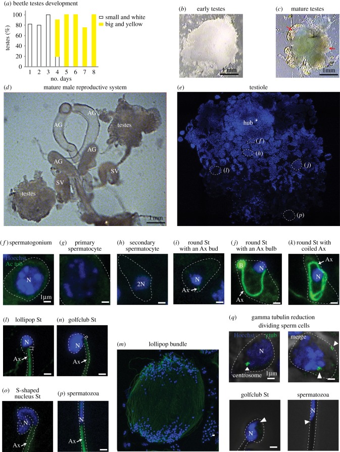
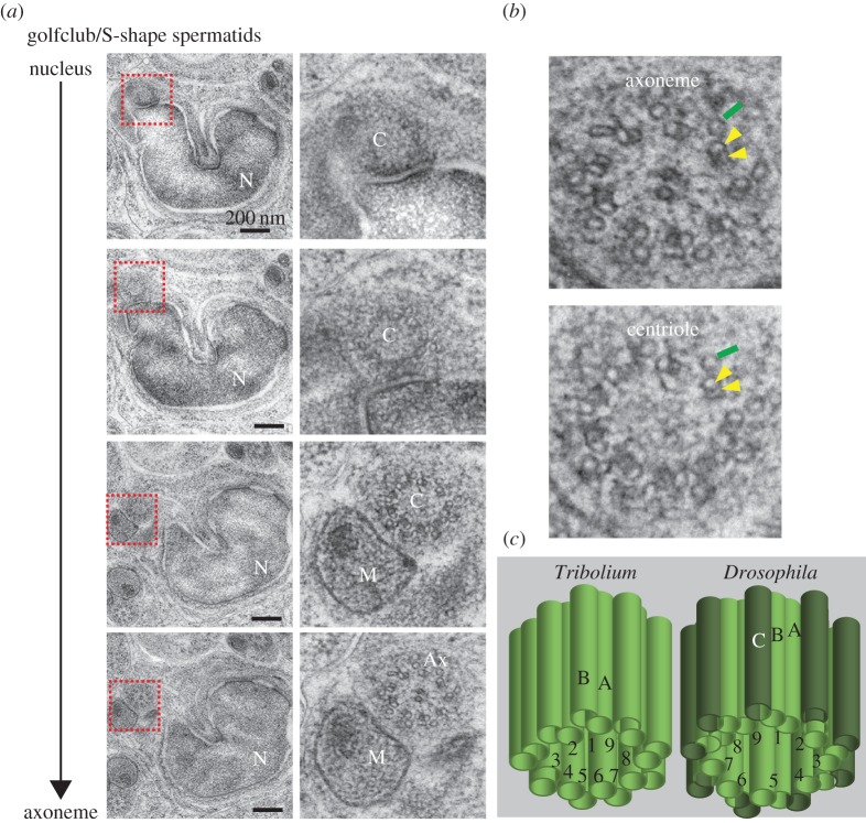
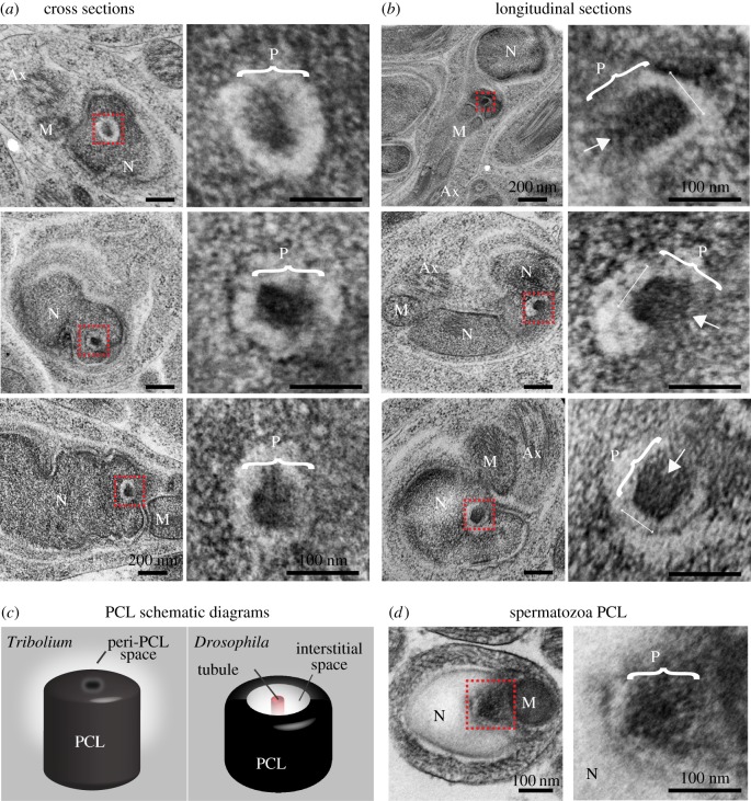
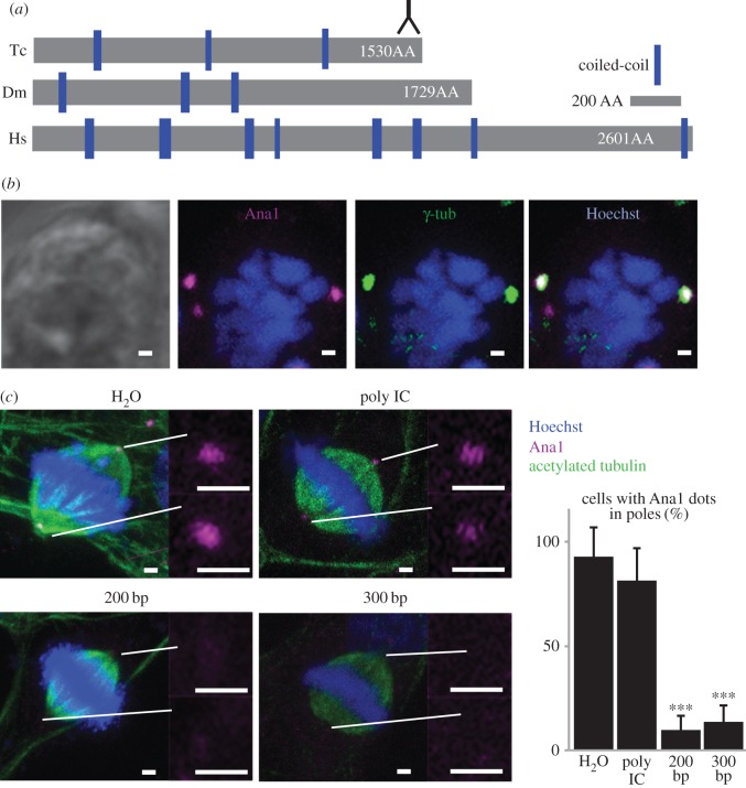
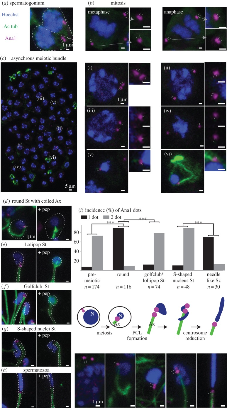
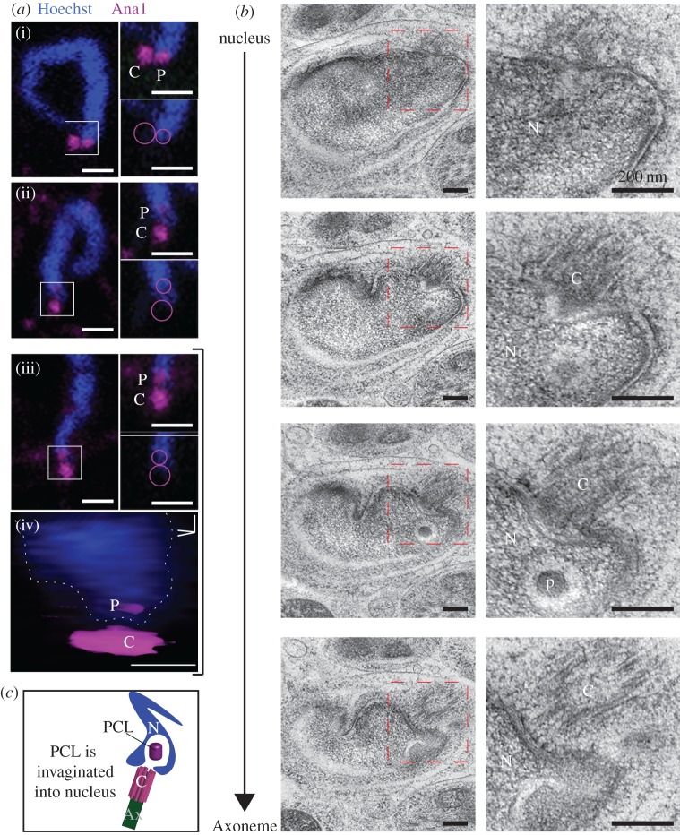
Typical centrioles are made of microtubules organized in ninefold symmetry. Most animal somatic cells have two centrioles for normal cell division and function. These centrioles originate from the zygote, but because the oocyte does not provide any centrioles, it is surprising that the zygotes of many animals are thought to inherit only one centriole from the sperm. Recently, in the sperm of Drosophila melanogaster, we discovered a second centriolar structure, the proximal centriole-like structure (PCL), which functions in the zygote. Whether the sperm of other insects has a second centriolar structure is unknown. Here, we characterized spermiogenesis in the red flour beetle, Tribolium castaneum Electron microscopy suggests that Tribolium has one microtubule-based centriole at the tip of the axoneme and a structure similar to the PCL, which lacks microtubules and lies in a cytoplasmic invagination of the nucleus. Immunostaining against the orthologue of the centriole/PCL protein, Ana1, also recognizes two centrioles near the nucleus during spermiogenesis: one that is microtubule-based at the tip of the axoneme, suggesting it is the centriole; and another that is more proximal and appears during early spermiogenesis, suggesting it is the PCL. Together, these findings suggest that Tribolium sperm has one microtubule-based centriole and one microtubule-lacking centriole.

Keywords: Ana1; PCL; Tribolium; atypical centrioles; centriole; sperm.


Figures

Figure 1.
Figure 1.
Tribolium spermiogenesis. (ac) As the beetles eclose from the pupa, their testes change (graph in a). They start as small white testes, surrounded by a big white cloud of fat (b). Around day 4, they change to large and yellow with no more than six clear lobes (testioles marked with red arrows) and very little fat (c). Though the early testes (b) appears equal in size to day 4 testes (c), in reality, the early testes fat fills the volume difference between the smaller testes and the larger ones on day 4. (d) The complete male reproductive system. (e) A single testiole stained with Hoechst (blue) to recognize the nuclei. Specific areas are presented in panels (f)–(p) as labelled. The Hoechst staining becomes lighter in later-stage sperm. (fp) Sperm cells at distinct stages are stained with Hoechst (blue) and anti-acetylated-tubulin (green). (q) Anti-γ-tubulin (green) staining is observed in dividing-stage sperm but is undetectable in late-stage sperm (golfclub spermatid and spermatozoa) at the expected site of the centriole (arrowhead). Abbreviations: AG, accessory gland; SV, seminal vesicle; Ax, axoneme; N, nucleus; B, bulb; St, spermatid. Scale bars, 1 µm.
Figure 2.
Figure 2.
The centriole of Tribolium spermatids has doublet microtubules. (a) High-pressure freezing (HPF), freeze substitution (FS) and serial section TEM analysis of Tribolium golfclub/S-shape spermatid showing the transition from a centriole lacking central microtubules to an axoneme with central microtubules. Abbreviations: Ax, axoneme; C, centriole; M, mitochondria; N, nucleus. (b) Magnification of axoneme and centriole cross-sections. Yellow arrowheads: the microtubules in the doublet microtubules; green line: accessory microtubule. (c) Model of the axoneme-attached centriole. The centriole is made of doublet microtubules labelled A and B in ninefold symmetry, which is different than Drosophila's sperm's axoneme-attached centriole's triplets, labelled A, B and C.
Figure 3.
Figure 3.
The 2nd centriole of Tribolium spermatids lack microtubules. (a,b) Representative HPF-FS TEM semi cross-section (a) and semi-longitudinal (b) analysis of Tribolium golfclub spermatids. Images on the right are insets of images on the left, signified by the red box. Brackets indicate the axis representing the PCL diameter. Arrows indicate the location of the barely detectable electron-dense central core. (c) A model of the second centriolar structure in spermatids. The structure of the PCL (P) is slightly different than Drosophila's PCL. (d) Representative TEM sections of the second centriolar structure in Tribolium spermatozoa. Abbreviations: Ax, axoneme; M, mitochondria; N, nucleus. Scale bars, 1 µm.
Figure 4.
Figure 4.
Tribolium Ana1. (a) Schematic comparison of Drosophila (Dm) Ana1, Tribolium (Tc) Ana1 and human CEP295 (Hs). Inversed ‘Y’ symbol indicates the region recognized by the antibody. (b) Colocalization of γ-tubulin and Ana1 in dividing TcA cells. (c) Ana1 labelling is reduced in TcA cells when treated with two different lengths of RNAi fragments against Ana1 (200 and 300 bp), but is not diminished significantly when treated with water (H2O) or a non-specific double-stranded RNAi (Poly IC). ***p < 0.001 by t-test. n = 3. Scale bars, 1 µm.
Figure 5.
Figure 5.
A 2nd centriole forms during Tribolium spermatogenesis. (ah) Anti-Ana1 labelling throughout Tribolium spermatogenesis. (c) As expected, an asynchronous meiotic bundle has spermatocytes, each with two centrosomes with a total of four centrioles (i–iii). Each secondary spermatocyte has one centriole at opposite poles (iv), and early spermatid stages have one centriole (v–vi). (dh) Anti-Ana1 dots are found at the base of the nucleus throughout spermiogenesis and this staining is abolished in the presence of a competitive peptide. (i) Quantification of Ana1 dots in different stages of sperm: one dot (black); two dots (grey). A schematic diagram showing the emergence of the second centriolar structure. p < 0.001 by chi-square test. Abbreviations: Ax, axoneme; St, spermatid; Sz, spermatozoa; N, nucleus. Thin lines indicate position of centriole and PCL. Scale bars, 1 µm.
Figure 6.
Figure 6.
The 2nd Ana-1 labelled centriole is the PCL. (a) Single 0.3 µm thick confocal sections showing that the proximal Ana1 dot is located inside the nucleus boundary in golfclub/S-shaped spermatid stage (i–iii). Circles mark the location of the centriole and PCL in the inset. Three-dimensional volume model of the PCL in an S-shaped spermatid iii (iv). Scale bars, 1 µm. (b) Serial section TEM analysis showing that only the PCL is found in the cytosolic invagination within the nucleus at the golfclub/S-shape spermatid stage. Scale bars, 200 nm. (c) A schematic diagram showing the location of the PCL in a cytosolic invagination within the nucleus. Abbreviations: Ax, axoneme; C, centriole; N, nucleus; P, PCL.

Similar articles

Cited by

References

    1. Bornens M. 2012. The centrosome in cells and organisms. Science 335, 422–426. (doi:10.1126/science.1209037) - DOI - PubMed
    1. Rodrigues-Martins A, Riparbelli M, Callaini G, Glover DM, Bettencourt-Dias M. 2007. Revisiting the role of the mother centriole in centriole biogenesis. Science 316, 1046–1050. (doi:10.1126/science.1142950) - DOI - PubMed
    1. Avidor-Reiss T, Gopalakrishnan J, Blachon S, Polyanovsky A. 2012. Centriole duplication and inheritance in Drosophila melanogaster. In The centrosome: cell and molecular mechanisms of functions and dysfunctions in disease (ed. Schatten H.), pp. 3–31. New York, NY: Humana Press.
    1. Fu J, Hagan IM, Glover DM. 2015. The centrosome and its duplication cycle. Cold Spring Harb. Perspect. Biol. 7, a015800. - PMC - PubMed
    1. Schatten G. 1994. The centrosome and its mode of inheritance: the reduction of the centrosome during gametogenesis and its restoration during fertilization. Dev. Biol. 165, 299–335. (doi:10.1006/dbio.1994.1256) - DOI - PubMed

LinkOut - more resources