. 2017 Aug;28(8):2353-2363.
doi: 10.1681/ASN.2016050585. Epub 2017 Mar 15.

Let-7 and MicroRNA-148 Regulate Parathyroid Hormone Levels in Secondary Hyperparathyroidism

Affiliations

Let-7 and MicroRNA-148 Regulate Parathyroid Hormone Levels in Secondary Hyperparathyroidism

Vitali Shilo et al. J Am Soc Nephrol. 2017 Aug.

Abstract

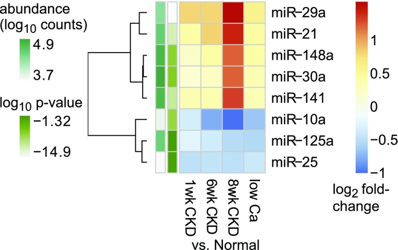
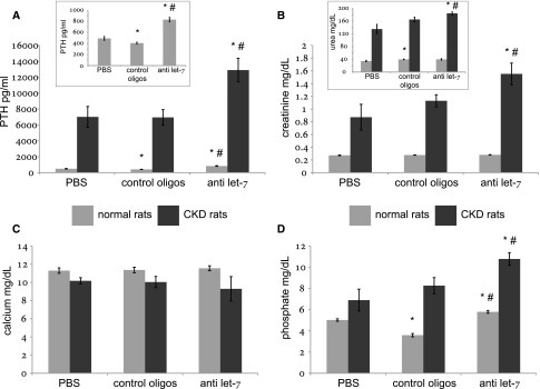
Secondary hyperparathyroidism commonly complicates CKD and associates with morbidity and mortality. We profiled microRNA (miRNA) in parathyroid glands from experimental hyperparathyroidism models and patients receiving dialysis and studied the function of specific miRNAs. miRNA deep-sequencing showed that human and rodent parathyroids share similar profiles. Parathyroids from uremic and normal rats segregated on the basis of their miRNA expression profiles, and a similar finding was observed in humans. We identified parathyroid miRNAs that were dysregulated in experimental hyperparathyroidism, including miR-29, miR-21, miR-148, miR-30, and miR-141 (upregulated); and miR-10, miR-125, and miR-25 (downregulated). Inhibition of the abundant let-7 family increased parathyroid hormone (PTH) secretion in normal and uremic rats, as well as in mouse parathyroid organ cultures. Conversely, inhibition of the upregulated miR-148 family prevented the increase in serum PTH level in uremic rats and decreased levels of secreted PTH in parathyroid cultures. The evolutionary conservation of abundant miRNAs in normal parathyroid glands and the regulation of these miRNAs in secondary hyperparathyroidism indicates their importance for parathyroid function and the development of hyperparathyroidism. Specifically, let-7 and miR-148 antagonism modified PTH secretion in vivo and in vitro, implying roles for these specific miRNAs. These findings may be utilized for therapeutic interventions aimed at altering PTH expression in diseases such as osteoporosis and secondary hyperparathyroidism.

Keywords: chronic kidney disease; gene expression; hyperparathyroidism; microRNA; mineral metabolism.


Figures

Figure 1.
Figure 1.
miRNA abundance is similar in normal human, mouse, and rat parathyroid glands. (A) Heat map representation of the most abundant miRNA sequence families in pooled normal specimens. The top 50 sequence families (not individual miRNAs) in humans are shown, with corresponding values from rats and mice. miRNA sequence families (rows) and species (columns) were hierarchically clustered applying Manhattan distance metric and Ward clustering method. The color scale represents abundance, where brighter shades correspond to higher values. (B) Venn diagram showing vast overlaps of top 50 miRNA sequence families between species. (C) Three-dimensional scatter plot showing interspecies expression correlations of top 25 miRNA sequence families (as determined in humans). Input data for these analyses (raw counts arranged in sequence families) are presented in Supplemental Tables 2, 5, and 8.
Figure 2.
Figure 2.
Principal component analysis (PCA) of miRNA profiles discriminates normal from hyperplastic glands. Plots depicting the main variables (PC1 and PC2) derived from PCAs which capture miRNA abundance levels into new variables. These new variables do not correlate with each other, however, each principal component correlates (or anticorrelates) with all miRNA, to different degrees. PCA plots of miRNA profiles (A) or differentially expressed miRNA (B) from normal rats and rats with SHP induced by either uremia or low-calcium diet. (C) PCA plot of miRNA profiles from normal deparaffinized human parathyroid glands and glands from ESRD patients with SHP. Ca, calcium.
Figure 3.
Figure 3.
Top differentially expressed miRNAs in experimental SHP show common patterns. Heat map showing the most abundant differentially expressed miRNA sequence families (derived from Supplemental Table 13, columns C–F). Color shades represent the fold change of the indicated miRNA in the indicated SHP model compared with control rats. Rows (miRNA families) were clustered applying Euclidean distance and complete clustering method. The side (row) annotations represent the ANOVA-like P values and the average abundance levels of the indicated miRNA families. Ca, calcium.
Figure 4.
Figure 4.
Evidence for delivery and functional target engagement of anti–let-7 oligonucleotides in parathyroid. Pooled total RNA from normal rats injected with anti–let-7 or control oligonucleotides underwent mRNA array analysis (Affymetrix GeneChip Rat Genome 230 2.0 Arrays). The fold-change values of protein coding genes included in the array were used to detect derepression of let-7 targets. (A) Sylamer analysis shows that two out of three words with highest peak enrichment in 3′utr of upregulated genes are reverse-complementary to the let-7 seed. (B) Empirical cumulative distribution function plot of mRNA log2 fold-changes showing a shift to the right (upregulation) among miranda-predicted let-7 targets compared with nontarget mRNAs: 11,191 nontarget mRNAs; 339 single-targeted mRNAs (P value 7.8e−05); 41 multitargeted mRNAs (P value 0.003). (C) Gene set enrichment analysis shows striking enrichment of predicted let-7 targets among genes upregulated by let-7 antagonizing oligonucleotides. Putative target genes according to this analysis are labeled as such in Supplemental Table 18.
Figure 5.
Figure 5.
Serum chemistry and PTH levels are altered in rats treated for 4 weeks with let-7 anti-miRs. Serum PTH (A), creatinine (B), urea (B; inset), calcium (C), and phosphate (D) levels in normal and CKD rats treated with let-7 anti-miRs, scrambled controls (“control oligos”), or PBS. *P<0.05 for the comparison with PBS-treated rats (within the same disease study group—normal/CKD); #P<0.05 for the comparison with control oligonucleotides-treated rats. Numbers of animals were six in each subgroup of normal rats and five in each subgroup of uremic rats.
Figure 6.
Figure 6.
PTH accumulation in vitro is augmented by let-7 anti-miRs. Mouse thyro-parathyroid blocks from normal mice were incubated in cell culture media in the presence of anti–let-7 oligonucleotides or scrambled controls (0.5 mg/ml), and medium was sampled for PTH ELISA at the indicated time points. *P<0.05 for the comparison with control oligonucleotides at the same time point.
Figure 7.
Figure 7.
Serum chemistry and PTH levels are altered in rats treated with miR-141/miR-148 antagonists. Serum PTH (top left panel), creatinine (top right panel), calcium (bottom right panel), and phosphate (bottom left panel) levels in CKD rats treated for 4 weeks with the indicated miRNA antagonists or scrambled control oligonucleotides. *P<0.05 for the comparison with control oligonucleotides. An interim analysis at 2 weeks is presented in Supplemental Figure 5.
Figure 8.
Figure 8.
PTH accumulation in vitro is diminished by miR-148 antagonism. Mouse thyro-parathyroid blocks from CKD mice were incubated in cell culture media in the presence of anti–miR-148 oligonucleotides or scrambled controls (0.5 mg/ml), and medium was sampled for PTH ELISA at the indicated time points. Overall accumulation was significantly lower in the presence of miR-148 anti-miRs (P<0.05 by repeated measures ANOVA).

References

    1. Bartel DP: MicroRNAs: Target recognition and regulatory functions. Cell 136: 215–233, 2009 - PMC - PubMed
    1. Friedman RC, Farh KK, Burge CB, Bartel DP: Most mammalian mRNAs are conserved targets of microRNAs. Genome Res 19: 92–105, 2009 - PMC - PubMed
    1. Farazi TA, Horlings HM, Ten Hoeve JJ, Mihailovic A, Halfwerk H, Morozov P, Brown M, Hafner M, Reyal F, van Kouwenhove M, Kreike B, Sie D, Hovestadt V, Wessels LFA, van de Vijver MJ, Tuschl T: MicroRNA sequence and expression analysis in breast tumors by deep sequencing. Cancer Res 71: 4443–4453, 2011 - PMC - PubMed
    1. Etheridge A, Lee I, Hood L, Galas D, Wang K: Extracellular microRNA: A new source of biomarkers. Mutat Res 717: 85–90, 2011 - PMC - PubMed
    1. Cheng CJ, Saltzman WM, Slack FJ: Canonical and non-canonical barriers facing antimiR cancer therapeutics. Curr Med Chem 20: 3582–3593, 2013 - PMC - PubMed