. 2017 Mar 15;9(381):eaai7514.
doi: 10.1126/scitranslmed.aai7514.

Staged induction of HIV-1 glycan-dependent broadly neutralizing antibodies

Affiliations

Staged induction of HIV-1 glycan-dependent broadly neutralizing antibodies

Mattia Bonsignori et al. Sci Transl Med. .

Abstract

A preventive HIV-1 vaccine should induce HIV-1-specific broadly neutralizing antibodies (bnAbs). However, bnAbs generally require high levels of somatic hypermutation (SHM) to acquire breadth, and current vaccine strategies have not been successful in inducing bnAbs. Because bnAbs directed against a glycosylated site adjacent to the third variable loop (V3) of the HIV-1 envelope protein require limited SHM, the V3-glycan epitope is an attractive vaccine target. By studying the cooperation among multiple V3-glycan B cell lineages and their coevolution with autologous virus throughout 5 years of infection, we identify key events in the ontogeny of a V3-glycan bnAb. Two autologous neutralizing antibody lineages selected for virus escape mutations and consequently allowed initiation and affinity maturation of a V3-glycan bnAb lineage. The nucleotide substitution required to initiate the bnAb lineage occurred at a low-probability site for activation-induced cytidine deaminase activity. Cooperation of B cell lineages and an improbable mutation critical for bnAb activity defined the necessary events leading to breadth in this V3-glycan bnAb lineage. These findings may, in part, explain why initiation of V3-glycan bnAbs is rare, and suggest an immunization strategy for inducing similar V3-glycan bnAbs.


Conflict of interest statement

Competing interests: M.B., R.R.M., M.A.M., H.-X.L., T.B., K.O.S., P.T.H., B.T.K., and B.F.H. have filed patent application(s) covering DH270 lineage antibodies and immunogens to induce such antibodies. B.A., H.-X.L., B.F.H., and S.J.D. have filed patent application(s) covering the V3 glycopeptide and its synthesis and use as an immunogen. All other authors declare that they have no competing interests.

Figures

Fig. 1
Fig. 1. DH270 lineage with time of appearance and neutralization by selected members
(A) Phylogenetic relationship of six monoclonal antibodies (mAbs) and 93 NGS VH(D)JH sequence reads in the DH270 clonal lineage. External nodes (filled circles) represent VH(D)JH nucleotide sequences of either antibodies retrieved from cultured and sorted memory B cells (labeled) or a curated data set of NGS VH(D)JH rearrangement reads (unlabeled). Coloring is by time of isolation. Samples from weeks 11, 19, 64, 111, 160, 186, and 240 were tested, and time points from which no NGS reads within the lineage were retrieved are reported in table S2. Internal nodes (open circles) represent inferred ancestral intermediate sequences. Units for branch-length estimates are nucleotide substitution per site. (B) Neutralization dendrograms display single mAb neutralization of a genetically diverse panel of 207 HIV-1 isolates. Coloring is by median inhibitory concentration (IC50). See also data set S1.
Fig. 2
Fig. 2. Heterologous breadth in the DH270 lineage
(A) Neutralizing activity of DH270.1, DH270.5, and DH270.6 bnAbs (columns) for 207 tier 2 heterologous viruses (rows). Coloring is by neutralization IC50 (in μg/ml). The first column displays the presence of a PNG site at position 332 (blue), at N334 (orange), or at neither one (black). The second column indicates the clade of each individual HIV-1 strain and is color-coded as indicated: clade A, green; clade B, blue; clade C, yellow; clade D, purple; CRF01, pink; clade G, cyan; others, gray. See data set S1. (B). Heterologous neutralization of all DH270 lineage antibodies for a 24-virus panel. Color coding for the presence of PNG sites, clade, and IC50 is the same as in (A). See fig. S1 and data set S3. (C) Covariation between VH mutation frequencies (x axis), neutralization breadth (y axis, top), and potency (y axis, bottom) of individual antibodies against viruses with a PNG site at position N332 from the larger (left) and smaller (right) pseudovirus panels. (D) Correlation between viral V1 loop length and DH270 lineage antibody neutralization. Top: Neutralization of 17 viruses (with N332; sensitive to at least one DH270 lineage antibody) by selected DH270 lineage antibodies from unmutated common ancestor (UCA) to mature bnAbs (x axis). Viruses are identified by their respective V1 loop lengths (y axis); for each virus, neutralization sensitivity is indicated by an open circle, and resistance is indicated by a solid circle. The P value is a Wilcoxon rank sum comparison of V1 length distributions between sensitive and resistant viruses. Bottom: Regression lines (IC50 for neutralization versus V1 loop length) for DH270.1 and DH270.6, with a P value based on Kendall’s τ.
Fig. 3
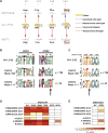
Fig. 3. A single disfavored mutation early during DH270 clonal development conferred neutralizing activity on the V3-glycan bnAb DH270 precursor antibodies
(A) Nucleotide alignment of DH270.IA4 and DH272 to VH1–2*02 sequence at the four VH positions that mutated from DH270.UCA to DH270.IA4. The mutated codons are highlighted in yellow. AID hotspots are indicated by red lines (solid, canonical; dashed, noncanonical); AID cold spots are indicated by blue lines (solid, canonical; dashed, noncanonical) (20). At position 169, DH270.IA4 retained positional conformity with DH272 but not identity conformity (red boxes). (B) Sequence logo plot of amino acids mutated from germline (top) in the NGS reads of the DH270 (middle) and DH272 (bottom) lineages at weeks 186 and 111 after transmission, respectively. Red asterisks indicate amino acids mutated in DH270.IA4. The black arrow indicates lack of identity conformity between the two lineages at amino acid position 57. (C) Sequence logo plot of nucleotide mutations (positions 165 to 173) in the DH270 and DH272 lineages at weeks 186 and 111 after transmission, respectively. The arrow indicates position 169. (D) Effect of reversion mutations on DH270.IA4 neutralization. Coloring is by IC50. WT, wild type. (E) Effect of G57R mutation on DH270.UCA auto-logous (top) and heterologous (bottom) neutralizing activity.
Fig. 4
Fig. 4. Cooperation among DH270, DH272, and DH475 N332-dependent V3-glycan neutralizing antibody lineages
(A) Neutralizing activity of DH272, DH475, and DH270 lineage antibodies (columns) against 90 autologous viruses isolated from CH848 over time (rows). Neutralization potency (IC50) is shown as indicated in the bar. For each pseudovirus, presence of a PNG site at N332 or N334, and V1 loop length are indicated in the right columns. See also data set S2. (B and C) Susceptibility of autologous viruses bearing selected immunotype-specific mutations to DH270.1 and to DH475 (B) or DH272 (C).
Fig. 5
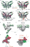
Fig. 5. Fab/scFv crystal structures and three-dimensional reconstruction of DH270.1 bound with the 92BR SOSIP.664 trimer
Superposition of backbone ribbon diagrams for the DH270 lineage members: UCA1 (gray), DH270.1 (green), and DH270.6(blue)alone(A), withtheDH272cooperating antibody (red) (B), with PGT128 (magenta) (C), and with PGT124 (orange) (D). Arrows indicate major differences in CDR regions. Top (E) and side (F) views of a fit of the DH270.1 Fab (green) and the BG505 SOSIP trimer (gray) onto a map obtained from negative-stain EM. Top (G) and side (H) views of the BG505 trimer [Protein Data Bank (PDB) ID: 5ACO] (28) (gray, with V1-V2 and V3 loops highlighted in red and blue, respectively) bound to PGT124 (PDB ID: 4R2G) (27) (orange), PGT128 (PDB ID: 3TYG) (17) (magenta), PGT135 (PDB ID: 4JM2) (22) (cyan), and DH270.1 (green), superposed. The arrows indicate the direction of the principal axis of each of the bnAb Fabs; the color of each arrow matches that of the corresponding bnAb. See also fig. S18.
Fig. 6
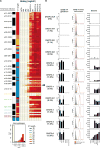
Fig. 6. DH270 lineage antibody binding to autologous CH848 Env components
(A) Binding of DH270 lineage antibodies (column) to 120 CH848 autologous gp120 Env glycoproteins (rows) grouped on the basis of time of isolation (w, week; d, day; black and white blocks). The last three rows show the neutralization profile of the three autologous viruses that lost the PNG at position N332 (blue blocks). V1 amino acid length of each virus is color-coded as indicated. Antibody binding is measured by enzyme-linked immunosorbent assay, expressed as log area under the curve (LogAUC), and color-coded on the basis of the categories shown in the histogram. The histogram shows the distribution of the measured values in each category. The black arrow indicated Env 10.17. Viruses isolated at and after week 186, which is the time of first evidence of DH270 lineage presence, are highlighted in different colors according to week of isolation. (B) Left: Binding to CH848 TF mutants with disrupted N301 and/or N332 glycan sites. Results are expressed as LogAUC. VH mutation frequency is shown in parentheses for each antibody (see also fig. S1A). Middle: Binding to CH848 Env trimer expressed on the cell surface of Chinese hamster ovary cells. Results are expressed as maximum percentage of binding and are representative of duplicate experiments. DH270 antibodies are in red. Palivizumab is the negative control (gray area). The curves indicate binding to the surface antigen on a scale of 0 to 100 (y axis); the highest peak between the test antibody and the negative control sets the value of 100. Right: Binding to free glycans measured on a microarray. Results are the average of background-subtracted triplicate measurements and are expressed in relative units (RU).

References

    1. Burton DR, Mascola JR. Antibody responses to envelope glycoproteins in HIV-1 infection. Nat Immunol. 2015;16:571–576. - PMC - PubMed
    1. Mascola JR, Haynes BF. HIV-1 neutralizing antibodies: Understanding nature’s pathways. Immunol Rev. 2013;254:225–244. - PMC - PubMed
    1. Walker LM, Huber M, Doores KJ, Falkowska E, Pejchal R, Julien JP, Wang SK, Ramos A, Chan-Hui PY, Moyle M, Mitcham JL, Hammond PW, Olsen OA, Phung P, Fling S, Wong CH, Phogat S, Wrin T, Simek MD, Koff WC, Wilson IA, Burton DR, Poignard P. Broad neutralization coverage of HIV by multiple highly potent antibodies. Nature. 2011;477:466–470. - PMC - PubMed
    1. Walker LM, Phogat SK, Chan-Hui PY, Wagner D, Phung P, Goss JL, Wrin T, Simek MD, Fling S, Mitcham JL, Lehrman JK, Priddy FH, Olsen OA, Frey SM, Hammond PW, Kaminsky S, Zamb T, Moyle M, Koff WC, Poignard P, Burton DR. Broad and potent neutralizing antibodies from an African donor reveal a new HIV-1 vaccine target. Science. 2009;326:285–289. - PMC - PubMed
    1. Doores KJ, Kong L, Krumm SA, Le KM, Sok D, Laserson U, Garces F, Poignard P, Wilson IA, Burton DR. Two classes of broadly neutralizing antibodies within a single lineage directed to the high-mannose patch of HIV envelope. J Virol. 2015;89:1105–1118. - PMC - PubMed

Publication types

Substances