Review
. 2017 Apr;97(2):839-887.
doi: 10.1152/physrev.00010.2016.

Autoantibodies to Synaptic Receptors and Neuronal Cell Surface Proteins in Autoimmune Diseases of the Central Nervous System

Affiliations
Review

Autoantibodies to Synaptic Receptors and Neuronal Cell Surface Proteins in Autoimmune Diseases of the Central Nervous System

Josep Dalmau et al. Physiol Rev. 2017 Apr.

Abstract

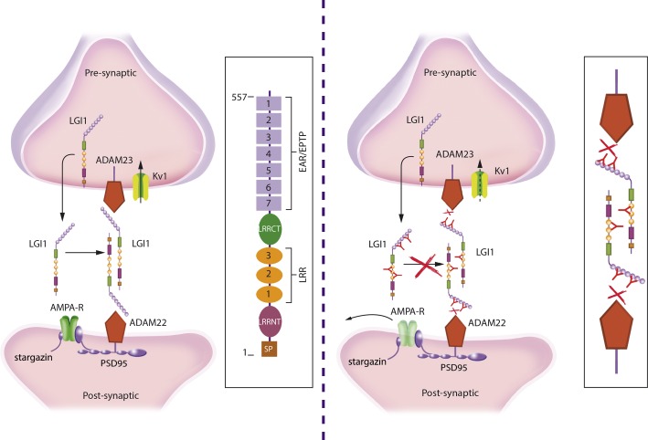
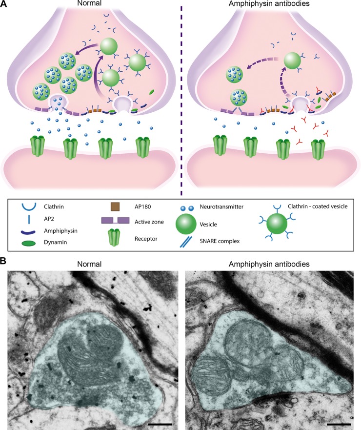
Investigations in the last 10 years have revealed a new category of neurological diseases mediated by antibodies against cell surface and synaptic proteins. There are currently 16 such diseases all characterized by autoantibodies against neuronal proteins involved in synaptic signaling and plasticity. In clinical practice these findings have changed the diagnostic and treatment approach to potentially lethal, but now treatable, neurological and psychiatric syndromes previously considered idiopathic or not even suspected to be immune-mediated. Studies show that patients' antibodies can impair the surface dynamics of the target receptors eliminating them from synapses (e.g., NMDA receptor), block the function of the antigens without changing their synaptic density (e.g., GABAb receptor), interfere with synaptic protein-protein interactions (LGI1, Caspr2), alter synapse formation (e.g., neurexin-3α), or by unclear mechanisms associate to a new form of tauopathy (IgLON5). Here we first trace the process of discovery of these diseases, describing the triggers and symptoms related to each autoantigen, and then review in detail the structural and functional alterations caused by the autoantibodies with special emphasis in those (NMDA receptor, amphiphysin) that have been modeled in animals.


Figures

FIGURE 1.
FIGURE 1.
Autoantibodies in classic paraneoplastic syndromes of the CNS and in novel autoimmune encephalopathies. A: in classic paraneoplastic syndromes of the CNS, the autoantibodies are directed against intracellular neuronal proteins that are also expressed by an underlying systemic tumor (onconeuronal proteins). These antibodies are useful diagnostic biomarkers, but there is no evidence they are pathogenic. Biopsy and autopsy studies of these patients show prominent inflammatory infiltrates of cytotoxic T-cells surrounding and indenting neurons, and causing neuronal degeneration (e.g., perforin or granzyme B cytotoxic mechanisms). In studies using live cultured neurons, the antibodies do not show binding to the target intracellular antigens. Patients with these syndromes rarely respond to treatments aimed to remove the antibodies or antibody-producing cells. Asterisks in A represent the intracellular location of the antigens. B: in the new category of autoantibodies against neuronal cell surface proteins or synaptic proteins [collectively called in this review autoimmune encephalopathies (AE)], the antibodies target epitopes exposed on the neuronal cell surface. Many patients with AE do not have an underlying tumor, and the autoantibodies have a direct pathogenic effect on the target neuronal proteins. Patients with these syndromes often respond to treatments aimed to remove the autoantibodies or antibody-producing cells.
FIGURE 2.
FIGURE 2.
Comparison of brain and neuronal reactivity of antibodies against a cell surface and an intracellular antigen. Coronal section of rat hippocampus immunolabeled with an autoantibody against a synaptic receptor (NMDA) from a patient with anti-NMDA receptor encephalitis (A), compared with the autoantibody against an intracellular neuronal protein (HuD) from a patient with small cell lung cancer and paraneoplastic encephalitis (B). Magnification of the reactivity is shown in C and D, respectively. Compared with the NMDA receptor autoantibody that shows intense reactivity with the neuropil of hippocampus, the HuD autoantibody does not react with the neuropil, and only shows intracellular staining after tissue permeabilization. In cultures of dissociated rat hippocampal neurons, only the NMDA receptor autoantibody reacts with the autoantigen in live nonpermeabilized neurons (E). The HuD autoantibody does not reach the target intracellular antigen in live neurons (F). Scale bars: A, B = 500 μm; C, D = 20 μm; E, F = 10 μm.
FIGURE 3.
FIGURE 3.
Rat brain immunostaining with autoantibodies of patients targeting neuronal cell surface and synaptic proteins. Sagittal and coronal sections of rat brain immunostained with 13 autoantibodies against neuronal cell surface and synaptic proteins. For DNER and mGluR1, which predominantly react with cerebellum, the coronal section has been replaced by a sagittal section of cerebellum. Autoantibodies against VGCC, dopamine 2R, and GlyR with patterns of immunostaining poorly visible with this technique have been excluded. Technique of immunostaining was reported in Ref. . All tissue sections have been mildly counterstained with hematoxylin. Scale bars: all panels = 2 mm. [From Dalmau et al. (56).]
FIGURE 4.
FIGURE 4.
Triggers of anti-NMDA receptor encephalitis and a proposed model of B-cell activation. The figure shows two identified triggers of the disorder: a tumor (usually ovarian teratoma) and much less frequently herpes simplex encephalitis. In ovarian teratoma, the nervous tissue present in the tumor contains neurons and NMDA receptors that are likely released by tumor-related necrotic changes, reaching the local, pelvic-abdominal lymph nodes (318). In cases of herpes simplex encephalitis, the prominent viral-induced inflammation, tissue necrosis, and neuronal degeneration may release the antigen which is transported to the local brain-draining deep cervical lymph nodes (an alternative route is through the venous sinuses). In the lymph nodes (pelvic-abdominal or deep cervical), the antigen is presented to naive B cells by antigen-presenting cells in cooperation with CD4+ T cells leading to generation of memory B cells and antibody-producing plasma cells. Activated memory B cells reach the brain through the bloodstream, crossing the choroidal plexus; in the brain the activated B cells undergo restimulation, antigen-driven affinity maturation, and differentiation into plasma cells. A smaller contribution would be the crossing of a leaky or disrupted BBB by autoantibodies. [From Dalmau et al. (56).]
FIGURE 5.
FIGURE 5.
Schematic representation of GluN1 and GluN2 subunits of the NMDA receptor and the main antibody-binding site. Antibodies from patients with anti-NMDA receptor encephalitis predominantly bind to an epitope region in the amino-terminal domain (ATD) of GluN1 that includes amino acids N368/G369 (106). This region is between a top and bottom lobe that confer a clamshell-like structure to the ATD. Point mutations at this region (N368/G369) abolish the reactivity of most patients' autoantibodies (106, 120). LBD, ligand binding domain; TMD, transmembrane domain; CTD, COOH-terminal domain.
FIGURE 6.
FIGURE 6.
Symptom development and time course of anti-NMDA receptor encephalitis. The graph shows the typical course of symptoms in a young adult with full-blown anti-NMDA receptor encephalitis. In children and young males, the symptom onset can be abnormal movements, seizures, or psychiatric symptoms. Otherwise, the progression of symptoms is remarkably similar in most patients. Milder forms of the disorder, without symptoms requiring intensive support care, are becoming more frequent as the disease is better known and diagnosed and treated earlier. [Modified from Kayser and Dalmau (164).]
FIGURE 7.
FIGURE 7.
Intrathecal synthesis of antibodies and brain infiltrates of plasma cells. A: comparison of antibody titers in CSF and serum of 53 patients with anti-NMDA receptor encephalitis; the total IgG was normalized between CSF and serum. The intensity of reactivity was measured by ELISA of HEK cell membranes expressing NMDA receptors. Rfu = relative fluorescence units. B and C: infiltrates of CD138+ cells (plasma cells and plasmablasts) in the brain biopsy of a patient with anti-NMDA receptor encephalitis. Note that CD+138 cells (brown cells) are present in perivascular (B), Virchow-Robin (V-R) (B), and interstitial spaces (C). In Virchow-Robin spaces, the CD+ 138 cells are adjacent to the tissue surface that delineates the spaces containing CSF and small vessels (v). Scale bars: 20 μm. [A from Dalmau et al. (57), with permission from Elsevier. B and C from Martinez-Hernandez et al. (215).]
FIGURE 8.
FIGURE 8.
MRI findings in limbic encephalitis compared with the surgical resection of the hippocampi of “Henry M.” MRI from a patient with autoimmune limbic encephalitis (A); the arrows point to the hippocampi which show increased signal intensity in fluid-attenuated inversion recovery (FLAIR) sequences, representing areas of inflammation and edema. The drawing in B shows in green the area of both hippocampi that was removed from patient “Henry M,” who was extensively studied over many years by Dr. Brenda Milner and whose findings were fundamental to understand the role of hippocampus in memory formation (281).
FIGURE 9.
FIGURE 9.
Schematic representation of the GABAb receptor, binding site, and effects of patients' autoantibodies. The GABAb receptor (A) is a heterodimer that comprises the B1 subunit, which has an extracellular domain that binds GABA or baclofen, and the B2 subunit that activates G proteins (Gi and Go) intracellularly. Both subunits are necessary for receptor function. The B1 subunit has two isoforms, B1a and B1b, that are present in presynaptic and postsynaptic GABAb receptors, respectively. The autoantibodies in GABAb receptor encephalitis predominantly target the NH2-terminal extracellular region of B1. Cultures of dissociated rat hippocampal neurons robustly express GABAb receptors. These neurons have numerous synapses with each other and spontaneously produce synaptic currents and action potentials. This electrical activity is powerfully attenuated by the application of baclofen. Treatment of the neurons with patient but not control CSF abrogates the effects of baclofen on neuronal excitability, suggesting that GABAb receptor antibodies may directly block GABAb receptor function (B).
FIGURE 10.
FIGURE 10.
LGI1 interactions at the synapse and proposed mechanism of dysfunction by LGI1 autoantibodies. LGI1 is a secreted neuronal glycoprotein that interacts with presynaptic ADAM23 and postsynaptic ADAM22 organizing a trans-synaptic protein complex that includes presynaptic Kv1.1 potassium channels and postsynaptic AMPA receptor (left panel; structure and main domains of LGI1 shown in the frame). Autoantibodies to LGI1 react with several different epitopes of the protein (right panel). It has been postulated that the antibodies interfere with the normal interactions of LGI1 probably decreasing the levels of the postsynaptic AMPA receptors and altering the function of the presynaptic Kv1 channels, leading to an increase of neuronal excitability (antibody-mediated disruption of interactions shown in the frame).
FIGURE 11.
FIGURE 11.
MRI of a patient with encephalitis and GABAa receptor antibodies. This disorder usually associates with cortical and subcortical FLAIR MRI abnormalities (A, B) that resolve along with the associated symptoms after immunotherapy (C, D).
FIGURE 12.
FIGURE 12.
Neuropathology of the IgLON5 syndrome. Autopsy studies of the brain of a patient with the IgLON5 syndrome demonstrating absence of inflammatory infiltrates (A and C), a neurofibrillary tangle (B), and neuronal deposits of hyperphosphorylated tau (D). Scale bar: A, C, D = 50 μm; B = 20 μm.
FIGURE 13.
FIGURE 13.
Cerebellar degeneration associated with DNER (Tr) antibodies. MRI of a patient with cerebellar ataxia associated with DNER antibodies; the arrow points to the cerebellum that shows prominent atrophy (A). The CSF of the patient showed robust reactivity with rodent cerebellum (B), including the Purkinje cells (inset) and molecular layer in a characteristic fine dotlike pattern representing the labeling of Purkinje cell dendrites.
FIGURE 14.
FIGURE 14.
Autoantibodies in stiff-person spectrum disorders. Schematic representation of an inhibitory synapse including the three main targets of autoantibodies in patients with stiff-person spectrum disorders: the presynaptic proteins GAD and amphiphysin and the postsynaptic glycine receptor (GlyR). Although autoantibodies to any of these proteins can result in a similar syndrome [stiff-person, stiff-limb, or progressive encephalomyelitis with rigidity and myoclonus (PERM)], GAD autoantibodies predominantly associate with nonparaneoplastic stiff-person syndrome, GlyR antibodies occur more frequently with nonparaneoplastic PERM, and amphiphysin antibodies usually associate with cancer-related stiff-person syndrome or PERM. GABAT, GABA transporter; GLYT, glycine transporter; VIAAT, vesicular inhibitory amino acid transporter.
FIGURE 15.
FIGURE 15.
Effects of anti-amphiphysin antibodies on spinal inhibitory pathways. A: spinal reflex pathways of antagonistic muscle groups: afferent fibers project to Ia interneurons (gray) mediating disynaptic reciprocal inhibition (recurrent inhibition by glycinergic transmission) onto motor neurons (MN) supplying antagonistic muscle groups. In addition, afferents also project to local interneurons via polysynaptic transmission. Last-order interneurons (black) mediate primary afferent depolarization (PAD) of afferent fibers by activation of GABAa receptors (axo-axonal synapses; presynaptic inhibition). B: examples of Hofmann (H)-reflex recording at higher frequency (10 Hz) after stimulation of a peripheral (tibial) nerve in rats. The first deflection is the anterograde muscle response (M), the second response is the H-reflex after monosynaptic transmission in the spinal cord. In normal conditions, the H-reflex is fully suppressed at high-frequency simulation (green trace). Note that upon long-term intrathecal application of human anti-amphiphysin antibodies, the suppression of the H-reflex is insufficient (red trace). C: H-reflex recordings after tibial nerve stimulation together with stimulation of a nerve supplying antagonistic muscles (peroneal nerve). Top trace shows simultaneous stimulation, and bottom trace represents recordings with a 50 ms preceding volley of peroneal nerve stimulation resulting in reduction of H-reflex amplitude (green traces). In rats with intrathecal application of anti-amphiphysin antibodies (red trace), GABAergic presynaptic inhibition is reduced as shown by absent H-reflex depression after heteronymous stimulation. D: time course of H-reflex inhibition after heteronymous stimulation. Depression of the H-reflex is most pronounced at interstimulus intervals (delay) of 25–100 ms demonstrating polysynaptic mechanisms of presynaptic inhibition. In rats with application of anti-amphiphysin antibodies, H-reflex inhibition is absent. E and F: determination of presynaptic inhibition by in vivo recording of dorsal root potentials (dorsal root L5 is cut on one side and afferent volleys are delivered by tibial nerve stimulation). Dorsal root potentials (long-lasting upward deflection as shown in F) are mediated by presynaptic GABAergic inhibition due to primary afferent depolarization. Dorsal root potential amplitude is severely reduced in rats after intrathecal treatment with anti-amphiphysin antibodies. [From Geis et al. (103), by permission of Oxford University Press.]
FIGURE 16.
FIGURE 16.
Anti-amphiphysin antibodies reduce vesicle release in GABAergic presynaptic terminals. A: vesicles in presynaptic terminals of primary neurons were loaded with FM-dyes (white). Subsequent stimulation results in reduction of fluorescence intensity indicating exocytosis of presynaptic vesicles. Presynaptic terminals were assigned to GABA or glutamatergic boutons by identification of vesicular GABA or glutamate transporter (VGAT and VGLUT, respectively) immunoreactivity (not shown). B: in control conditions, GABA and glutamatergic terminals of neurons preincubated with control IgG show regular patterns of vesicle release. In neurons pretreated with anti-amphiphysin antibodies, initial fluorescence intensity as a measure for vesicle content is reduced in GABAergic terminals and vesicle release is slowed. Schematic depicts the situation of reduced vesicle loading in GABAergic synapses that result in reduced synaptic transmission upon stimulation. [From Geis et al. (103), by permission of Oxford University Press.]
FIGURE 17.
FIGURE 17.
Proposed model of activity-induced synaptic dysfunction due to amphiphysin antibodies. Antibodies to amphiphysin interact with their target antigen at the step of vesicle recycling (A). Binding of antibodies to the amphiphysin SH3 domain blocks the interaction with dynamin and other endocytic proteins (right panel in A). This leads to defective endocytosis with a reduction of clathrin-coated intermediates and subsequently to a reduced number of presynaptic vesicles filled with neurotransmitter and available for exocytosis. Blocking of endocytic function induces an accumulation of adaptor proteins (AP2, AP180) at the cell membrane. B: electron microscopy images of spinal presynaptic boutons (light blue) of rats after intrathecal passive-transfer of IgG from a healthy subject (normal) or anti-amphiphysin antibodies and continuous stimulation of Ia afferent fibers. Vesicle pool and GABA immunoreactivity (postembedding immunogold stain, black dots) are depleted in the synapse of an animal treated with pathogenic amphiphysin antibodies. Scale bar: 200 nm.
FIGURE 18.
FIGURE 18.
Patient's NMDA receptor autoantibodies cause a specific reduction of the density of cell surface and synaptic NMDA receptor clusters. Cultures of dissociated rat hippocampal neurons immunolabeled to demonstrate the cell surface NMDA receptors (green clusters) after being exposed for 24 h to control CSF (without NMDA receptor antibodies) or CSF from a patient with high titers NMDA receptor antibodies (A). The framed dendrites are shown at higher magnification in B, which demonstrate the density of cell surface NMDA receptor clusters (green), PSD95 (red), and synaptic NMDA receptor clusters defined by the colocalization of NMDA receptors with PSD95 (yellow). Neurons exposed to patients' CSF showed a significant reduction of cell surface NMDA receptors (not shown) and synaptic NMDA receptors compared with neurons exposed to control CSF or not treated; no effects were noted on PSD95 (C and D). ***P < 0.001. For additional information, see Refs. , 234. Each circle indicates the number of clusters per 20 μm length in a different dendrite. Scale bars: A = 20 μm; B = 10 μm.
FIGURE 19.
FIGURE 19.
Cerebroventricular infusion of patients' NMDA receptor antibodies causes memory and behavioral deficits in mice. Mice underwent placement of bilateral ventricular catheters connected to subcutaneous osmotic pumps that during 14 days continuously infused CSF from patients with high-titer NMDA receptor antibodies or control CSF (without antibodies). During and after the infusion, cohorts of animals underwent multiple behavioral studies or were killed to determine the effects of the antibodies. Animals infused with patients' but not control CSF showed a progressive accumulation of brain-bound IgG that was maximal on day 18 (A and B) in a pattern identical to that seen by direct brain immunostaining with patients' NMDA receptor antibodies (compare A with panel NMDA receptor of Figure 3). Extraction and characterization of the brain-bound IgG confirmed it to be NMDA receptor antibodies (not shown). Quantitative analysis of the NMDA receptor clusters in the hippocampus (representative squares in B) showed a significant reduction of cell surface and synaptic NMDA receptors, but not PSD95, in animals infused with patients' antibodies (C–E; gray columns: patients' CSF, white columns: control CSF). These effects were maximal on day 18. The reduction of NMDA receptors in hippocampus of mice infused with patients' antibodies was accompanied by severe memory impairment (novel object recognition test) and anhedonic behaviors (indifference to sugar-containing water) (gray circles in F and G) that did not occur in animals infused with control CSF (white circles in F and G). All antibody effects, from reduction of NMDA receptors to memory and behavioral deficits, recovered 10 days after stopping the antibody infusion. *P < 0.05; $$P < 0.01; *** $$$P < 0.001; for experimental details, see Ref. . Scale bars: A = 2 mm; B = 200 μm. [From Planaguma et al. (258), by permission of Oxford University Press.]
FIGURE 20.
FIGURE 20.
Patients' NMDA receptor antibodies cause severe impairment of long-term synaptic plasticity in the hippocampus of mice that is partially prevented by ephrin-B2. Experiments conducted with acute sections of mice chronically treated with ventricular infusion of CSF of patients with anti-NMDA receptor encephalitis, or control CSF, with or without soluble ephrin-B2 added to the CSF. The Schaffer collateral pathway was stimulated, and field potentials were recorded in the CA1 region of the hippocampus; long-term potentiation (LTP) was induced by theta-burst stimulation (TBS). A: example traces of individual recordings showing average traces of baseline recording before LTP induction (black traces) and after LTP (red traces). Slope and peak amplitude of fEPSPs are increased following TBS in mice infused with control CSF (Ct CSF) and Ct CSF + ephrin-B2, whereas manifestation of LTP is strongly impaired in the animals infused with patients' CSF antibodies (Pt CSF). In mice infused with Pt CSF + ephrin-B2, there is an increase of slope compared with mice infused with Pt CSF without ephrin-B2. Note that initial peak amplitude of fEPSP may vary within individual recordings. B: time course of fEPSP recordings demonstrating robust changes in fEPSP slope in the Ct CSF (n = 7 recordings, blue open circles) and Ct CSF + ephrin-B2 group (n = 7, cyan open squares) which is stable throughout the recording period after TBS (arrow). In animals chronically infused with Pt CSF (n = 7, red solid circles) the induction of synaptic LTP is markedly impaired. Recordings from the Pt CSF + ephrin-B2 group (n = 5, gray solid squares) show partially resolved effects on synaptic plasticity following LTP induction. C: quantitative analysis of LTP-induced changes in fEPSPs in the plateau interval after TBS depicted compared with each individual baseline value (slope increase as median values ± SE in the consolidation phase during the last 50 min of each recording, starting 15 min after TBS). Chronic application of Pt CSF results in marked reduction of LTP (13.3 ± 4.1% slope increase vs. 73.6 ± 19.3% and 68.3 ± 14.7% in Ct CSF and Ct CSF + ephrin-B2, respectively). Coadministration of soluble ephrin-B2 improved fEPSP potentiation to levels of 33.7 ± 5.5%. Significance of treatment effect was assessed by two-way ANOVA (P < 0.0001 for treatment group) and by post hoc analysis with Bonferroni correction (***P < 0.001). [From Planaguma et al. (257), with permission from John Wiley and Sons, Inc.]
FIGURE 21.
FIGURE 21.
Proposed model of antibody-mediated disruption of NMDA receptors leading to neuropsychiatric disease. Specific binding of patients' antibodies to the GluN1 subunit of the NMDA receptor alters the normal interaction between NMDA receptor and EphB2 displacing them from synaptic to extrasynaptic sites before the NMDA receptors are internalized (A). Inset in A shows the normal cross-talk between EphB2 and the NMDA receptor at the extracellular and intracellular levels (the latter via kinase signaling; phosphorylation sites in both receptors shown with green circles). The internalization of NMDA receptors caused by patients' antibodies leads to reduced NMDA receptor-mediated synaptic currents (144, 234), impaired long-term potentiation (257, 348), and the syndrome characterized by encephalopathy (usually with EEG alterations) (285), memory deficits, and other neuropsychiatric manifestations (58).

References

    1. Abou-Zeid E, Boursoulian LJ, Metzer WS, Gundogdu B. Morvan syndrome: a case report and review of the literature. J Clin Neuromuscul Dis 13: 214–227, 2012. - PubMed
    1. Abraham WC. Metaplasticity: tuning synapses and networks for plasticity. Nat Rev Neurosci 9: 387, 2008. - PubMed
    1. Adams C, Diadori P, Schoenroth L, Fritzler M. Autoantibodies in childhood post-varicella acute cerebellar ataxia. Can J Neurol Sci 27: 316–320, 2000. - PubMed
    1. Adang LA, Lynch DR, Panzer JA. Pediatric anti-NMDA receptor encephalitis is seasonal. Ann Clin Transl Neurol 1: 921–925, 2014. - PMC - PubMed
    1. Alamowitch S, Graus F, Uchuya M, Rene R, Bescansa E, Delattre JY. Limbic encephalitis and small cell lung cancer. Clinical and immunological features. Brain 120: 923–928, 1997. - PubMed

Publication types

MeSH terms