. 2017 Apr 28;292(17):7011-7022.
doi: 10.1074/jbc.M116.774356. Epub 2017 Mar 15.

Selective export of autotaxin from the endoplasmic reticulum

Affiliations

Selective export of autotaxin from the endoplasmic reticulum

Lin Lyu et al. J Biol Chem. .

Abstract

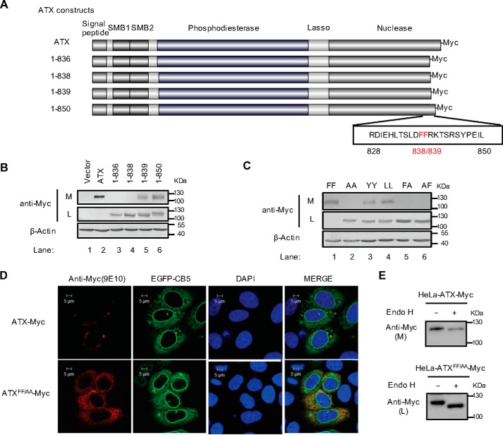
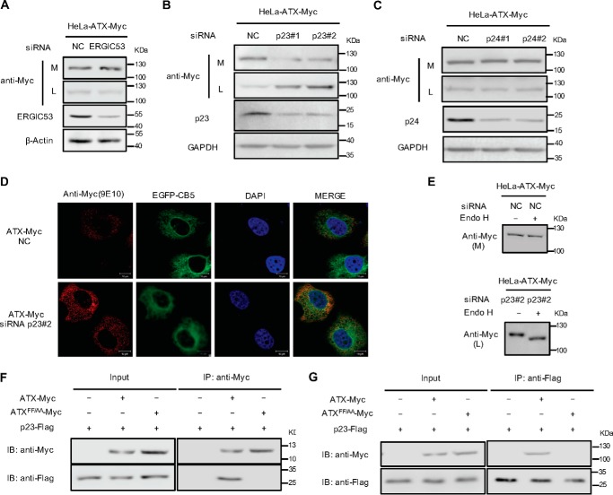
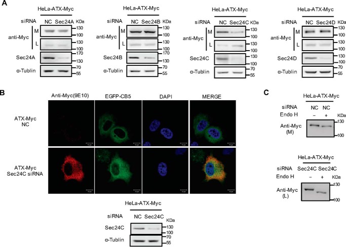
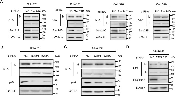
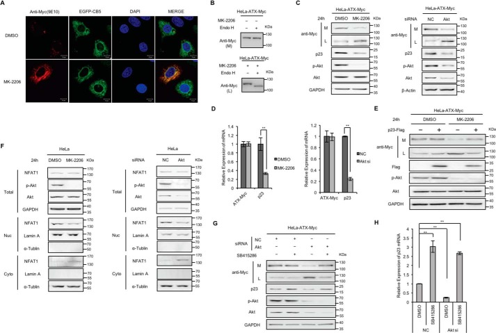
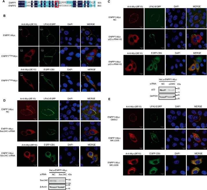
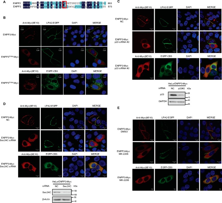
Autotaxin (ATX) or ectonucleotide pyrophosphatase/phosphodiesterase 2 (ENPP2) is a secretory glycoprotein and functions as the key enzyme for lysophosphatidic acid generation. The mechanism of ATX protein trafficking is largely unknown. Here, we demonstrated that p23, a member of the p24 protein family, was the protein-sorting receptor required for endoplasmic reticulum (ER) export of ATX. A di-phenylalanine (Phe-838/Phe-839) motif in the human ATX C-terminal region was identified as a transport signal essential for the ATX-p23 interaction. Knockdown of individual Sec24 isoforms by siRNA revealed that ER export of ATX was impaired only if Sec24C was down-regulated. These results suggest that ATX is selectively exported from the ER through a p23, Sec24C-dependent pathway. In addition, it was found that AKT signaling played a role in ATX secretion regulation to facilitate ATX ER export by enhancing the nuclear factor of activated T cell-mediated p23 expression. Furthermore, the di-hydrophobic amino acid motifs (FY) also existed in the C-terminal regions of human ENPP1 and ENPP3. Such a p23, Sec24C-dependent selective ER export mechanism is conserved among these ENPP family members.

Keywords: COPII; ENPP family; ER export; Sec24C; autotaxin; endoplasmic reticulum (ER); lysophospholipid; p23; protein sorting; receptor.


Conflict of interest statement

The authors declare that they have no conflicts of interest with the contents of this article

Figures

Figure 1.
Figure 1.
Identification of a di-phenylalanine (838FF839) motif essential for ATX secretion. A, schematic representation of the wild-type ATX and the truncated ATX mutants with Myc tag at the C terminus. The number of amino acid residues in each ATX protein is indicated, and the di-phenylalanine (838FF839) motif is labeled in red. B, wild-type ATX and the truncated ATX mutants with C-terminal Myc tag were expressed in HeLa cells as indicated. The levels of ATX-Myc or ATX mutant with Myc tag in cell lysates (L) and culture medium (M) were detected by immunoblotting with anti-Myc antibody. C, wild-type ATX (FF) and the indicated ATX mutants, in which the FF motif was replaced by two alanines (AA), two tyrosines (YY), two leucines (LL), or AF, were expressed in HeLa cells as indicated. The levels of ATX-Myc or ATX mutant with Myc tag in cell lysates and culture medium were detected by immunoblotting with anti-Myc antibody. D, plasmid pcDNA3-ATX-Myc or pcDNA3-ATXFF/AA-Myc, in which the FF motif was replaced by AA, was co-transfected with pEGFP-N1-CB5 into HeLa cells. Cells were fixed and permeabilized 48 h after transfection. ATX-Myc and ATXFF/AA-Myc were visualized by confocal microscopy with anti-Myc (9E10) monoclonal antibody (red). Endoplasmic reticulum was labeled by EGFP-CB5 (green). Nuclei were counterstained with DAPI (blue). E, Endo H treatment of secreted ATX-Myc and intracellular ATXFF/AA-Myc. The concentrated (∼30-fold) serum-free conditional culture medium of the cells transfected with pcDNA3-ATX-Myc and the lysate of the cells transfected with pcDNA3-ATXFF/AA-Myc were treated with Endo H and then analyzed by Western blotting with anti-Myc antibody. Data are representative of three independent experiments.
Figure 2.
Figure 2.
Identification of cargo receptor involved in ER export of ATX. A, HeLa cells were transfected with pcDNA3-ATX-Myc. Twenty four hours after transfection, cells were transfected with siRNA against ERGIC53. Forty eight hours after siRNA transfection, ATX-Myc protein levels in cell lysates (L) and culture medium (M) were detected by immunoblotting with anti-Myc antibody. B and C, HeLa cells were transfected with pcDNA3-ATX-Myc. Twenty four hours after transfection, cells were transfected with nonspecific control siRNA (NC) or siRNAs (#1 and #2) against p23 (B) or siRNAs (#1 and #2) against p24 (C). Forty eight hours after siRNA transfection, ATX-Myc protein levels in cell lysates (L) and culture medium (M) were detected by immunoblotting with anti-Myc antibody. D, HeLa cells were co-transfected with pcDNA3-ATX-Myc and pEGFP-N1-CB5 and then treated with nonspecific control siRNA (NC) or p23 siRNA (#2) for 48 h. ATX-Myc was visualized by confocal microscopy with anti-Myc (9E10) monoclonal antibody (red). Endoplasmic reticulum was labeled by EGFP-CB5 (green). Nuclei were counterstained with DAPI (blue). E, HeLa cells were transfected with pcDNA3-ATX-Myc. Twenty four hours after plasmid transfection, cells were transfected with nonspecific control siRNA (NC) or p23 siRNA (#2) for 48 h. The concentrated (∼30-fold) serum-free conditional culture medium of the control siRNA-treated cells and the lysate of p23 siRNA-treated cells were treated with Endo H and then analyzed by Western blotting with anti-Myc antibody. F, HeLa cells were co-transfected with the plasmid driving the expression of p23-FLAG together with pcDNA3-ATX-Myc or pcDNA3-ATXFF/AA-Myc as indicated. Cells were collected 48 h after transfection. The whole-cell lysates were immunoprecipitated with anti-Myc antibody attached to agarose, and then the immunocomplex was detected by Western blotting using anti-Myc and anti-FLAG as indicated. G, whole-cell lysates mentioned in F were immunoprecipitated with anti-FLAG antibody attached to agarose, and then the immunocomplex was detected by Western blotting using anti-Myc and anti-FLAG antibody as indicated. Data are representative of three independent experiments.
Figure 3.
Figure 3.
Identification of Sec24 isoform involved in ER export of ATX. A, HeLa cells were transfected with pcDNA3-ATX-Myc. Twenty four hours after transfection, cells were transfected with nonspecific control siRNA (NC) or siRNA against the indicated Sec24 isoform. Forty eight hours after siRNA transfection, the ATX-Myc protein levels in cell lysates (L) and culture medium (M) were detected by immunoblotting with anti-Myc antibody. B, HeLa cells were co-transfected with pcDNA3-ATX-Myc and pEGFP-N1-CB5 and then treated with nonspecific control siRNA or Sec24C siRNA for 48 h. ATX-Myc was visualized by confocal microscopy with anti-Myc (9E10) monoclonal antibody (red). Endoplasmic reticulum was labeled by EGFP-CB5 (green). Nuclei were counterstained with DAPI (blue). C, HeLa cells were transfected with pcDNA3-ATX-Myc. Twenty four hours after transfection, cells were transfected with nonspecific control siRNA or Sec24C siRNA for 48 h. The concentrated (∼30-fold) serum-free conditional culture medium of the control siRNA-treated cells and the lysate of Sec24C siRNA-treated cells were treated with Endo H and then analyzed by Western blotting with anti-Myc antibody. Data are representative of three independent experiments.
Figure 4.
Figure 4.
Effects of Sec24 isoform, p23, or ERGI53 knockdown on the secretion of endogenous ATX. A, Colo320 cells were transfected with nonspecific control siRNA (NC) or siRNA against the indicated Sec24 isoform. Forty eight hours after transfection, ATX protein levels in cell lysates (L) and culture medium (M) were detected by immunoblotting with anti-ATX antibody. B and C, Colo320 cells were transfected with nonspecific control siRNA or siRNAs (#1 and #2) against p23 (B) or siRNAs (#1 and #2) against p24 (C). Forty eight hours after siRNA transfection, ATX protein levels in cell lysates and culture medium were detected by immunoblotting with anti-ATX antibody. D, Colo320 cells were transfected with the nonspecific control siRNA or siRNA against ERGIC53. Forty eight hours after transfection, ATX protein levels in cell lysates and culture medium were detected by immunoblotting with anti-ATX antibody. Data are representative of three independent experiments.
Figure 5.
Figure 5.
Effects of AKT inhibitor treatment and AKT knockdown on ATX secretion. A, HeLa cells were co-transfected with pcDNA3-ATX-Myc and pEGFP-N1-CB5 and then treated with or without AKT inhibitor MK-2206 (5 μm) for 24 h. ATX-Myc was visualized by confocal microscopy with anti-Myc (9E10) monoclonal antibody (red). Endoplasmic reticulum was labeled by EGFP-CB5 (green). Nuclei were counterstained with DAPI (blue). B, HeLa cells were transfected with pcDNA3-ATX-Myc and then treated with or without AKT inhibitor MK-2206 (5 μm) for 24 h. The concentrated (∼30-fold) serum-free conditional culture medium of the control cells and the lysate of AKT inhibitor-treated cells were treated with Endo H and then analyzed by Western blotting with anti-Myc antibody. C, HeLa cells with exogenous expression of ATX-Myc were treated with MK-2206 (5 μm) for 24 h or treated with siRNA against AKT for 48 h. Then ATX-Myc protein levels in cell lysates (L) and culture medium (M) were detected by immunoblotting with anti-Myc antibody. p23, AKT, and phosphorylated AKT (p-AKT) levels in cell lysates were detected by immunoblotting. D, HeLa cells with exogenous expression of ATX-Myc were treated with MK-2206 (5 μm) for 24 h or treated with siRNA against AKT for 48 h. ATX-Myc and p23 mRNA levels were detected by quantitative reverse transcription PCR (qRT-PCR). E, HeLa cells with exogenous expression of ATX-Myc and FLAG-p23 were treated with or without AKT inhibitor MK-2206 (5 μm) for 24 h. ATX-Myc protein levels in cell lysates and culture medium were detected by immunoblotting with anti-Myc antibody. FLAG-p23 levels in cell lysates were detected by immunoblotting with anti-FLAG antibody. F, HeLa cells were treated with MK-2206 (5 μm) for 24 h or treated with siRNA against AKT for 48 h. Then, NFAT1 levels in nuclear fraction (Nuc), cytoplasmic fraction (Cyto), and total cell lysates (Total) were detected by Western blotting. Lamin A and α-tubulin were used as the nuclear and cytoplasm markers, as indicated. G and H, HeLa cells with exogenous expression of ATX-Myc were transfected with nonspecific control siRNA (NC) or siRNA against AKT. Thirty two hours after transfection, cells were treated with or without GSK3β inhibitor SB415286 (25 μm) for 16 h. Then, ATX-Myc protein levels in cell lysates and culture medium, and p23 levels in cell lysates were detected by immunoblotting (G). p23 mRNA levels were detected by qRT-PCR (H). The p value was derived from analysis of variance. **, p < 0.005. Data are representative of three independent experiments.
Figure 6.
Figure 6.
Roles of di-hydrophobic amino acid motif (FY), p23, Sec24C, and AKT in the cellular translocation of ENPP1. A, amino acid sequence alignment of ENPP1 and ENPP2 (ATX) C-terminal regions. The FF/FY motif is indicated in a red rectangular box. B, di-hydrophobic amino acid motif (FY) is essential for the translocation of ENPP1. pEGFP-C1-LPA2 or pEGFP-N1-CB5 was co-transfected with pcDNA3-ENPP1-Myc or pcDNA3-ENPP1FY/AA-Myc as indicated into HeLa cells. Cells were fixed and permeabilized 48 h after transfection. C, effects of p23 knockdown on the translocation of ENPP1. HeLa cells were treated with nonspecific control siRNA (NC) or p23 siRNA#2 for 24 h and then transfected with pEGFP-C1-LPA2 or pEGFP-N1-CB5 together with pcDNA3-ENPP1-Myc. Cells were fixed and permeabilized 24 h after transfection. D, effects of Sec24C knockdown on the translocation of ENPP1. HeLa cells were treated with nonspecific control siRNA (NC) or Sec24C siRNA for 24 h and then transfected with pEGFP-C1-LPA2 or pEGFP-N1-CB5 together with pcDNA3-ENPP1-Myc. Cells were fixed and permeabilized 24 h after transfection. E, HeLa cells were transfected with pEGFP-C1-LPA2 or pEGFP-N1-CB5 together with pcDNA3-ENPP1-Myc and then treated with or without AKT inhibitor MK-2206 (5 μm). Cells were fixed and permeabilized 24 h after inhibitor treatment. ENPP1-Myc and ENPP1FY/AA-Myc were visualized by confocal microscopy with anti-Myc (9E10) monoclonal antibody (red). LPA2-EGFP was used as a marker of cell membrane (green). Endoplasmic reticulum was labeled by EGFP-CB5 (green). The nuclei were counterstained with DAPI (blue). Data are representative of three independent experiments.
Figure 7.
Figure 7.
Roles of di-hydrophobic amino acid motif (FY), p23, Sec24C, and AKT in the cellular translocation of ENPP3. A, amino acid sequence alignment of ENPP3 and ENPP2 (ATX) C-terminal regions. The FF/FY motif is indicated in red rectangular box. B, di-hydrophobic amino acid motif (FY) is essential for the translocation of ENPP3. pEGFP-C1-LPA2 or pEGFP-N1-CB5 was co-transfected with pcDNA3-ENPP3-Myc or pcDNA3-ENPP3 FY/AA-Myc as indicated into HeLa cells. Cells were fixed and permeabilized 48 h after transfection. C, effects of p23 knockdown on the translocation of ENPP3. HeLa cells were treated with nonspecific control siRNA (NC) or p23 siRNA#2 for 24 h and then transfected with pEGFP-C1-LPA2 or pEGFP-N1-CB5 together with pcDNA3-ENPP3-Myc. Cells were fixed and permeabilized 24 h after transfection. D, effects of Sec24C knockdown on the translocation of ENPP3. HeLa cells were treated with nonspecific control siRNA (NC) or Sec24C siRNA for 24 h, and then transfected with pEGFP-C1-LPA2 or pEGFP-N1-CB5 together with pcDNA3-ENPP3-Myc. Cells were fixed and permeabilized 24 h after transfection. E, HeLa cells were transfected with pEGFP-C1-LPA2 or pEGFP-N1-CB5 together with pcDNA3-ENPP3-Myc and then treated with or without AKT inhibitor MK-2206 (5 μm). Cells were fixed and permeabilized 24 h after inhibitor treatment. ENPP3-Myc and ENPP3FY/AA-Myc were visualized by confocal microscopy with anti-Myc (9E10) monoclonal antibody (red). LPA2-EGFP was used as a marker of cell membrane (green). Endoplasmic reticulum was labeled by EGFP-CB5 (green). The nuclei were counterstained with DAPI (blue). Data are representative of three independent experiments.

Similar articles

Cited by

References

    1. Yuelling L. M., and Fuss B. (2008) Autotaxin (ATX): a multi-functional and multi-modular protein possessing enzymatic lyso-PLD activity and matricellular properties. Biochim. Biophys. Acta 1781, 525–530 - PMC - PubMed
    1. Pua T. L., Wang F. Q., and Fishman D. A. (2009) Roles of LPA in ovarian cancer development and progression. Future Oncol. 5, 1659–1673 - PubMed
    1. Willier S., Butt E., and Grunewald T. G. (2013) Lysophosphatidic acid (LPA) signalling in cell migration and cancer invasion: a focussed review and analysis of LPA receptor gene expression on the basis of more than 1700 cancer microarrays. Biol. Cell 105, 317–333 - PubMed
    1. Zaslavsky A., Singh L. S., Tan H., Ding H., Liang Z., and Xu Y. (2006) Homo- and hetero-dimerization of LPA/S1P receptors, OGR1 and GPR4. Biochim. Biophys. Acta 1761, 1200–1212 - PubMed
    1. Im D. S. (2010) Pharmacological tools for lysophospholipid GPCRs: development of agonists and antagonists for LPA and S1P receptors. Acta Pharmacol. Sin. 31, 1213–1222 - PMC - PubMed

Publication types

Substances

LinkOut - more resources