. 2017 Apr 28;292(17):6978-6986.
doi: 10.1074/jbc.M117.780742. Epub 2017 Mar 15.

Folding of a single domain protein entering the endoplasmic reticulum precedes disulfide formation

Affiliations

Folding of a single domain protein entering the endoplasmic reticulum precedes disulfide formation

Philip J Robinson et al. J Biol Chem. .

Abstract

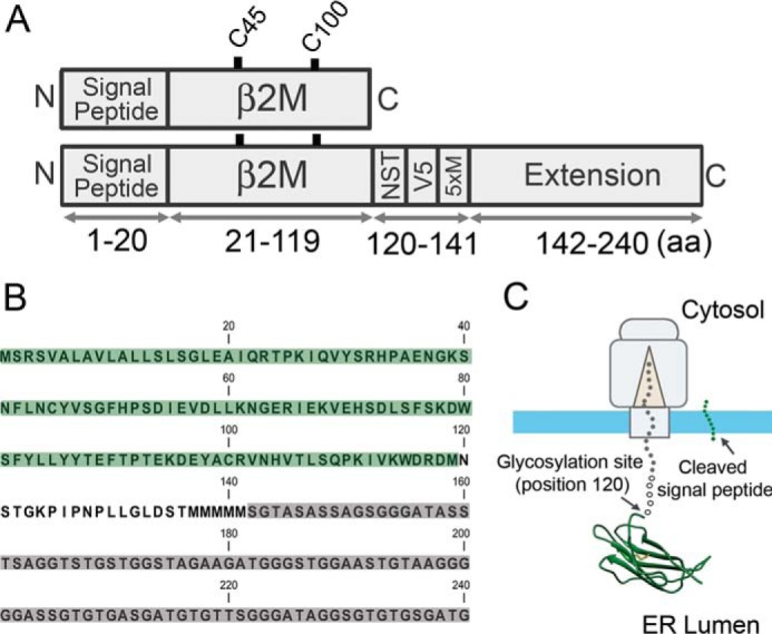
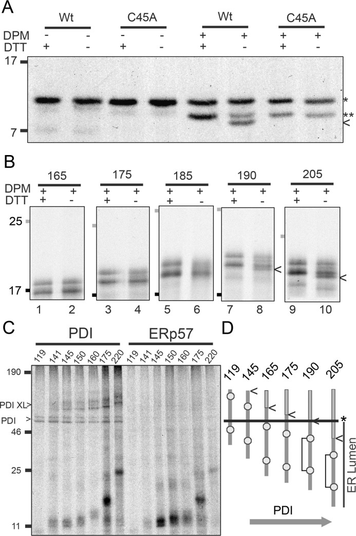
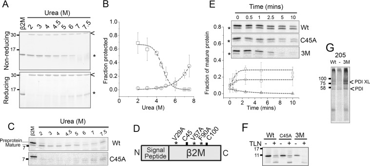
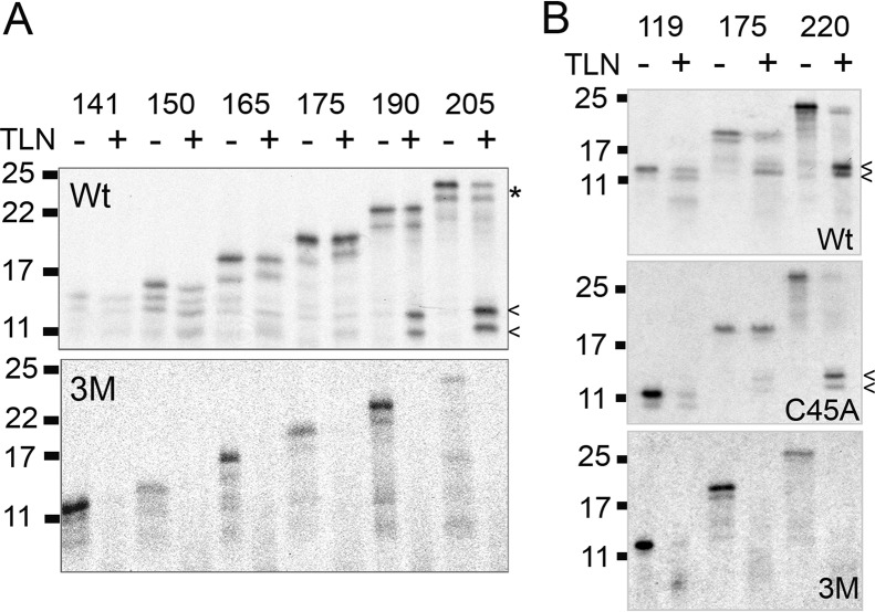
The relationship between protein synthesis, folding, and disulfide formation within the endoplasmic reticulum (ER) is poorly understood. Previous studies have suggested that pre-existing disulfide links are absolutely required to allow protein folding and, conversely, that protein folding occurs prior to disulfide formation. To address the question of what happens first within the ER, that is, protein folding or disulfide formation, we studied folding events at the early stages of polypeptide chain translocation into the mammalian ER using stalled translation intermediates. Our results demonstrate that polypeptide folding can occur without complete domain translocation. Protein disulfide isomerase (PDI) interacts with these early intermediates, but disulfide formation does not occur unless the entire sequence of the protein domain is translocated. This is the first evidence that folding of the polypeptide chain precedes disulfide formation within a cellular context and highlights key differences between protein folding in the ER and refolding of purified proteins.

Keywords: ER; disulfide; disulfide formation; endoplasmic reticulum; protein disulfide isomerase; protein folding; protein translocation; β2-microglobulin.


Conflict of interest statement

The authors declare that they have no conflict of interest

Figures

Figure 1.
Figure 1.
Primary structure of the extended β2M construct and anticipated folding during translocation. A, schematic representation of β2M with and without the extension showing the cysteine residues (Cys-45 and Cys-100) and the organization of key elements including the glycosylation site (NST), V5 epitope (V5), and methionine residues (Met5). B, the complete amino acid sequence of the extended β2M construct with the β2M domain highlighted in green, the additional elements (NST, V5, and Met5) in white, and the extension in gray. C, model of the ribosome-Sec complex displaying the extended β2M construct. The N-terminal β2M domain folds to the native structure once ER exposed, whereas the C terminus is attached to the cytosolic ribosome. 5xM, Met5; aa, amino acid.
Figure 2.
Figure 2.
Translation and translocation of extended β2M intermediates. Autoradiographs of radiolabeled, immunoisolated translation product generated from stalled intermediates of between 165 and 220 amino acids. Gels were run under reducing conditions. A and B, translations were performed (A) in the absence of DPMs and (B) in the presence of DPMs. An example of signal peptide cleavage (*) and glycosylation (arrow) are highlighted for the 220 sample. C, the translation was performed in the presence of DPMs and treated with RNase A on completion. These data are representative of three independent repeats. D, model showing the approximate extension length required to span the ribosome-Sec complex as estimated from the glycosylation data.
Figure 3.
Figure 3.
Intrachain disulfide formation and PDI interactions. Disulfide formation is assessed by SDS-PAGE analysis of immunoisolated, radiolabeled translation product under reducing (+DTT) and nonreducing (−DTT) gel conditions. A, autoradiographs show results for released, β2M (Wt, wild-type) and the single cysteine mutant (C45A) in the absence and presence of DPMs, with pre-protein (*) and mature protein (**) highlighted. Under nonreducing conditions oxidized β2MWt is detected by a shift in the mature protein (arrow). B, autoradiographs of stalled, extended β2M intermediates between 165 and 205 amino acids translated in the presence of DPMs. Molecular weight markers represent 17 (black) and 25 (gray). Arrows indicate the shift associated with disulfide formation for the 190 and 205 stalled intermediates under nonreducing conditions. Results shown are representative of five repeats (165–190 samples) and three repeats (205 sample). C, PDI interactions are detected through cysteine-specific cross-linking, followed by immunoisolation using a PDI antibody. Autoradiograph of a reducing gel shows a range of stalled intermediates (aa 119–220) with PDI-specific cross-links detected for intermediates from 141–220 (PDI XL). PDI expressed in the translation from endogenous RNA was also immunoisolated (PDI). The same samples were immunoisolated with an ERp57 antibody and no cross-links were detected. Data are representative of three independent repeats. D, diagram summarizing the timing of disulfide formation and PDI interactions relative to the entry of β2M into the ER. The arrow indicates the C-terminal end of the β2M sequence (position 120) and the asterisk shows the Sec-ER boundary.
Figure 4.
Figure 4.
The disulfide bond is not required for folding during native synthesis but stabilizes the final fold. A, Coomassie Blue stained SDS-PAGE showing proteolysis of purified β2M (21–119) (*) by thermolysin (arrow) across a range of urea concentrations, under nonreducing and reducing conditions. B, refolding curves of reduced (circles) and oxidized (squares) β2M produced by quantification of gel bands following treatment (mean ± S.D. for n = 3). C, autoradiographs of thermolysin-induced proteolysis of refolded, released β2M (Wt) compared with the single cysteine mutant (C45A), produced through in vitro translation in the presence of DPMs. Data are representative of four independent experiments. D, schematic representation of β2M showing the location of the destabilizing mutations relative to the cysteine residues. E, proteolysis time courses of released β2MWt (squares), β2MC45A (circles), and β2M3M (triangles) produced through in vitro translation in the presence of DPMs. The inset shows representative autoradiographs of these time courses with the plot showing degradation of the mature protein (*) calculated from gel quantification of mature protein and subsequent normalization to total protein (pre-protein plus mature protein) (mean ± S.D. for n = 3). F, thermolysin (TLN) digestion of nondenatured, released β2M translation product (Wt, C45A, or 3M) synthesized in the absence of DPMs. Image is representative of two independent experiments. G, PDI interactions detected by cross-linking to Wt or the 3M mutant, (−) = no template. Data are representative of three independent experiments. All gels in this figure were run under reducing conditions. Trend lines in panels B and E are for guidance only.
Figure 5.
Figure 5.
Co-translational folding of extended β2M to protease resistant conformations proceeds as the nascent chain emerges. Autoradiographs showing stalled translation intermediates of extended β2M produced in the presence of SP cells, assessed for protease susceptibility by thermolysin (TLN) treatment. A and B, digestion profiles of translation intermediates between (A) 141–205 and (B) 119–220 amino acids in length, for wild-type (Wt), 3M mutant (3M) and the single cysteine mutant (C45A). Arrows indicate lower molecular weight resistant bands and the asterisk highlights an example of undigested material for the 205 intermediate. The gels were run under reducing conditions and are representative of three or more independent experiments.

Similar articles

Cited by

References

    1. Fass D. (2012) Disulfide bonding in protein biophysics. Annu. Rev. Biophys. 41, 63–79 - PubMed
    1. Hudson D. A., Gannon S. A., and Thorpe C. (2015) Oxidative protein folding: from thiol-disulfide exchange reactions to the redox poise of the endoplasmic reticulum. Free Radic. Biol. Med. 80, 171–182 - PMC - PubMed
    1. Camacho C. J., and Thirumalai D. (1995) Modeling the role of disulfide bonds in protein folding: entropic barriers and pathways. Proteins 22, 27–40 - PubMed
    1. Wallis A. K., and Freedman R. B. (2013) Assisting oxidative protein folding: how do protein disulphide-isomerases couple conformational and chemical processes in protein folding? Top. Curr. Chem. 328, 1–34 - PubMed
    1. Zhang G., and Ignatova Z. (2011) Folding at the birth of the nascent chain: coordinating translation with co-translational folding. Curr. Opin. Struct. Biol. 21, 25–31 - PubMed

Publication types

Associated data

LinkOut - more resources