. 2017 May 30;135(22):2163-2177.
doi: 10.1161/CIRCULATIONAHA.116.023877. Epub 2017 Mar 15.

Distinct Regulatory Effects of Myeloid Cell and Endothelial Cell NAPDH Oxidase 2 on Blood Pressure

Affiliations

Distinct Regulatory Effects of Myeloid Cell and Endothelial Cell NAPDH Oxidase 2 on Blood Pressure

Can Martin Sag et al. Circulation. .

Abstract

Background: Hypertension caused by increased renin-angiotensin system activation is associated with elevated reactive oxygen species production. Previous studies implicate NADPH oxidase (Nox) proteins as important reactive oxygen species sources during renin-angiotensin system activation, with different Nox isoforms being potentially involved. Among these, Nox2 is expressed in multiple cell types, including endothelial cells, fibroblasts, immune cells, and microglia. Blood pressure (BP) is regulated at the central nervous system, renal, and vascular levels, but the cell-specific role of Nox2 in BP regulation is unknown.

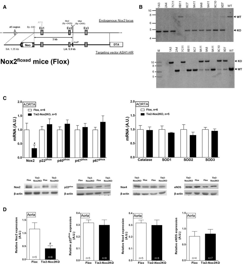
Methods: We generated a novel mouse model with a floxed Nox2 gene and used Tie2-Cre, LysM Cre, or Cdh5-CreERT2 driver lines to develop cell-specific models of Nox2 perturbation to investigate its role in BP regulation.

Results: Unexpectedly, Nox2 deletion in myeloid but not endothelial cells resulted in a significant reduction in basal BP. Both Tie2-CreNox2 knockout (KO) mice (in which Nox2 was deficient in both endothelial cells and myeloid cells) and LysM CreNox2KO mice (in which Nox2 was deficient in myeloid cells) had significantly lower BP than littermate controls, whereas basal BP was unaltered in Cdh5-CreERT2 Nox2KO mice (in which Nox2 is deficient only in endothelial cells). The lower BP was attributable to an increased NO bioavailability that dynamically dilated resistance vessels in vivo under basal conditions without a change in renal function. Myeloid-specific Nox2 deletion had no effect on angiotensin II-induced hypertension, which, however, was blunted in Tie2-CreNox2KO mice, along with preservation of endothelium-dependent relaxation during angiotensin II stimulation.

Conclusions: We identify a hitherto unrecognized modulation of basal BP by myeloid cell Nox2, whereas endothelial cell Nox2 regulates angiotensin II-induced hypertension. These results identify distinct cell-specific roles for Nox2 in BP regulation.

Keywords: NADPH oxidase; angiotensin II; blood pressure; mice.


Figures

Figure 1.
Figure 1.
Generation of Tie2-Nox2 knockout (KO) mice. A, Targeting strategy for generation of Nox2flox mice (Flox). The endogenous Nox2 locus is shown at the top and the targeting vector at the bottom. LoxP sites are represented by blue triangles; FRT sites, by double red triangles. After successful targeting, the neomycin (Neo) cassette was excised with the FRT sites. Cre-mediated recombination deletes a 3-kilobase fragment of Nox2, including the transcriptional initiation site and the first 2 exons. B, Southern blot analysis of genomic DNA from selected ES cell clones screened for 5′-homologous (top) and 3′-homologous (bottom) recombination. The clones 1A3 and 7C11 were used for blastocyst injection to generate Nox2flox mice. WT indicates wild-type. C, mRNA levels of Nox2, accessory subunits, and antioxidant genes in Tie2-Nox2KO and Flox control aortas. D, Protein levels of Nox2, p22phox, Nox4, and endothelial nitric oxide synthase (eNOS) in Tie2-Nox2KO and control aortas. Representative immunoblots are shown above and mean data below. #P<0.05 vs Flox.
Figure 2.
Figure 2.
Tie2-Nox2 knockout (KO) mice have reduced basal blood pressure. A, Representative telemetric blood pressure (BP) traces in Tie2-Nox2KO mice and Flox controls. B, Mean data for BP, heart rate, and activity level. #P<0.05 vs Flox.
Figure 3.
Figure 3.
Renal function, vascular remodeling, and in vitro vascular function in Tie2-Nox2 knockout (KO) mice.A, Renal function assessed by response to a short-term saline challenge. Changes in urine osmolarity, volume, and sodium and potassium concentrations are shown. RM-TW-ANOVA indicates repeated-measures 2-way ANOVA; and N.S., not significant between groups. B, Representative histological sections of aortas from Tie2-Nox2KO and Flox mice (×40 magnification) and mean intima-media area and thickness. C, Concentration-response curves for response of aortic rings to the nitric oxide donor sodium nitroprusside (SNP), phenylephrine (PE), and acetylcholine (ACh). D, Concentration-response curves for response of mesenteric arteries to ACh.
Figure 4.
Figure 4.
Tie2-Nox2 knockout (KO) mice demonstrate blunted in vitro and in vivo responses to angiotensin II (AngII). A, Top Left, Superoxide levels in Tie2-Nox2KO and Flox aortas at baseline and after AngII stimulation, assessed by high-performance liquid chromatography of the specific dihydroethidium oxidation product 2-hydroxyethidium (EOH). *P<0.05 vs control conditions (2-way-ANOVA). #P<0.05 vs Flox. Top Right, Effect of AngII pretreatment on vasodilation of aortic rings to acetylcholine (ACh). Bottom, Effect of MnTMPyP on ACh responses in AngII-treated rings from Flox mice (left) and Tie2-Nox2KO mice (right). B, Effect of long-term AngII infusion (1.1 mg·kg−1·d−1) on telemetric blood pressure (BP) and heart rate in Tie2-Nox2KO and Flox mice. RM-TW-ANOVA indicates repeated-measures 2-way ANOVA. *Significant interaction. #Significant difference between genotypes.
Figure 5.
Figure 5.
Tie2-Nox2 knockout (KO) mice have increased basal nitric oxide (NO) bioavailability in vivo. A, Blood pressure (BP) response to Nω-nitro-l-arginine methyl ester (L-NAME) in Tie2-Nox2KO and Flox mice, assessed by telemetry. *Significant interaction as tested by repeated-measures 2-way ANOVA (RM-TW-ANOVA). #Significant difference between genotypes. B and C, Representative magnetic resonance images of the carotid artery in Tie2-Nox2KO and Flox mice at baseline (B) and after L-NAME treatment (C). Scale bar, 1 mm. D through F, Mean data for diastolic area of the carotid artery at baseline (D), after acetylcholine (ACh) treatment (E), and after L-NAME treatment (F). *P<0.05 vs control conditions (TW-ANOVA). #P<0.05 vs Flox.
Figure 6.
Figure 6.
LysM-Cre-Nox2 knockout (KO) mice have reduced basal blood pressure (BP) and increased vascular nitric oxide (NO) bioavailability. A, Cre-mediated recombination in bone marrow–derived cells from Tie2-Nox2KO mice. WT indicates wild-type. B, Functionally deficient reactive oxygen species production in phorbol ester (PMA)–stimulated bone marrow cells and circulating mononuclear cells from Tie2-Nox2KO mice, assessed by flow cytometry in cells loaded with dihydroethidium. C, Reduced basal BP (telemetry) in LysM-Cre-Nox2KO mice. #P<0.05 vs Flox. D, In vivo response to Nω-nitro-l-arginine methyl ester (L-NAME) in LysM-Cre-Nox2KO mice and controls. #Significance vs Flox as tested by repeated-measures 2-way ANOVA (RM-TW-ANOVA). E. Increased aortic NO levels in LysM-Cre-Nox2KO under basal conditions, as assessed by electron paramagnetic resonance (EPR). Representative EPR spectra are shown on the right. #P<0.05 vs Flox.
Figure 7.
Figure 7.
Cdh5-CreERT2-Nox2 knockout (KO) mice have unaltered basal blood pressure (BP). A, Telemetric BP and heart rate at baseline. B, BP responses to Nω-nitro-l-arginine methyl ester (L-NAME) in Cdh5-CreERT2-Nox2KO and Flox mice. L-NAME increased BP in both groups, but there was no significant difference between groups by repeated-measures 2-way ANOVA (RM-TW-ANOVA). C, Comparable aortic nitric oxide (NO) levels in Cdh5-CreERT2-Nox2KO and controls under basal conditions, as assessed by electron paramagnetic resonance (EPR). Representative EPR spectra are shown on the right.

Comment in

Similar articles

Cited by

References

    1. Laursen JB, Rajagopalan S, Galis Z, Tarpey M, Freeman BA, Harrison DG. Role of superoxide in angiotensin II-induced but not catecholamine-induced hypertension. Circulation. 1997;95:588–593. - PubMed
    1. Higashi Y, Sasaki S, Nakagawa K, Matsuura H, Oshima T, Chayama K. Endothelial function and oxidative stress in renovascular hypertension. N Engl J Med. 2002;346:1954–1962. doi: 10.1056/NEJMoa013591. - PubMed
    1. Dröge W. Free radicals in the physiological control of cell function. Physiol Rev. 2002;82:47–95. doi: 10.1152/physrev.00018.2001. - PubMed
    1. Burgoyne JR, Mongue-Din H, Eaton P, Shah AM. Redox signaling in cardiac physiology and pathology. Circ Res. 2012;111:1091–1106. doi: 10.1161/CIRCRESAHA.111.255216. - PubMed
    1. Lonn E, Bosch J, Yusuf S, Sheridan P, Pogue J, Arnold JM, Ross C, Arnold A, Sleight P, Probstfield J, Dagenais GR HOPE and HOPE-TOO Trial Investigators. Effects of long-term vitamin E supplementation on cardiovascular events and cancer: a randomized controlled trial. JAMA. 2005;293:1338–1347. doi: 10.1001/jama.293.11.1338. - PubMed

MeSH terms