Review
. 2017 Apr;30(2):503-528.
doi: 10.1128/CMR.00074-16.

BK Polyomavirus: Clinical Aspects, Immune Regulation, and Emerging Therapies

Affiliations
Review

BK Polyomavirus: Clinical Aspects, Immune Regulation, and Emerging Therapies

George R Ambalathingal et al. Clin Microbiol Rev. 2017 Apr.

Abstract

BK polyomavirus (BKV) causes frequent infections during childhood and establishes persistent infections within renal tubular cells and the uroepithelium, with minimal clinical implications. However, reactivation of BKV in immunocompromised individuals following renal or hematopoietic stem cell transplantation may cause serious complications, including BKV-associated nephropathy (BKVAN), ureteric stenosis, or hemorrhagic cystitis. Implementation of more potent immunosuppression and increased posttransplant surveillance has resulted in a higher incidence of BKVAN. Antiviral immunity plays a crucial role in controlling BKV replication, and our increasing knowledge about host-virus interactions has led to the development of improved diagnostic tools and clinical management strategies. Currently, there are no effective antiviral agents for BKV infection, and the mainstay of managing reactivation is reduction of immunosuppression. Development of immune-based therapies to combat BKV may provide new and exciting opportunities for the successful treatment of BKV-associated complications.

Keywords: B cell responses; T cell immunity; T cells; immunotherapy; natural killer cells; pathogenesis; transplant; virus.


Figures

FIG 1
FIG 1
Phylogenetic tree showing the distances of relationships between the human polyomaviruses discovered to date.
FIG 2
FIG 2
Cryo-electron microscopy structure of BKV, showing an external view, minor capsid proteins, and genome organization. (A) External view of the virion at a contour level of 0.022. The electron density maps were sharpened using a negative B factor correction (B = −456 and −804 Å2). (B) View of a 40-Å-thick slab through the unsharpened/unmasked virion map, shown at a contour level of 0.0034. Pyramidal density below each VP1 penton and two shells of electron density adjacent to the inner capsid layer can be seen. The density within 6 Å of the fitted coordinates for SV40 VP1 is colored gray. The remaining density is colored in a radial color scheme. Densities for VP2 and VP3 are colored blue and green, and those for packaged dsDNA are yellow and pink. (C) Enlarged view of the pyramidal density beneath a single VP1 penton of the virion, shown at a contour level of 0.0032. Strands of dsDNA wrapped around a human histone octamer (PDB entry 1AOI) are shown, indicating that the two shells of density have comparable spacings. A discrete connective density between the pyramidal density and internal shells is also apparent. (Adapted from reference [published under a Creative Commons {CC BY} license].)
FIG 3
FIG 3
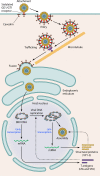
Schematic outline of BKV entry into host cells. For caveola-mediated entry of BKV into host cells, attachment of VP1 to the ganglioside receptors GD1 and GT1 initializes the internalization of BKV through caveola formation. The caveola encapsulating the virus then translocates in the cytoplasm with the help of microtubules. The virus then fuses with the endoplasmic reticulum (ER), where the VP1 layer disassembles. The virus then enters the host nucleus and lies episomally. In the nucleus, the early coding regions are transcribed first, which regulates the transcription of late coding regions.
FIG 4
FIG 4
Genome stucture of BKV. The transcription of both early and late coding regions proceeds in a bidirectional way from the origin of replication (ORI) within the noncoding control region (NCCR). The transcriptional splicing regions are represented by dashed lines. The late coding regions encode structural proteins (VP1, VP2, and VP3), while the early coding regions transcribe the tumorigenic proteins (LTA and STA). The late coding regions also encode the nonimmunogenic agnoprotein. The expression of miRNA complementary to the 3′ end of LTA has been shown to be involved in replication control of BKV.
FIG 5
FIG 5
Identified risk factors for BKV infection and/or reactivation in transplant recipients. These risk factors are associated with either the immunological, age, sex, or metabolic status of the recipient and donor or the physiological/clinical profile of the transplanted organ. Each of these risk factors can directly or indirectly increase the risk of BKV infection and/or reactivation.
None
None
None
None
None

References

    1. Gardner SD, Field AM, Coleman DV, Hulme B. 1971. New human papovavirus (B.K.) isolated from urine after renal transplantation. Lancet i:1253–1257. - PubMed
    1. Bennett SM, Broekema NM, Imperiale MJ. 2012. BK polyomavirus: emerging pathogen. Microbes Infect 14:672–683. doi:10.1016/j.micinf.2012.02.002. - DOI - PMC - PubMed
    1. Chatterjee M, Weyandt TB, Frisque RJ. 2000. Identification of archetype and rearranged forms of BK virus in leukocytes from healthy individuals. J Med Virol 60:353–362. - PubMed
    1. Knowles WA. 2006. Discovery and epidemiology of the human polyomaviruses BK virus (BKV) and JC virus (JCV). Adv Exp Med Biol 577:19–45. doi:10.1007/0-387-32957-9_2. - DOI - PubMed
    1. Boukoum H, Nahdi I, Sahtout W, Skiri H, Segondy M, Aouni M. 2016. BK and JC virus infections in healthy patients compared to kidney transplant recipients in Tunisia. Microb Pathog 97:204–208. doi:10.1016/j.micpath.2016.06.015. - DOI - PubMed

MeSH terms

Substances