. 2017 May 1;28(9):1271-1283.
doi: 10.1091/mbc.E17-01-0008. Epub 2017 Mar 15.

Deubiquitinating enzymes Ubp2 and Ubp15 regulate endocytosis by limiting ubiquitination and degradation of ARTs

Affiliations

Deubiquitinating enzymes Ubp2 and Ubp15 regulate endocytosis by limiting ubiquitination and degradation of ARTs

Hsuan-Chung Ho et al. Mol Biol Cell. .

Abstract

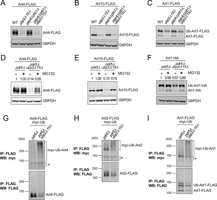
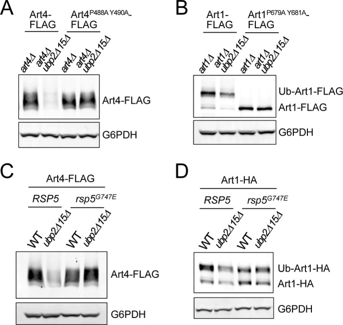
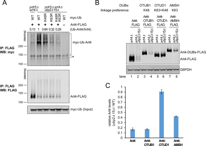
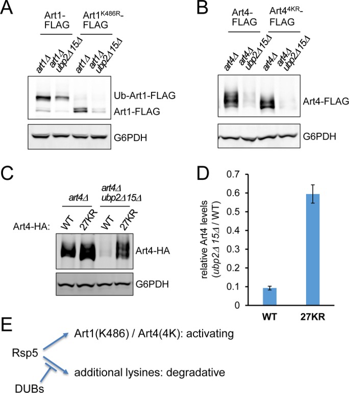
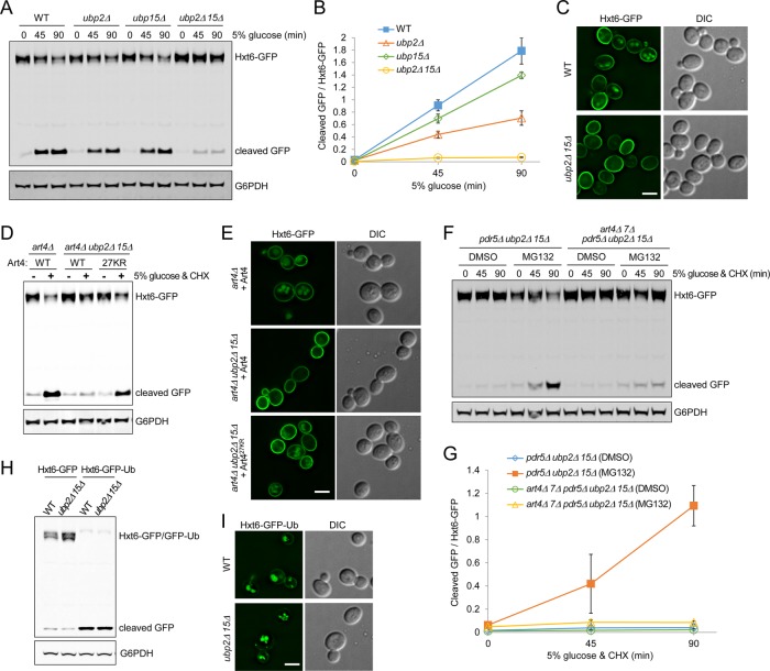
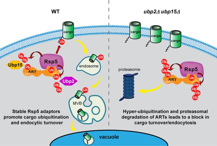
Endocytic down-regulation of cell-surface proteins is a fundamental cellular process for cell survival and adaptation to environmental stimuli. Ubiquitination of cargo proteins serves as the sorting signal for downstream trafficking and relies on the arrestin-related trafficking adaptor (ART)-Rsp5 ubiquitin ligase adaptor network in yeast. Hence proper regulation of the abundance and activity of these ligase-adaptor complexes is critical for main-tenance of optimal plasma membrane protein composition. Here we report that the stability of ARTs is regulated by the deubiquitinating enzymes (DUBs) Ubp2 and Ubp15. By counteracting the E3 ubiquitin ligase Rsp5, Ubp2 and Ubp15 prevent hyperubiquitination and proteasomal degradation of ARTs. Specifically, we show that loss of both Ubp2 and Ubp15 results in a defect in Hxt6 endocytosis associated with Art4 instability. Our results uncover a novel function for DUBs in the endocytic pathway by which Ubp2 and Ubp15 positively regulate the ART-Rsp5 network.


Figures

FIGURE 1:
FIGURE 1:
Ubp2 and Ubp15 maintain the abundance of Rsp5 adaptor proteins. (A) Protein extracts of wild-type (WT) or the indicated yeast mutant strains expressing plasmid-encoded Art4-FLAG were analyzed by SDS–PAGE and immunoblot. (B–I) Protein extracts of WT, ubp2Δ, ubp15Δ, and ubp2Δ ubp15Δ yeast cells expressing chromosomally tagged ARTs were analyzed by SDS–PAGE and immunoblot. Ub-Art1, K486-dependent ubiquitinated form of Art1 (Lin et al., 2008). *Nonspecific band. The numbers above each lane indicate the protein abundance of ARTs determined by the LI-COR quantification and normalized with the loading control G6PDH.
FIGURE 2:
FIGURE 2:
Ubp2 and Ubp15 regulate the degradation of ARTs. (A) art4Δ and art4Δ ubp2Δ ubp15Δ yeast cells carrying Art4-FLAG, Art4-UL36-FLAG, or Art4-UL36C40S-FLAG expression vector. Protein extracts were analyzed by SDS–PAGE and immunoblot. (B) Immunoblot analysis of chromosomal Art4-FLAG in WT and ubp2Δ ubp15Δ yeast cells carrying functional or catalytically dead Ubp2/Ubp15 expression vector. (C) WT and ubp2Δ ubp15Δ yeast cells expressing chromosomal Art4-FLAG or ubp2Δ ubp15Δ cells overexpressing (OE) Art4-FLAG (driven by the CYC1 promoter inserted into ART4 locus) were treated with 100 μg/ml CHX to stop protein synthesis. Protein extracts from the indicated time points were analyzed by SDS–PAGE and immunoblot. (D) Art4 levels (relative to t = 0) as in C were quantified by the LI-COR system. Data from three independent experiments are presented as mean ± SD. (E, F) similar to C and D, except that yeast strains with chromosomal Art1-FLAG were examined.
FIGURE 3:
FIGURE 3:
Ubp2 and Ubp15 prevent hyperubiquitination and proteasomal degradation of ARTs. (A–C) Protein extracts of indicated yeast strains expressing plasmid-encoded Art4-FLAG (A), Art10-FLAG (B), or Art1-FLAG (C) were analyzed by SDS–PAGE and immunoblot. (D–F) pdr5Δ and pdr5Δ ubp2Δ ubp15Δ cells expressing plasmid-encoded Art4-FLAG (D), Art10-FLAG (E), or Art1-HA (F) were mock-treated (dimethyl sulfoxide [DMSO]) or treated with MG132 (25 μg/ml) for 60 min, and protein extracts were analyzed by SDS–PAGE and immunoblot. The numbers above each lane indicate the protein abundance of ARTs determined by the LI-COR quantification and normalized with the loading control G6PDH. (G–I) pdr5Δ and pdr5Δ ubp2Δ ubp15Δ cells expressing plasmid-encoded Art4-FLAG (G), Art2-FLAG (H), or Art1-FLAG (I) and myc-ubiquitin (overexpression controlled by copper-inducible promoter) were treated with MG132 (25 μg/ml) for 60 min. Samples were immunoprecipitated using anti-FLAG antibody under denaturing conditions and analyzed by SDS–PAGE and immunoblot. *Nonspecific.
FIGURE 4:
FIGURE 4:
ART degradation in ubp2Δ ubp15Δ is mediated by E3 ubiquitin ligase Rsp5. (A) Protein extracts of art4Δ and art4Δ ubp2Δ ubp15Δ yeast cells expressing plasmid-encoded Art4-FLAG or Art4P488A Y490A-FLAG were analyzed by SDS–PAGE and immunoblot. (B) Protein extracts of art1Δ and art1Δ ubp2Δ ubp15Δ yeast cells expressing plasmid-encoded Art1-FLAG or Art1P679A Y681A-FLAG were analyzed by SDS–PAGE and immunoblot. (C, D) Protein extracts of RSP5, RSP5 ubp2Δ ubp15Δ, rsp5G747E, and rsp5G747E ubp2Δ ubp15Δ cells expressing plasmid-encoded Art4-FLAG (C) or Art1-HA (D) were analyzed by SDS–PAGE and immunoblot.
FIGURE 5:
FIGURE 5:
Art4 hyperubiquitination and degradation in ubp2Δ ubp15Δ depends on the formation of K63-linked polyubiquitin chains. (A) pdr5Δ art4Δ and pdr5Δ art4Δ ubp2Δ ubp15Δ yeast cells carrying Art4-FLAG or empty vector, as well as WT, K48R, K63R, or K48R K63R myc-ubiquitin expression vector. Cells were treated with MG132 (25 μg/ml) for 60 min. Samples were immunoprecipitated using anti-FLAG antibody under denaturing conditions and analyzed by SDS–PAGE and immunoblot. The numbers above each lane indicate the relative ratio of myc-Ub-Art4 vs. total Art4 determined by the LI-COR quantification. To compare more accurately the levels of myc-Ub conjugated Art4, stacking gel was included for the analysis. *Nonspecific. (B) art4Δ and art4Δ ubp2Δ ubp15Δ yeast cells carrying Art4-FLAG, Art4-OTUB1-FLAG, Art4-OTUD1-FLAG, or Art4-AMSH-FLAG expression vector. Protein extracts were analyzed by SDS–PAGE and immunoblot. (C) Relative Art4/Art4-OTUB1, Art4-OTUD1, and Art4-AMSH levels (art4Δ ubp2Δ ubp15Δ cells versus art4Δ cells) as in B were quantified using the LI-COR system. Data from at least three independent experiments are presented as mean ± SD.
FIGURE 6:
FIGURE 6:
ART degradation in ubp2Δ ubp15Δ is not mediated by ubiquitination at the activating lysine residues. (A) Protein extracts of art1Δ and art1Δ ubp2Δ ubp15Δ yeast cells expressing plasmid-encoded Art1-FLAG or Art1K486R-FLAG were analyzed by SDS–PAGE and immunoblot. (B) Protein extracts of art4Δ and art4Δ ubp2Δ ubp15Δ yeast cells expressing plasmid-encoded Art4-FLAG or Art4K(235/245/264/267)R-FLAG were analyzed by SDS–PAGE and immunoblot. (C) Protein extracts of art4Δ and art4Δ ubp2Δ ubp15Δ cells expressing plasmid-encoded Art4-HA or Art427KR-HA were analyzed by SDS–PAGE and immunoblot. (D) Relative Art4 levels (art4Δ ubp2Δ ubp15Δ cells vs. art4Δ cells) as in C were quantified using the LI-COR system. Data from three independent experiments are presented as mean ± SD. (E) Model for functionally distinct ubiquitinations of ARTs. Rsp5 ubiquitinates Art1 at K486 and Art4 at K235/K245/K264/K267, which are required for their activation. In the absence of Ubp2 and Ubp15, additional lysines on ARTs are ubiquitinated, causing their degradation.
FIGURE 7:
FIGURE 7:
Ubp2 and Ubp15 promote Hxt6 endocytic down-regulation by stabilizing Art4. (A–C) The indicated strains expressing chromosomal Hxt6-GFP were grown to mid log phase in medium containing 0.05% glucose. Hxt6 endocytosis was stimulated with 5% glucose. (A) Protein extracts at the indicated time points were analyzed by SDS–PAGE and immunoblot. The cleaved GFP signals indicate the product of Hxt6-GFP degradation in the vacuole. (B) The ratio of cleaved GFP vs. full-length Hxt6-GFP as in A was determined by the quantification using the LI-COR system. Data from three independent experiments are presented as mean ± SD. (C) Samples were analyzed by fluorescence microscopy after 90 min of stimulation. Scale bar, 2.5 μm. (D, E) art4Δ and art4Δ ubp2Δ ubp15Δ cells expressing chromosomal Hxt6-GFP and plasmid-encoded Art4 or Art427KR were grown in medium containing 0.05% glucose. Hxt6 endocytosis was stimulated with 5% glucose and 50 μg/ml CHX. (D) Protein extracts after 3.5 h of stimulation were analyzed by SDS–PAGE and immunoblot. (E) Samples were analyzed by fluorescence microscopy after 3 h of stimulation. Scale bar, 2.5 μm. (F) pdr5Δ ubp2Δ ubp15Δ and art4Δ art7Δ pdr5Δ ubp2Δ ubp15Δ yeast cells expressing chromosomal Hxt6-GFP were grown to early log phase in medium containing 0.05% glucose and then mock-treated (DMSO) or treated with MG132 (25 μg/ml) for 90 min. Hxt6 endocytosis was stimulated with 5% glucose and 50 μg/ml CHX. Protein extracts from the indicated time points were analyzed by SDS–PAGE and immunoblot. (G) The ratio of cleaved GFP vs. full-length Hxt6-GFP as in F was determined by the quantification using the LI-COR system. Data from three independent experiments are presented as mean ± SD. (H) WT and ubp2Δ ubp15Δ cells expressing plasmid-encoded Hxt6-GFP or Hxt6-GFP-Ub were grown to mid log phase in medium containing 0.05% glucose. Protein extracts were analyzed by SDS–PAGE and immunoblot. (I) Similar to H, WT and ubp2Δ ubp15Δ cells expressing plasmid-encoded Hxt6-GFP-Ub were analyzed by fluorescence microscopy. Scale bar, 2.5 μm.
FIGURE 8:
FIGURE 8:
Model for Ubp2 and Ubp15 preventing Rsp5-mediated ART hyperubiquitination. In WT cells, Ubp2 and Ubp15 counteract Rsp5 activity and prevent ART hyperubiquitination. This is achieved most likely by physical interactions between Ubp2 and Rsp5, as well as between Ubp15 and ARTs. We propose that ART-Rsp5 complexes are direct targets of Ubp2 and/or Ubp15. Ubp2 and Ubp15 may regulate different steps of an ART-Rsp5 loading cycle, with Ubp2 engaging ART-Rsp5 complexes by binding to Rsp5, and Ubp15 engaging ARTs. Whether Ubp15 interacts with ARTs in complexes with Rsp5 or with ARTs that have dissociated from Rsp5 is unclear. ART-Rsp5 promotes PM cargo ubiquitination, leading to endocytic down-regulation. In ubp2Δ ubp15Δ cells, ARTs are hyperubiquitinated by Rsp5. At least several ARTs exhibit enhanced protein degradation through the proteasome, leading to reduced ART abundance and defective cargo turnover. The degradative hyperubiquitination of Art4 occurs most likely at N- and C-terminal regions. Regions of degradative ubiquitination on other ARTs have not been determined. Although the model illustrates that Ubp2 and Ubp15 target different residues/regions, it is unknown how these DUBs recognize substrates or whether motif specificity exists. Ub, ubiquitin; red arrow, Rsp5 E3 ligase activity.

Similar articles

Cited by

References

    1. Alvaro CG, Aindow A, Thorner J. Differential phosphorylation provides a switch to control how alpha-arrestin rod1 down-regulates mating pheromone response in saccharomyces cerevisiae. Genetics. 2016;203:299–317. - PMC - PubMed
    1. Alvaro CG, O’Donnell AF, Prosser DC, Augustine AA, Goldman A, Brodsky JL, Cyert MS, Wendland B, Thorner J. Specific alpha-arrestins negatively regulate Saccharomyces cerevisiae pheromone response by down-modulating the G-protein-coupled receptor Ste2. Mol Cell Biol. 2014;34:2660–2681. - PMC - PubMed
    1. Amerik AY, Hochstrasser M. Mechanism and function of deubiquitinating enzymes. Biochim Biophys Acta. 2004;1695:189–207. - PubMed
    1. Amerik AY, Li SJ, Hochstrasser M. Analysis of the deubiquitinating enzymes of the yeast Saccharomyces cerevisiae. Biol Chem. 2000a;381:981–992. - PubMed
    1. Amerik AY, Nowak J, Swaminathan S, Hochstrasser M. The Doa4 deubiquitinating enzyme is functionally linked to the vacuolar protein-sorting and endocytic pathways. Mol Biol Cell. 2000b;11:3365–3380. - PMC - PubMed

MeSH terms

Substances

LinkOut - more resources