. 2017 May 1;198(9):3705-3718.
doi: 10.4049/jimmunol.1601932. Epub 2017 Mar 15.

Identification of C3b-Binding Small-Molecule Complement Inhibitors Using Cheminformatics

Affiliations

Identification of C3b-Binding Small-Molecule Complement Inhibitors Using Cheminformatics

Brandon L Garcia et al. J Immunol. .

Abstract

The complement system is an elegantly regulated biochemical cascade formed by the collective molecular recognition properties and proteolytic activities of more than two dozen membrane-bound or serum proteins. Complement plays diverse roles in human physiology, such as acting as a sentry against invading microorganisms, priming of the adaptive immune response, and removal of immune complexes. However, dysregulation of complement can serve as a trigger for a wide range of human diseases, which include autoimmune, inflammatory, and degenerative conditions. Despite several potential advantages of modulating complement with small-molecule inhibitors, small-molecule drugs are highly underrepresented in the current complement-directed therapeutics pipeline. In this study, we have employed a cheminformatics drug discovery approach based on the extensive structural and functional knowledge available for the central proteolytic fragment of the cascade, C3b. Using parallel in silico screening methodologies, we identified 45 small molecules that putatively bind C3b near ligand-guided functional hot spots. Surface plasmon resonance experiments resulted in the validation of seven dose-dependent C3b-binding compounds. Competition-based biochemical assays demonstrated the ability of several C3b-binding compounds to interfere with binding of the original C3b ligand that guided their discovery. In vitro assays of complement function identified a single complement inhibitory compound, termed cmp-5, and mechanistic studies of the cmp-5 inhibitory mode revealed it acts at the level of C5 activation. This study has led to the identification of a promising new class of C3b-binding small-molecule complement inhibitors and, to our knowledge, provides the first demonstration of cheminformatics-based, complement-directed drug discovery.


Figures

Figure 1
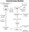
Figure 1. Cheminformatics workflow schematic
Small molecules that putatively bind C3b near functionally important “hot-spots” were identified using an in silico docking approach. Target binding boxes were defined on C3b corresponding to a region of the Ba domain contact site of the C3b/fB interface (PDB: 2XWJ) (30) or the entirety of the C3c/compstatin interface (PDB: 2QKI) (31). Next, 4.27 x 10⁶ unique drug-like compounds originating from the ZINC database “Leads Now” subset were docked into each set of coordinates using AutoDock Vina (32). Results were ranked according to calculated binding energies and compounds exhibiting +4.5 S.D. from the average value were triaged. This compound subset was then assessed for favorable predicted physicochemical properties (i.e. xLogP/solubility), structural diversity, and commercial availability. An alternative ligand-based method (ChemVassa) was used to identify and dock small molecules with chemical and structural similarity to SCIN-B. Here, the C3c/SCIN-B crystal structure (PDB: 3T4A), guided by previously obtained mutagenesis-based C3b affinity data (25, 34, 35), was used to calculate a ChemVassa information signature. A prioritized set of compounds from each screen were obtained via commercial synthesis and evaluated for direct binding to C3b (SPR) and complement inhibitory activity (ELISA). Validated hits were queried against the entire set of purchasable compounds in the ZINC database and those exhibiting high similarity were rescreened following an identical workflow.
Figure 2
Figure 2. In silico screening of small molecules which bind C3b near functional “hot-spots”
(A) AP C3 proconvertases are formed upon interaction of surface-attached C3b (grey) with fB (cyan oval). Subsequent cleavage of fB by the serine protease fD (red) results in active AP convertases (C3bBb) and release of the Ba fragment (open cyan triangle). Compounds capable of disrupting the C3b/fB protein-protein interface hold potential as complement inhibitors. A site formed between the CCP3 domain of the Ba fragment and C3b was selected as the target region for the fB-based screen (PDB: 2XWJ). The C3b/C3c contact residues used to guide each screen have been highlighted in yellow (middle panels) while the final Autodock Vina binding box site selection is shown on C3b in red (bottom panels). (B) Compstatin (orange pentagon) is a synthetic cyclic peptide which potently inactivates complement by binding C3 and preventing its cleavage by AP or CP/LP (i.e. C4b2a) C3 convertases. The co-crystal structure of C3c/compstatin (PDB: 2QKI) was used to define binding box coordinates for compound docking. (C) SCINs (purple cylinder) are multifunctional C3b-binding complement evasion proteins expressed by S. aureus which act at the level of AP C3 convertases. To identify potential small molecule mimics of SCIN, ChemVassa was used to calculate a chemical information signature for the primary C3b interaction site formed by residues on the second α-helix of SCIN-B (PDB: 3T4A). Compounds in the ZINC database with closely matched information signatures were subsequently docked onto C3b using Autodock Vina.
Figure 3
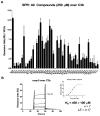
Figure 3. SPR measurements of direct C3b-binding
SPR was used to measure direct interaction of small molecules with C3b. (A) Each compound was injected at 250 μM over a C3b biosensor in duplicate. Binding response was obtained as detailed in the Materials and Methods section and is expressed as RU/100 Da. Cp40 (100 μM) was injected to derive an experimental maximal signal for this surface (13 ± 0.27 RU/100 Da). Compounds fb-2-2, fb-2-13, and fb-2-19 exhibited abnormal sensorgram shape and were removed from further analysis. These compounds are denoted with an asterisk (*). (B) Representative sensorgrams and fit using free Rmax from a variable concentration injection series of cmp-5 over C3b. A dissociation constant (KD) was obtained by steady-state analysis using Biacore T200 Evaluation Software and KD is reported as the mean ± S.D. from seven independent injection series performed across five separate C3b biosensors. LE = ligand efficiency computed as ΔG/Nheavy atoms where ΔG = −RT x ln (KD,SPR).
Figure 4
Figure 4. The effect of fB-2 compounds on the fB/C3b interaction
(A) The docking positions of the fB-2 compound library are shown in the context of the fB/C3b crystal structure (PDB: 2XWJ). C3b is shown in surface representation (grey), while a cartoon rendering of fB is shown with the Ba fragment colored cyan and the Bb fragment colored green. Docking of the fB-2 library predicts that each compound binds to a deep cleft overlapping the fB Ba fragment binding site near the CCP-2/CCP-3 interface. (B) To evaluate the ability of fB-2 compounds to interfere with the Ba/C3b interaction, we developed an AlphaScreen luminescent microbead competition assay which uses a recombinant form of Ba encompassing CCP domains 1–3 in conjunction with site-specifically biotinylated C3b. To validate the competitive assay format a variable concentration series of full-length fB (KD = 340 nM, dashed line) or untagged Ba(1–3) (KD = 4,700 nM, solid line) was used. Control wells lacking competitor have been treated as 100% response. (C) The ability of each fB-2 compound (250 μM) to interfere with the Ba/C3b interaction was assessed in duplicate competition experiments. Compounds 2–4, 2–9, 2–10, 2–18, 2–20, and 2–21 exhibited significant competition at this concentration relative to wells containing DMSO vehicle control (*p ≤ 0.05, **p ≤ 0.01) (DG) To determine the effect of fB-2 library compounds on the full-length fB/C3b interaction, we developed an LE-SPR-based competition assay which utilizes site-specifically biotinylated C3b to generate a biosensor where C3b is oriented in a homogenous and physiologically relevant orientation. (D) Native fB using conventional SPR or (E) dye-labeled B23-fB using LE-SPR were first evaluated in a variable concentration injection series. Each experiment was performed in duplicate and KD’s were derived using 1:1 Langmuir kinetic models of binding (fits overlaid as red lines). (F) A representative LE-SPR competition experiment where fixed 500 nM B23-fB was co-injected with selected fB-2 compounds (500 μM) over the C3b biosensor. Only compound fB-20 showed weak but statistically significant competition (***p ≤ 0.001). Each competition experiment was performed in duplicate and the residual binding signal of each injection is reported in (G) as the percent (%) signal relative to DMSO vehicle control. Additional details for all experiments shown here are provided in the Materials and Methods section.
Figure 5
Figure 5. Evaluation of complement inhibitory activities of all compounds
(A) The ability of each compound (250 μM) to inhibit complement under conditions selective for the AP was evaluated in an ELISA where an anti-MAC antibody was used for detection. Cp40 (10 μM) was used as a positive control. Data is normalized to signal produced by a 2.5% (v/v) DMSO vehicle control. Data are represented as the mean ± S.D. of three independent experiments. Only cmp-5 exhibited significant inhibition at this concentration. (B) The compstatin library was evaluated using a CP-specific ELISA where MAC detection was used. Cp40 (10 μM) was used as a positive control. Only cmp-5 exhibited significant inhibition at this concentration (****p ≤ 0.0001).
Figure 6
Figure 6. Structure-based similarity search yields inhibitory cmp-5 analogues
(A) Compound cmp-5 is characterized by a central aniline group linked to a pyrazol moiety and an isobenzofuran ring. The purchasable subset of the ZINC database was queried for compounds that possessed ≥ 50% structural similarity to cmp-5, which yielded a total of 2,514 compounds. This compound set was docked onto C3b using the coordinates for the compstatin binding box, scored, and ranked as outlined in Figures 1 & 2. This resulted in a total of 17 compounds with equal or more favorable predicted C3b-binding energies, of which 11 were obtained commercially. Structural similarity measurements relative to cmp-5 were obtained by calculating Tanimoto coefficients (italicized) (73, 74). (B) A CP ELISA assay was used to assess the complement inhibitory activity of each cmp-5 analogue. Several cmp-5 analogues significantly inhibit MAC formation (cmp5-2, -5, -6, -8, -14, and -15; ****p ≤ 0.0001).
Figure 7
Figure 7. Cmp-5 analogues interfere with C3b/compstatin binding
(A) The lowest energy docking pose for cmp-5/C3b was superimposed onto the C3c/compstatin crystal structure (PDB: 2QKI). Cmp-5 (red) overlaps a significant region of the compstatin (cyan) binding site on C3b (grey surface) (note, C3c has been hidden for clarity). (B, C) To assess whether cmp-5 or cmp-5 analogues can compete with compstatin for C3b binding we performed LE-SPR-based competition experiments (see Fig. S3A-C for assay validation). A fixed concentration of B23-TRX-4W9A (500 nM) was co-injected with 500 μM of each compound or with vehicle control (5% (v/v) DMSO). As a positive control for competition, 65 μM of compstatin peptide (Tocris) was also co-injected. The presence of compounds cmp-5, -2, -3, -7, -14, and the compstatin peptide all result in the reduction of specific C3b-binding by B23-TRX-4W9A. Each compound was tested on a minimum of two independent C3b surfaces (***p ≤ 0.001, ****p ≤ 0.0001). Panel (B) depicts representative epigrams while panel (C) reports the residual binding of B23-TRX-4W9A in the presence of each competitor relative to DMSO controls.
Figure 8
Figure 8. Compound cmp-5 inhibits MAC deposition in a dose-dependent manner and prevents cleavage of C5
(A) Dose-dependent inhibition of complement by cmp-5 was assessed by a CP ELISA assay where the deposition of C3b, C4b, and MAC in 1% (v/v) serum were monitored. While cmp-5 had no effect on C3b or C4b deposition at cmp-5 concentrations up to 625 μM, an IC50 of 530 μM for cmp-5 was measured when MAC deposition was detected. Detection of (B) C3b or (C) C4b deposition using a CP ELISA in the presence of 250 μM cmp-5 analogues. (D) MAC deposition was measured in the presence of a fixed concentration of cmp-5 (500 μM) or 2.5% (v/v) DMSO vehicle control under varying serum concentrations (0–4% (v/v)). An EC50 of 0.88% (v/v) serum (DMSO control) vs. 1.43% (v/v) serum (cmp-5) was calculated (MAC deposition). (E) A C5a capture ELISA was used to determine the relative amount of C5a produced in the corresponding CP ELISA reactions presented in panel B. An EC50 of 1.55% (v/v) serum (DMSO control) vs. 2.04% (v/v) serum (cmp-5) was observed, indicating a correlation of lower MAC deposition with less C5 cleavage. Data are represented as the mean ± S.D. of three or four independent experiments. All fits were obtained by 4-parameter variable slope nonlinear regression analysis using GraphPad Prism v5.04. (F) An LE-SPR-based competition assay was used to assess whether compstatin or cmp-5 compounds can interfere with C5/C3b binding (see Fig. S3D–F for corresponding assay validation experiments and representative epigrams). A fixed concentration of B23-C5 (250 nM) was co-injected with either unlabeled C5 (250 nM) (i.e. “cold” C5), TRX-4W9A (12 μM), or compstatin peptide (65 μM) in a running buffer of HBS-T. Each of these analytes reduces the B23-C5 specific LE-SPR signal. The presence of 500 μM cmp-5, cmp5-2, and cmp5-14 did not affect the B23-C5-associated LE-SPR signal relative to DMSO vehicle control. Each competition experiment was performed between two and four times (*p ≤ 0.05, ***p ≤ 0.001, ****p ≤ 0.0001). Injections of 250 nM B23-C5 were treated as the control signal for “cold” C5, TRX-4W9A, and compstatin. In contrast, each of the small molecule compounds has been normalized to injections of 250 nM B23-C5 in the presence of vehicle only control (5% (v/v) DMSO).

References

    1. Ricklin D, Hajishengallis G, Yang K, Lambris JD. Complement: a key system for immune surveillance and homeostasis. Nat Immunol. 2010;11:785–797. - PMC - PubMed
    1. Morgan BP, Gasque P. Extrahepatic complement biosynthesis: where, when and why? Clin Exp Immunol. 1997;107:1–7. - PMC - PubMed
    1. Kolev M, Le Friec G, Kemper C. Complement--tapping into new sites and effector systems. Nat Rev Immunol. 2014;14:811–820. - PubMed
    1. Barnum SR. Complement biosynthesis in the central nervous system. Crit Rev Oral Biol Med. 1995;6:132–146. - PubMed
    1. Cardone J, Le Friec G, Vantourout P, Roberts A, Fuchs A, Jackson I, Suddason T, Lord G, Atkinson JP, Cope A, Hayday A, Kemper C. Complement regulator CD46 temporally regulates cytokine production by conventional and unconventional T cells. Nat Immunol. 2010;11:862–871. - PMC - PubMed

Publication types

MeSH terms