Review
. 2017 Mar 15;474(7):1127-1147.
doi: 10.1042/BCJ20160762.

Chemical approaches to targeted protein degradation through modulation of the ubiquitin-proteasome pathway

Affiliations
Review

Chemical approaches to targeted protein degradation through modulation of the ubiquitin-proteasome pathway

Ian Collins et al. Biochem J. .

Abstract

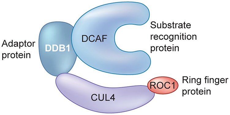
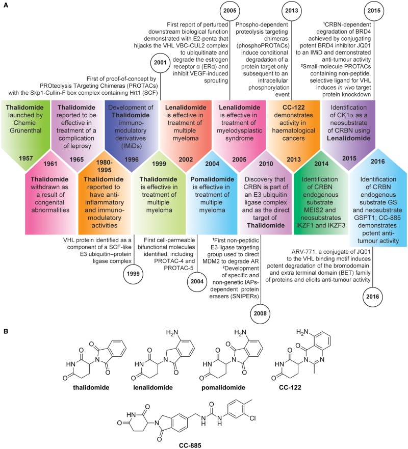
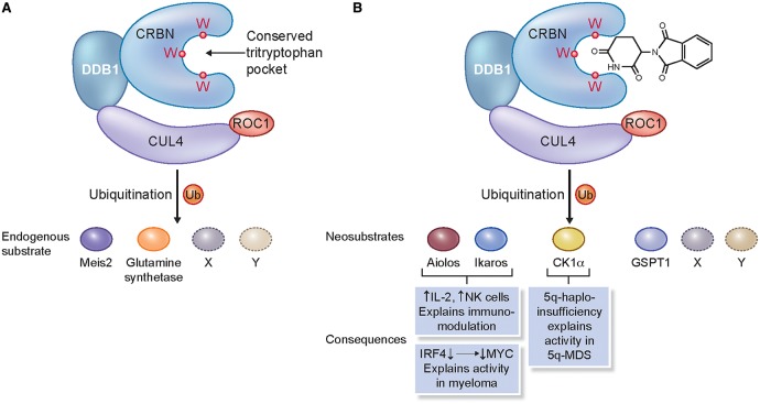
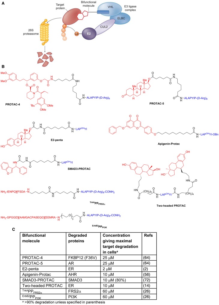
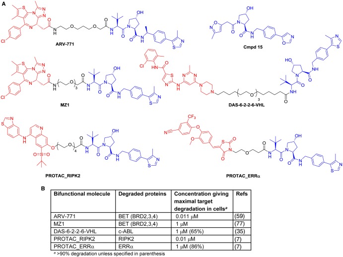
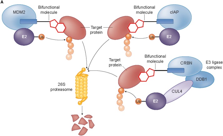
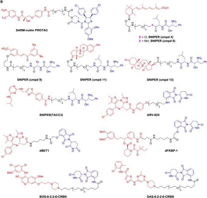
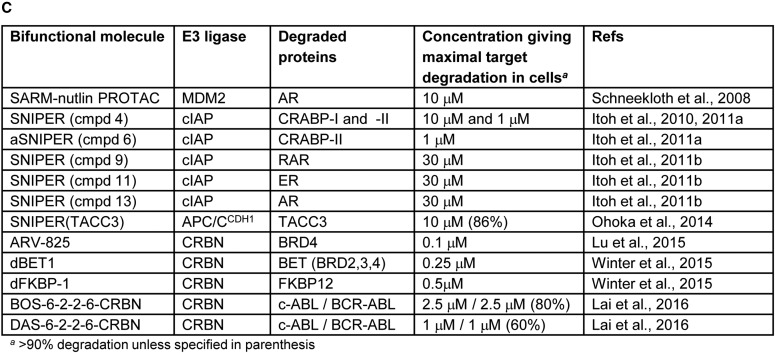
Manipulation of the ubiquitin-proteasome system to achieve targeted degradation of proteins within cells using chemical tools and drugs has the potential to transform pharmacological and therapeutic approaches in cancer and other diseases. An increased understanding of the molecular mechanism of thalidomide and its analogues following their clinical use has unlocked small-molecule modulation of the substrate specificity of the E3 ligase cereblon (CRBN), which in turn has resulted in the advancement of new immunomodulatory drugs (IMiDs) into the clinic. The degradation of multiple context-specific proteins by these pleiotropic small molecules provides a means to uncover new cell biology and to generate future drug molecules against currently undruggable targets. In parallel, the development of larger bifunctional molecules that bring together highly specific protein targets in complexes with CRBN, von Hippel-Lindau, or other E3 ligases to promote ubiquitin-dependent degradation has progressed to generate selective chemical compounds with potent effects in cells and in vivo models, providing valuable tools for biological target validation and with future potential for therapeutic use. In this review, we survey recent breakthroughs achieved in these two complementary methods and the discovery of new modes of direct and indirect engagement of target proteins with the proteasome. We discuss the experimental characterisation that validates the use of molecules that promote protein degradation as chemical tools, the preclinical and clinical examples disclosed to date, and the future prospects for this exciting area of chemical biology.

Keywords: chemical biology; chemical tools; ubiquitin ligases; ubiquitin–proteasome system.


Conflict of interest statement

Raj Chopra is a former employee of Celgene Corporation which has a commercial interest in IMiDs.

Figures

Figure 1.
Figure 1.. An example of an E3 ligase complex.
Components of the CUL-RING ligase 4 (CRL4). CUL4, Cullin-RING ligase 4; DCAF, DDB1- and CUL4-associated factor; DDB1, DNA damage-binding protein 1; ROC1, RING-box protein 1.
Figure 2.
Figure 2.. Timelines for the exploitation of E3 ligases for drug discovery and chemical biology.
(A) Timeline (block arrows) of the development of immunomodulatory drugs (IMiDs), the discovery of CRBN, and its substrates. A timeline (circles) of key steps in the development of bifunctional molecules hijacking E3 ligases described in this review is shown in parallel. (B) Structures of published IMiDs.
Figure 3.
Figure 3.. The consequences of perturbation of CUL4CRBN E3 ligase function.
(A) The role of CRL4CRBN in mediating protein homeostasis of endogenous substrates (Meis2 and glutamine synthetase); the conserved tritryptophan pocket that binds IMiDs is highlighted. (B) Known protein substrates of consequence (for example, Aiolos, Ikaros, CK1α, and GSPT1) whose rate of degradation is altered in response to the binding of thalidomide to the tritryptophan pocket of CRL4CRBN; as yet undiscovered substrates are denoted by X and Y.
Figure 4.
Figure 4.. Selected bifunctional molecules hijacking the VHL E3 ligase using peptide motifs to target VHL.
(A) Cartoon showing the complexes involved in VHL-dependent ubiquitination (ELBC, elongin B–elongin C heterodimer; CUL2, cullin2; E2, E2 ubiquitin ligase; Ub, ubiquitin). (B) Structures of bifunctional molecules showing the affinity groups targeting proteins for degradation (red), linker motifs (black), and peptide motifs targeting the VHL E3 ligase (blue). Y denotes sites of intracellular phosphorylation. POH, hydroxyproline. (C) Proteins targeted for degradation by selected bifunctional molecules and the concentrations used in cellular assays where maximal target depletion was observed (AHR, aryl hydrocarbon receptor; AR, androgen receptor; ER, estrogen receptor; FKBP12, FK506-binding protein 12; FRS2α, fibroblast growth factor receptor substrate 2; PI3K, phosphatidylinositol-4,5-bisphosphate 3-kinase; SMAD3, SMAD family member 3).
Figure 5.
Figure 5.. Selected bifunctional molecules hijacking the VHL E3 ligase using small-molecule VHL inhibitors.
(A) Structures of bifunctional molecules hijacking the VHL E3 ligase, showing the affinity groups targeting proteins for degradation (red), linker motifs (black), and VHL E3 ligase-targeting motif (blue). (B) Proteins targeted for degradation by selected bifunctional molecules and the concentrations used in cellular assays where maximal target depletion was observed (BET, bromodomain and extra-terminal family of proteins; BRD4, bromodomain-containing protein 4, member of the BET family; c-ABL, Abelson tyrosine kinase; RIPK, receptor-interacting serine/threonine-protein kinase 1; ERRα, estrogen-related receptor α).
Figure 6.
Figure 6.. Selected bifunctional molecules hijacking the MDM2, cIAP1, and CRBN E3 ligases.
(A) Cartoon showing the complexes involved in MDM2-, cIAP-, and CRBN-dependent ubiquitination (cIAP, cellular inhibitor of apoptosis protein; CRBN, cereblon; CUL4, cullin 4; DDB1, DNA damage-binding protein 1; E2, E2 ubiquitin ligase; MDM2, mouse double minute 2 homologue; Ub, ubiquitin). (B) Structures of bifunctional molecules showing the affinity groups targeting proteins for degradation (red), linker motifs (black), and small-molecule motifs that recruit the E3 ligases (blue). (C) Proteins targeted for degradation by selected bifunctional molecules, the E3 ligases recruited, and the concentrations used in cellular assays where maximal target depletion was observed (APC/CCDH1, anaphase-promoting complex/cyclosome in complex with CDH1; AR, androgen receptor; BCR-ABL, breakpoint cluster region — Abelson kinase fusion; BRD4, bromodomain 4; c-ABL, Abelson murine leukaemia viral oncogene cellular homologue; CRBP, cellular retinoic acid-binding protein; ER, estrogen receptor; FKBP12, FK506-binding protein 12; RAR, retinoic acid receptor; TACC3, transforming acidic coiled-coil-3).
Figure 7.
Figure 7.. Selected bifunctional molecules directing target degradation through binding of HSP70 or the 20S proteasome.
(A) Cartoons showing the complexes involved in (i) HSP70-dependent protein degradation mediated by a hydrophobic adamantyl tag (HSP70, heat shock protein 70; Ub, ubiquitin) and (ii) direct recruitment of the 20S proteasome by Boc3Arg tags. (B) Structures of bifunctional molecules that direct target protein degradation through binding of HSP70 (SARD279) or the 20S proteasome (EA-B3A, TMP-B3A) showing the affinity groups targeting proteins for degradation (red), linker motifs (black), and small-molecule motifs targeting degradation machinery (blue). (C) Mode of action and proteins targeted for degradation, and the concentrations used in cellular assays where maximal target depletion was observed (AR, androgen receptor; eDHFR, E. coli dihydrofolate reductase; GST-α1, glutathione S-transferase).

Similar articles

Cited by

References

    1. Angers S., Li T., Yi X., MacCoss M.J., Moon R.T. and Zheng N. (2006) Molecular architecture and assembly of the DDB1-CUL4A ubiquitin ligase machinery. Nature 443, 590–593 doi: 10.1038/nature05175 - DOI - PubMed
    1. Bargagna-Mohan P., Baek S.-H., Lee H., Kim K. and Mohan R. (2005) Use of PROTACS as molecular probes of angiogenesis. Bioorg. Med. Chem. Lett. 15, 2724–2727 doi: 10.1016/j.bmcl.2005.04.008 - DOI - PMC - PubMed
    1. Bartlett J.B., Dredge K. and Dalgleish A.G. (2004) The evolution of thalidomide and its IMiD derivatives as anticancer agents. Nat. Rev. Cancer 4, 314–322 doi: 10.1038/nrc1323 - DOI - PubMed
    1. Bett J.S. (2016) Proteostasis regulation by the ubiquitin system. Essays Biochem. 60, 143–151 doi: 10.1042/EBC20160001 - DOI - PubMed
    1. Bhogaraju S., and Dikic I. (2014) A peek into the atomic details of thalidomide's clinical effects. Nat. Struct. Mol. Biol. 21, 739–740 doi: 10.1038/nsmb.2882 - DOI - PubMed

MeSH terms