. 2017 Apr 28;91(10):e00211-17.
doi: 10.1128/JVI.00211-17. Print 2017 May 15.

Proteolytic Cleavage of Bovine Adenovirus 3-Encoded pVIII

Affiliations

Proteolytic Cleavage of Bovine Adenovirus 3-Encoded pVIII

Amit Gaba et al. J Virol. .

Abstract

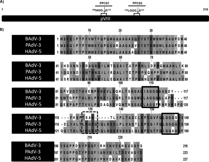
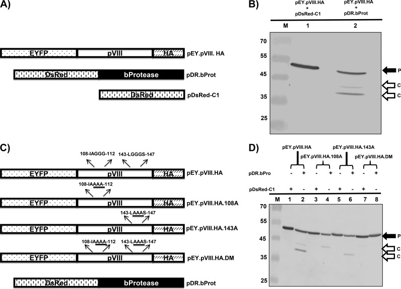
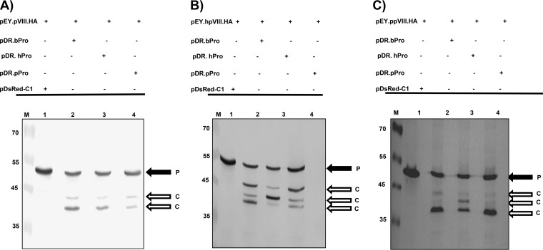
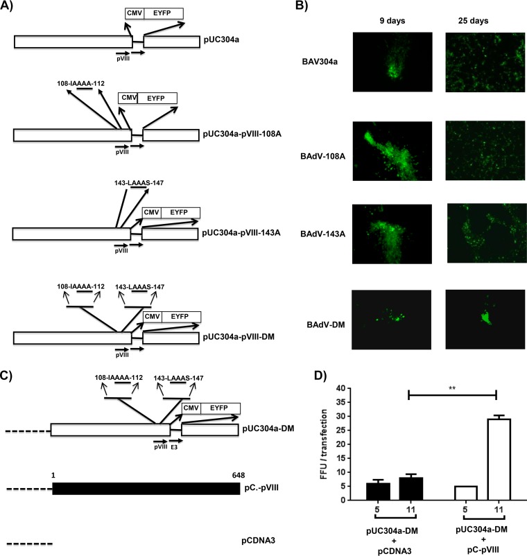
Proteolytic maturation involving cleavage of one nonstructural and six structural precursor proteins including pVIII by adenovirus protease is an important aspect of the adenovirus life cycle. The pVIII encoded by bovine adenovirus 3 (BAdV-3) is a protein of 216 amino acids and contains two potential protease cleavage sites. Here, we report that BAdV-3 pVIII is cleaved by adenovirus protease at both potential consensus protease cleavage sites. Usage of at least one cleavage site appears essential for the production of progeny BAdV-3 virions as glycine-to-alanine mutation of both protease cleavage sites appears lethal for the production of progeny virions. However, mutation of a single protease cleavage site of BAdV-3 pVIII significantly affects the efficient production of infectious progeny virions. Further analysis revealed no significant defect in endosome escape, genome replication, capsid formation, and virus assembly. Interestingly, cleavage of pVIII at both potential cleavage sites appears essential for the production of stable BAdV-3 virions as BAdV-3 expressing pVIII containing a glycine-to-alanine mutation of either of the potential cleavage sites is thermolabile, and this mutation leads to the production of noninfectious virions.IMPORTANCE Here, we demonstrated that the BAdV-3 adenovirus protease cleaves BAdV-3 pVIII at both potential protease cleavage sites. Although cleavage of pVIII at one of the two adenoviral protease cleavage sites is required for the production of progeny virions, the mutation of a single cleavage site of pVIII affects the efficient production of infectious progeny virions. Further analysis indicated that the mutation of a single protease cleavage site (glycine to alanine) of pVIII produces thermolabile virions, which leads to the production of noninfectious virions with disrupted capsids. We thus provide evidence about the requirement of proteolytic cleavage of pVIII for production of infectious progeny virions. We feel that our study has significantly advanced the understanding of the requirement of adenovirus protease cleavage of pVIII.

Keywords: BAdV-3; adenovirus protease; pVIII; proteolytic cleavage; thermolabile.


Figures

FIG 1
FIG 1
Consensus protease cleavage sites of pVIII. (A) Schematic diagram of BAdV-3 pVIII showing the two potential protease cleavage sites (PPCS), PPCS1 (amino acids 108 to 112) and PPCS2 (amino acids 143 to 147). Arrows depict the site of cleavage. (B) Conservation of PPCS in pVIII protein. The PPCS conserved among pVIII of BAdV-3, PAdV-3, and HAdV-5 are indicated by boxed in solid lines, and PPCS conserved among PAdV-3 and HAdV-5 only are boxed in dotted lines. Sequences were downloaded from UniProt (BAdV-3, UniProt accession number O92788; PADV-3, Q83453; HAdV-5, P24936) and aligned with T-Coffee (42). The figure was created using JalView (43).
FIG 2
FIG 2
Cleavage of BAdV-3 pVIII protein in transfected cells. (A) Schematic diagram of plasmid DNAs. The names of the plasmids are depicted on the right of diagrams. The EYFP (enhanced yellow fluorescent protein)-pVIII (BAdV-3), bProtease (BAdV-3), and DsRed DNA plasmids are depicted. (B) Cleavage of BAdV-3 pVIII in transfected cells. Proteins from the lysates of 293T cells cotransfected with the indicated plasmid DNAs were separated by 12% SDS-PAGE, transferred to nitrocellulose membrane, and analyzed by Western blotting using anti-GFP antibody. P, uncleaved EY.pVIII.HA fusion protein; C, cleaved product. The molecular mass markers (lane M) in kilodaltons are shown on the left. (C) Schematic diagram of plasmid DNAs showing potential protease cleavage sites. The origin of the DNA is depicted. The amino acid sequences of potential protease cleavage sites are depicted above the diagrams, and glycine-to-alanine substitutions are underlined. (D) Cleavage of BAdV-3 pVIII mutant proteins in transfected cells. Proteins from the lysates of 293T cells cotransfected with the indicated plasmid DNAs were separated by 12% SDS-PAGE, transferred to nitrocellulose membranes, and analyzed by Western blotting using anti-GFP antibody. The molecular mass markers (M) in kilodaltons are shown on the left. P, uncleaved fusion protein; C, cleaved product.
FIG 3
FIG 3
Cleavage of pVIII by different adenoviral proteases in transfected cells. Proteins from the lysates of 293T cells cotransfected with the indicated plasmid DNAs were separated by 12% SDS-PAGE, transferred to nitrocellulose membrane, and analyzed by Western blotting using anti-GFP antibody. The cleavage of pVIII of BAdV-3 pVIII (A), HAdV-5 (B), and PAdV-3 (C) by adenoviral proteases is represented. The molecular mass markers (lanes M) are shown (in kilodaltons) on the left of each panel. P, uncleaved fusion protein; C, cleaved product.
FIG 4
FIG 4
Isolation and characterization of mutant BAdV-3. (A). Schematic representation of plasmids. The BAdV-3 sequence is represented by an open box. The thin line represents the deleted E3 region (44). The alanines substituted for glycines are underlined. The numbers represent the amino acids of BAdV-3 pVIII; the human cytomegalovirus (CMV) immediate early promoter and EYFP (enhanced yellow fluorescent protein) are also indicated. Horizontal arrows indicate the direction of transcription. (B) Direct fluorescence. A monolayer of VIDO DT1 cells (22) was transfected with 4 to 6 μg of plasmid DNA of pUC304a (BAV304a), pUC304a-pVIII-108A (BAdV-108A), pUC304a-pVIII-143A (BAdV-143A), or pUC304a-pVIII-DM (BAdV-DM) and visualized at the indicated times posttransfection for the expression of EYFP and development of cytopathic effects using a TCS SP5 (Leica) fluorescence microscope. An increase in green fluorescence indicates that virus production could be seen after the 9th day posttransfection. (C). Schematic representation of plasmid pUC304a-DM. The dotted line represents the plasmid sequence. The BAdV-3 sequence is represented by an open box. The thin line represents the deleted E3 region (44). The alanines substituted for glycines are underlined. The numbers represent the amino acids of BAdV-3 pVIII; the human cytomegalovirus (CMV) immediate early promoter and EYFP (enhanced yellow fluorescent protein) are also shown. Horizontal arrows indicate the direction of transcription. The filled box represents the nucleotide sequence of BAdV-3 pVIII. The dotted line represents the nucleotide sequence of plasmid DNA. (D) Complementation assay. The VIDO DT1 cells (22) were cotransfected with the indicated plasmids, and fluorescent focus forming units were counted at the indicated day posttransfection (x axis). Values represent averages from two independent replicates, and error bars indicate the standard deviations. Statistical differences among the groups were calculated using an unpaired t test. **, P < 0.01.
FIG 5
FIG 5
Virus growth. (A) CsCl purification. Virus was purified from infected cell lysates by CsCl density gradient purification. Mature virion bands after double density gradient centrifugation are shown (B). Western blotting. Proteins from purified BAV304a, BAdV-108A, or BAdV-143A were separated on 4 to 20% gradient SDS-PAGE gels, transferred to nitrocellulose, and probed by Western blotting using anti-pVIIIb serum (17). (C) Virus titer. The infected MDBK cells were harvested at the indicated times postinfection and freeze-thawed, and virus was titrated on MDBK cells.
FIG 6
FIG 6
Analysis of mutant BAdV-3 viruses. (A) Virus infectivity. The MDBK cells were infected with equivalent amounts of infectious particles of the indicated viruses in triplicate. The infected cells were visualized for the expression of GFP at 18 h postinfection by a TCS SP5 (Leica) fluorescence microscope. (B) Viral genome replication. The MDBK cells were infected with equivalent amounts of infectious particles of the indicated virus in triplicate. At indicated times postinfection, the infected cells were collected, and genomic DNA was isolated. The viral genome copy number was determined by quantitative PCR and divided by actin copy number for normalization. For comparison, the normalized genome copy number values for each virus at each time point were compared to the value of wild-type virus at 6 hpi. (C) Subcellular distribution of BAdV-3. Monolayers of MDBK cells (1 × 10⁶ cells/well) were incubated with 1.4 × 10⁷ purified virions at 4°C. After 1 h of incubation, the cells were incubated at 37°C for 30 min. Finally, the cells were processed and visualized by transmission electron microscopy: frames 1 to 3, BAV304a; frames 4 to 6, BAdV-108A; frames 7 to 9, BAdV-143A. Boxed areas are enlarged, as indicated. (D) For each virus, 10 cells were selected randomly, and virus particles in endosomes, cytoplasm, and at plasma membrane were counted. Error bars indicate standard errors of the means of three independent experiments.
FIG 7
FIG 7
Thermostability of mutant virions. (A) Purified virions (105 TCID50) grown in MDBK cells were incubated at various temperatures for 3 days in PBS containing 10% glycerol, and the residual viral infectivity was determined by titration on MDBK cells. (B) Purified virions (105 TCID50) grown in MDBK cells were incubated at 37°C for the indicated periods of time in PBS containing 10% glycerol, and the residual viral infectivity was determined by titration on MDBK cells.
FIG 8
FIG 8
Analysis of viral protein expression in infected cells. Proteins from the lysates of MDBK cells were separated by 4 to 20% gradient SDS-PAGE gels, transferred to nitrocellulose membranes, and probed by Western blotting using anti-DNA binding protein (DBP) (36), anti-pVII (37), anti-V (38), anti-hexon (39), and anti-100K (9) serum followed by Alexa Fluor 680-conjugated goat anti-rabbit antibody (Invitrogen). The results were analyzed by using an Odyssey Infrared Imaging System. Values represent averages from two independent replicates, and error bars indicate standard deviations. All data were analyzed using GraphPad Prism, version 6 (GraphPad Software, Inc., La Jolla, CA, USA). Statistical differences among the groups were calculated using an unpaired t test, and significance is indicated as follows: *, P < 0.05; **, P < 0.01, ***, P < 0.001; ****, P < 0.0001.
FIG 9
FIG 9
Analysis of viral protein incorporation in purified virions. Proteins from virions of BAV304a, BAdV-108A, or BAdV-143A subjected to two round of CsCl purification were separated by 4 to 20% gradient SDS-PAGE gels, transferred to nitrocellulose, and probed by Western blotting using protein-specific antiserum. All data were analyzed using GraphPad Prism, version 6 (GraphPad Software, Inc., La Jolla, CA, USA). Statistical differences among the groups were calculated using an unpaired t test. **, P < 0.01.
FIG 10
FIG 10
Electron microscopic analysis. (A) Purified BAV304a, BAdV-108A, or BAdV-143A is shown, as indicated, at a magnification of ×30,000. The arrows indicate enlargements of selected boxed regions of each virus (magnification, ×1,000,000). (B) MDBK cells infected with BAV304a, BAdV-108A, or BAdV-143A, as indicated, are shown at a magnification of ×10,000. The arrows indicate the enlargements of selected boxed regions at a magnification of ×30,000.
FIG 10
FIG 10
Electron microscopic analysis. (A) Purified BAV304a, BAdV-108A, or BAdV-143A is shown, as indicated, at a magnification of ×30,000. The arrows indicate enlargements of selected boxed regions of each virus (magnification, ×1,000,000). (B) MDBK cells infected with BAV304a, BAdV-108A, or BAdV-143A, as indicated, are shown at a magnification of ×10,000. The arrows indicate the enlargements of selected boxed regions at a magnification of ×30,000.

Similar articles

Cited by

References

    1. Berk AJ. 2007. Adenoviridae: the viruses and their replication, p 2355–2395. In Knipe DM, Howley PM, Griffin DE, Lamb RA, Martin MA, Roizman B, Straus SE (ed), Fields virology, 5th ed Lippincott Williams & Wilkins, Philadelphia, PA.
    1. Campos SK, Barry MA. 2007. Current advances and future challenges in adenoviral vector biology and targeting. Curr Gene Ther 7:189–204. doi:10.2174/156652307780859062. - DOI - PMC - PubMed
    1. Tatsis N, Ertl HC. 2004. Adenoviruses as vaccine vectors. Mol Ther 10:616–629. doi:10.1016/j.ymthe.2004.07.013. - DOI - PMC - PubMed
    1. Webster A, Russell S, Talbot P, Russell Wt, Kemp G. 1989. Characterization of the adenovirus proteinase: substrate specificity. J Gen Virol 70:3225–3234. doi:10.1099/0022-1317-70-12-3225. - DOI - PubMed
    1. Weber J. 1976. Genetic analysis of adenovirus type 2 III. Temperature sensitivity of processing viral proteins. J Virol 17:462–471. - PMC - PubMed

Publication types

LinkOut - more resources