. 2017 May 12;91(11):e02416-16.
doi: 10.1128/JVI.02416-16. Print 2017 Jun 1.

African Swine Fever Virus NP868R Capping Enzyme Promotes Reovirus Rescue during Reverse Genetics by Promoting Reovirus Protein Expression, Virion Assembly, and RNA Incorporation into Infectious Virions

Affiliations

African Swine Fever Virus NP868R Capping Enzyme Promotes Reovirus Rescue during Reverse Genetics by Promoting Reovirus Protein Expression, Virion Assembly, and RNA Incorporation into Infectious Virions

Heather E Eaton et al. J Virol. .

Abstract

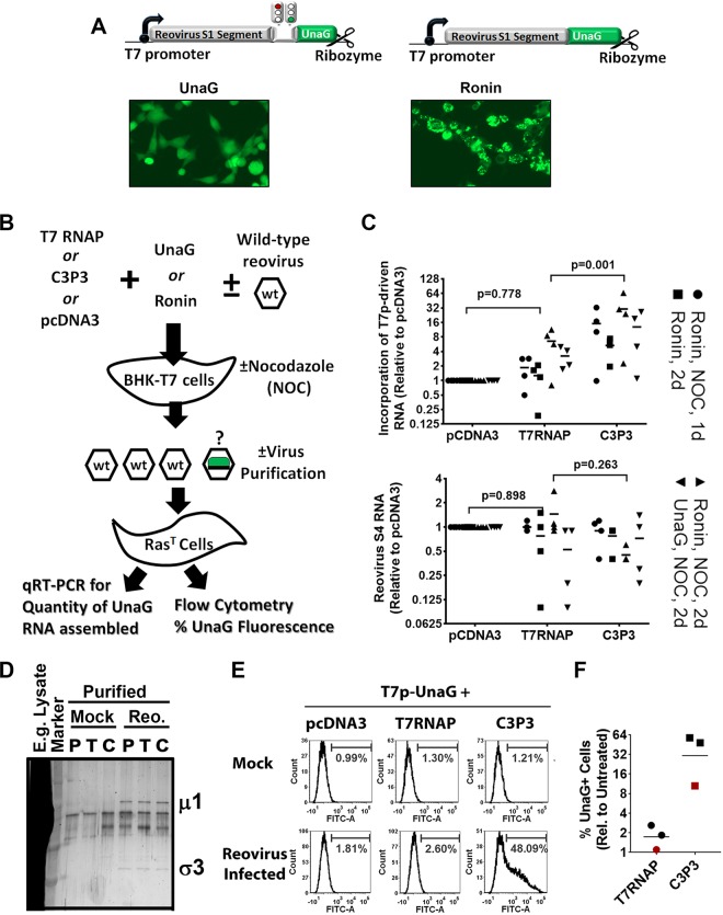
Reoviruses, like many eukaryotic viruses, contain an inverted 7-methylguanosine (m7G) cap linked to the 5' nucleotide of mRNA. The traditional functions of capping are to promote mRNA stability, protein translation, and concealment from cellular proteins that recognize foreign RNA. To address the role of mRNA capping during reovirus replication, we assessed the benefits of adding the African swine fever virus NP868R capping enzyme during reovirus rescue. C3P3, a fusion protein containing T7 RNA polymerase and NP868R, was found to increase protein expression 5- to 10-fold compared to T7 RNA polymerase alone while enhancing reovirus rescue from the current reverse genetics system by 100-fold. Surprisingly, RNA stability was not increased by C3P3, suggesting a direct effect on protein translation. A time course analysis revealed that C3P3 increased protein synthesis within the first 2 days of a reverse genetics transfection. This analysis also revealed that C3P3 enhanced processing of outer capsid μ1 protein to μ1C, a previously described hallmark of reovirus assembly. Finally, to determine the rate of infectious-RNA incorporation into new virions, we developed a new recombinant reovirus S1 gene that expressed the fluorescent protein UnaG. Following transfection of cells with UnaG and infection with wild-type virus, passage of UnaG through progeny was significantly enhanced by C3P3. These data suggest that capping provides nontraditional functions to reovirus, such as promoting assembly and infectious-RNA incorporation.IMPORTANCE Our findings expand our understanding of how viruses utilize capping, suggesting that capping provides nontraditional functions to reovirus, such as promoting assembly and infectious-RNA incorporation, in addition to enhancing protein translation. Beyond providing mechanistic insight into reovirus replication, our findings also show that reovirus reverse genetics rescue is enhanced 100-fold by the NP868R capping enzyme. Since reovirus shows promise as a cancer therapy, efficient reovirus reverse genetics rescue will accelerate production of recombinant reoviruses as candidates to enhance therapeutic potency. NP868R-assisted reovirus rescue will also expedite production of recombinant reovirus for mechanistic insights into reovirus protein function and structure.

Keywords: C3P3; NP868R; capping; reovirus; reverse genetics.


Figures

FIG 1
FIG 1
The African swine fever virus capping enzyme, NP868R, promotes expression from T7 RNA polymerase-driven plasmids. (A) Diagram of constructs used for panels B to F. (B) BHK-T7 cells were either untransfected or cotransfected with two plasmids, as indicated. The cells were assessed for green fluorescence (GFP) by microscopy at 48 hpt. Similarity in cell density and health was confirmed by DIC microscopy. (C to F) Cells were cotransfected with T7p-GFP and a secondary plasmid as indicated. Live cells were assessed by flow cytometry at 48 hpt. The proportion of GFP+ cells was measured with a marker set to generate 1 to 2% GFP+ cells for the pcDNA3 negative control. MFI was calculated based on a marker that spanned the entire spectrum. The proportion of GFP+ cells and the MFI were graphed relative to C3P3-transfected cells, which were set to 100% to standardize among experiments. Experiments were conducted in BHK-T7 (C and D), BHK21 (E), and HEK293T and COS-7 (F) cells. (D and E) One-way analysis of variance (ANOVA) was used with Bonferroni's multiple-comparison test relative to C3P3. *, P < 0.05; **, P < 0.01; ***, P < 0.001; ns, not significant; n = 3. The error bars indicate standard deviations (SD).
FIG 2
FIG 2
The African swine fever virus capping enzyme, NP868R, is associated with reduced mRNA levels but a 10-fold increase in protein expression from T7 RNA polymerase-driven plasmids. BHK-T7 cells were left untransfected (UT); transfected with pcDNA3 only; or transfected with T7p-GFP, T7p-S4, and T7p-M2 in combination with either T7RNAP, C3P3, or pcDNA3, as indicated. (A) Levels of M2, S4, or GFP plasmids were measured by quantitative PCR and are presented relative to T7RNAP-cotransfected conditions. No pairs were significantly different by two-way ANOVA and Tukey's multiple-comparison test (n = 3). (B) Western blot analysis for levels of GFP, σ3, μ1, and β-actin proteins. (C) Densitometric analysis for intensities of GFP, σ3, and μ1 proteins standardized to β-actin levels and normalized to T7RNAP-transfected conditions (set to 1). P values relative to T7RNAP were calculated by one-way ANOVA with Dunnett's multiple-comparison test (n = 4). ns, P > 0.05. (D) Levels of M2, S4, and GFP mRNA were assessed by qRT-PCR. Statistics as for panel C (n = 3). Horizontal lines indicate means.
FIG 3
FIG 3
The African swine fever virus capping enzyme, NP868R, promotes reovirus production from the T7 RNA polymerase-driven reverse genetics system 100-fold. BHK-T7 cells were transfected with the four reovirus reverse genetics plasmids that together express all 10 reovirus genes flanked by T7 polymerase and ribozyme sequences. The cells were cotransfected with various combinations of T7 RNA polymerase, NP868R, LZip-T7RNAP, LZip-NP868R, and C3P3, as indicated. Lysates of transfected BHK-T7 cells were then assessed for titers of rescued reovirus by standard plaque assay on L929 mouse fibroblasts. One-way ANOVA and Bonferroni's multiple-comparison test were used to compare conditions for 3 to 8 independent experiments. The error bars indicate SD.
FIG 4
FIG 4
NP868R capping enzyme promotes modest accumulation of reovirus RNA and proteins but greatly enhances the levels of progeny reovirus production. BHK-T7 cells were transfected with T7 RNA polymerase (T7) or C3P3, along with the four reverse genetics plasmids that carry all 10 reovirus genes. As a control for macromolecular synthesis in the absence of virus rescue, C3P3 was transfected with only the S4 and M2 reovirus genes, with pcDNA3 used to equalize relative plasmid ratios. Lysates were collected at 1 through 5 days posttransfection (D.P.T.). All plots of absolute levels (A and C, left, and D and E, top) are plotted as means ± SD for 3 independent experiments. All plots for relative levels between T7 or C3P3 conditions (A and C, right, and D and E, bottom) show means and P values above each time point as analyzed by column statistics and one-sample t tests relative to the hypothetical value of 1. Similarly filled or open symbols represent duplicates of 3 independent experiments. (A) Levels of S4 plasmid were quantified by qRT-PCR and plotted as means and SD for each condition (left) or as relative plasmid levels between T7 and C3P3 conditions (right). (B) Reovirus rescue with C3P3 and all reovirus genes was conducted in the presence or absence of neutralizing antibodies in the media of BHK-T7 cells. Cell lysates collected at 1 to 5 days were subjected to Western blot analysis with reovirus polyclonal and β-actin-specific antibodies. The locations of reovirus structural proteins μ1 and μ1 cleavage to produce μ1C (encoded by M2), σ3 (encoded by S4), and β-actin loading control are indicated. (C) Levels of reovirus proteins μ1, μ1C, and σ3 were also evaluated, in the absence of neutralizing antibodies, among the three conditions (T7 plus all reovirus segments, C3P3 plus all reovirus segments, and C3P3 plus S4 and M2). (i) Western blot analysis as for panel B showing one duplicate each from two independent experiments. (ii) Levels of total μ1/μ1C quantified by densitometry and plotted as means ± SD for each condition (left) or as relative protein levels between T7 and C3P3 conditions (right). (iii) Levels of μ1-to-μ1C processing formulated as 100 × [μ1C/(μ1 + μ1C)] and plotted as means ± SD for each condition (left) or as relative protein levels between T7 and C3P3 conditions (right). (D) Levels of S4 RNA quantified by qRT-PCR and plotted as means and SD for each condition (top) or as relative plasmid levels between T7 and C3P3 conditions (bottom). (E) Reovirus titers quantified by plaque assay on L929 cells and plotted as means and SD for each condition (top) or as relative plasmid levels between T7 and C3P3 conditions (bottom).
FIG 5
FIG 5
Reovirus RNAs are encapsidated more efficiently when coupled with NP868R-mediated capping. (A) Reovirus S1 genome segment modified to express the UnaG green fluorescent protein as a fusion protein with the N-terminal half of σ1 (Ronin construct) or as an independent protein alongside σ1-N (UnaG construct). Both the Ronin and UnaG constructs were used to produce recombinant reovirus through reverse genetics, and the microscopy images show that the two recombinant viruses could produce green fluorescence. (B) Diagram of the experiment performed to generate the data for panels C and D. BHK-T7 cells were transfected with pcDNA3, T7RNAP, or C3P3 and either the UnaG or Ronin reporter plasmid for 1 to 2 days and were then infected with wild-type reovirus in the presence or absence of NOC. One, 2, or 3 days postinfection, the transfected/infected BHK-T7 lysates were used to infect Ras-transformed NIH 3T3 mouse fibroblasts (RasT) and were assessed for UnaG RNA and fluorescence. (C) (Top) Expression of UnaG in RasT cells was assessed using qRT-PCR for four independent experiments, each containing infection with wild-type reovirus and nocodazole, at 1or 2 days (d) posttransfection, as indicated. (Bottom) Reovirus S4 transcript levels were monitored to determine if the UnaG or Ronin reporter construct affected reovirus replication. RNA levels under pcDNA3 versus T7RNAP and C3P3 versus T7RNAP conditions were compared using two-way ANOVA and Tukey's multiple-comparison posttest. (D) Silver staining confirmed that reovirus particles were successfully purified from BHK-T7 cells transfected with pcDNA3 (P), T7RNAP (T), or C3P3 (C) and either mock or reovirus infected, as indicated. One example lysate (E.g. Lysate) of C3P3-transfected and reovirus-infected BHK-T7 cells before purification is provided to demonstrate the extent of purification. The locations of reovirus major structural proteins μ1 and σ3 are indicated. (E) RasT cells treated with purified virions from BHK-T7 cells transfected with pcDNA3, T7RNAP, or C3P3 and either mock or reovirus infected (as indicated) were subjected to flow cytometric analysis to measure levels of UnaG protein fluorescence. FITC, fluorescein isothiocyanate. (F) Results from three independent experiments similar to that in panel E using either purified virions (black symbols) or unpurified lysates (red symbols).
FIG 6
FIG 6
Model in which RNA capping promotes reovirus protein expression and, most notably, encapsidation of reovirus RNA to produce infectious virions.

Similar articles

Cited by

References

    1. Ramanathan A, Robb GB, Chan SH. 2016. mRNA capping: biological functions and applications. Nucleic Acids Res 44:7511–7526. doi: 10.1093/nar/gkw551. - DOI - PMC - PubMed
    1. Decroly E, Ferron F, Lescar J, Canard B. 2011. Conventional and unconventional mechanisms for capping viral mRNA. Nat Rev Microbiol 10:51–65. doi: 10.1038/nrmicro2675. - DOI - PMC - PubMed
    1. Schoenberg DR, Maquat LE. 2009. Re-capping the message. Trends Biochem Sci 34:435–442. doi: 10.1016/j.tibs.2009.05.003. - DOI - PMC - PubMed
    1. Picard-Jean F, Tremblay-Letourneau M, Serra E, Dimech C, Schulz H, Anselin M, Dutilly V, Bisaillon M. 2013. RNA 5′-end maturation: a crucial step in the replication of viral genomes. In Romanowski V. (ed), Current issues in molecular virology: viral genetics and biotechnological applications. In Tech, Rijeka, Croatia. doi: 10.5772/56166. - DOI
    1. Izaurralde E, Lewis J, McGuigan C, Jankowska M, Darzynkiewicz E, Mattaj IW. 1994. A nuclear cap binding protein complex involved in pre-mRNA splicing. Cell 78:657–668. doi: 10.1016/0092-8674(94)90530-4. - DOI - PubMed

Publication types

MeSH terms