. 2017 Mar 15;7(1):191.
doi: 10.1038/s41598-017-00301-w.

MKL1 defines the H3K4Me3 landscape for NF-κB dependent inflammatory response

Affiliations

MKL1 defines the H3K4Me3 landscape for NF-κB dependent inflammatory response

Liming Yu et al. Sci Rep. .

Abstract

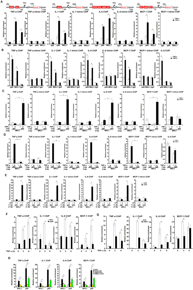
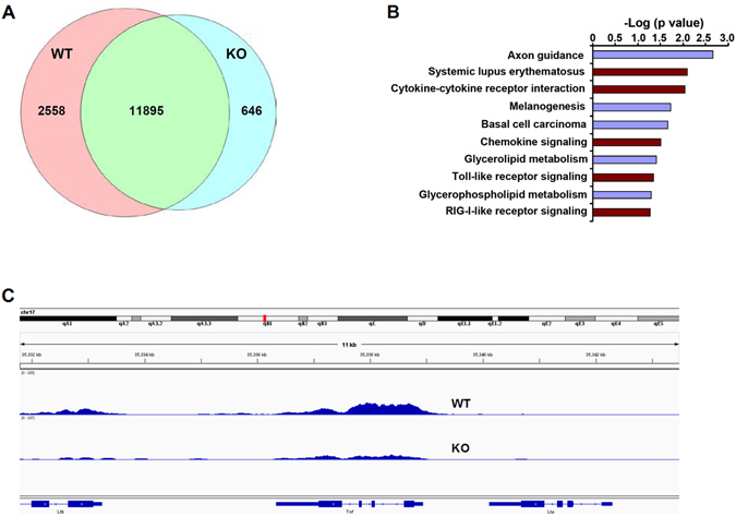
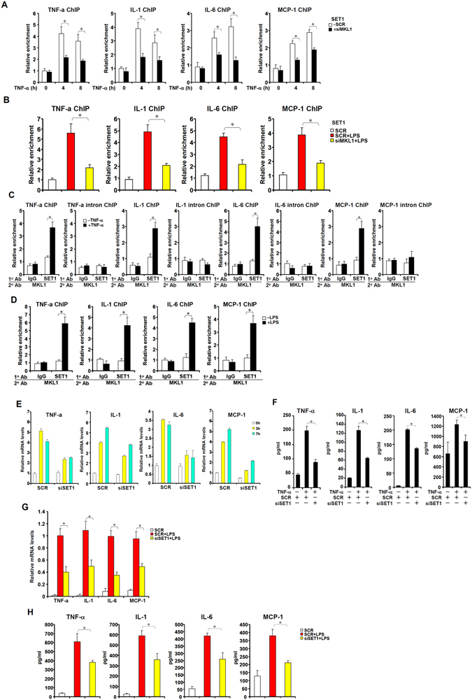
Macrophage-dependent inflammatory response is considered a pivotal biological process that contributes to a host of diseases when aberrantly activated. The underlying epigenetic mechanism is not completely understood. We report here that MKL1 was both sufficient and necessary for p65-dependent pro-inflammatory transcriptional program in immortalized macrophages, in primary human and mouse macrophages, and in an animal model of systemic inflammation (endotoxic shock). Extensive chromatin immunoprecipitation (ChIP) profiling and ChIP-seq analyses revealed that MKL1 deficiency erased key histone modifications synonymous with transactivation on p65 target promoters. Specifically, MKL1 defined histone H3K4 trimethylation landscape for NF-κB dependent transcription. MKL1 recruited an H3K4 trimethyltransferase SET1 to the promoter regions of p65 target genes. There, our work has identified a novel modifier of p65-dependent pro-inflammatory transcription, which may serve as potential therapeutic targets in treating inflammation related diseases.


Conflict of interest statement

The authors declare that they have no competing interests.

Figures

Figure 1
Figure 1
MKL1 enhances Rel-A/p65 dependent transcription. (A) FLAG-MKL1 and V5-p65 were co-transfected into HEK293 cells with indicated promoter constructs. Data are expressed as relative luciferase unit (RLU). (B) FLAG-MKL1 was co-transfected into HEK293 cells with indicated promoter constructs followed by treatment with TNF-α (10 ng/ml) for 6 hours. Data are expressed as RLU. (C) FLAG-MKL1 was co-transfected into cells with a generic κB reporter construct followed by treatment with TNF-α (10 ng/ml) for 6 hours. Data are expressed as RLU. (D,E) THP-1 cells were transfected FLAG-MKL1 followed by treatment with TNF-α (10 ng/ml) for 6 hours. mRNA (D) and protein (E) levels of pro-inflammatory mediators were measured by qPCR and ELISA. (F,G) THP-1 cells were transfected FLAG-MKL1 followed by treatment with LPS (100 ng/ml) for 6 hours. mRNA (F) and protein (G) levels of pro-inflammatory mediators were measured by qPCR and ELISA.
Figure 2
Figure 2
MKL1 deficiency dampens Rel-A/p65 dependent transcription. (A,B) THP-1 cells were transfected with indicated siRNAs followed by treatment with TNF-α (10 ng/ml) for 6 hours. mRNA (A) and protein (B) levels of pro-inflammatory mediators were measured by qPCR and ELISA. (C,D) THP-1 cells were transfected with indicated siRNAs followed by treatment with LPS (100 ng/ml) for 6 hours. mRNA (C) and protein (D) levels of pro-inflammatory mediators were measured by qPCR and ELISA. (E) WT or MKL KO MEF cells were transfected with a generic κB reporter construct followed by treatment with TNF-α (10 ng/ml) for 6 hours. Data are expressed as RLU. (FI) MEF cells were treated with TNF-α (10 ng/ml). mRNA (F) and protein (G) levels of pro-inflammatory mediators were measured by qPCR and ELISA. (H) Heat map and (I) Go analysis.
Figure 3
Figure 3
A reciprocal interplay between MKL1 and p65 on the chromatin. (A,B) THP-1 cells (A) or differentiated primary human macrophages (B) were treated with TNF-α (10 ng/ml) for 6 hours. ChIP assays were performed with indicated antibodies. Precipitated DNA was amplified with indicated primers shown with conserved binding motifs. κBRE, NF-κB response element; SRE, serum response element; TSS, transcription start site (C,D) THP-1 cells (C) and differentiated primary human macrophages (D) were treated with or without TNF-α (10 ng/ml) for 3 hours. Re-ChIP assays were performed with indicated antibodies. (E) THP-1 cells were treated with or without LPS (100 ng/ml) for 6 hours. Re-ChIP assays were performed with indicated antibodies. (E,F) THP-1 cells were transfected with indicated siRNA followed by treatment with TNF-α (10 ng/ml) for 6 hours. ChIP assays were performed with anti-MKL1 (E) or anti-p65 (F). (H) THP-1 cells were transfected with indicated siRNA followed by treatment with LPS (100 ng/ml) for 6 hours. ChIP assays were performed with anti-MKL1 or anti-p65.
Figure 4
Figure 4
MKL1 deficiency cripples inflammatory response in mice. (A) C57/BL6 mice were injected with LPS (25 mg/kg). Peritoneal macrophages were isolated 6 hours after injection and ChIP assays were performed with anti-MKL1. (B,C) Sex- and age-matched WT and MKL1 KO male mice were injected with LPS (25 mg/kg). Survival (B) and body temperature (C) were monitored following injection. N = 10 mice for each group. (DG) Mice were sacrificed 4 hours after injection. Circulating levels of pro-inflammatory mediators were measured by ELISA. N = 6 mice for each group.
Figure 5
Figure 5
MKL1 influences the chromatin structure of pro-inflammatory genes. (AC) THP-1 cells were transfected with indicated siRNA followed by treatment with TNF-α (10 ng/ml). ChIP assays were performed with anti-monomethyl H3K4 (A), anti-dimethyl H3K4 (B), and anti-trimethyl H3K4 (C). (DF) THP-1 cells were transfected with indicated siRNA followed by treatment with LPS (100 ng/ml) for 6 hours. ChIP assays were performed with anti-monomethyl H3K4 (D), anti-dimethyl H3K4 (E), and anti-trimethyl H3K4 (F). (G,H) WT or KO BMDMs were treated with TNF-α (10 ng/ml). ChIP assays were performed with anti-dimethyl H3K4 (G) and anti-trimethyl H3K4 (H).
Figure 6
Figure 6
MKL1 influences global H3K4Me3 levels in BMDMs. Wild type (WT) or MKL1 deficient (KO) BMDMs were treated with TNF-α (10 ng/ml) for 3 hours. ChIP was performed with anti-H3K4Me3. (A) Venn diagram illustrating the overlap of binding peaks identified in WT and KO BMBM ChIP-seq data sets. (B) Pathway analysis of significantly enriched H3K4Me3 tags found within ±2 kb relative to the TSS of the nearest gene. (C) Graphical view of H3K4Me3 ChIP-seq binding peaks (tag densities) around the TSS of TNF-α.
Figure 7
Figure 7
MKL1 recruits SET1 to activate the transcription of p65 target genes. (A) THP-1 cells were transfected with indicated siRNA followed by treatment with TNF-α (10 ng/ml) and harvested at indicated time points. ChIP assays were performed with anti-SET1. (B) THP-1 cells were transfected with indicated siRNA followed by treatment with LPS (100 ng/ml) for 6 hours. ChIP assays were performed with anti-SET1. (C) THP-1 cells were treated with or without TNF-α (10 ng/ml) for 3 hours. Re-ChIP assays were performed with indicated antibodies. (D) THP-1 cells were treated with or without LPS (100 ng/ml) for 6 hours. Re-ChIP assays were performed with indicated antibodies. (E) THP-1 cells were transfected with indicated siRNA followed by treatment with TNF-α (10 ng/ml) and harvested at indicated time points. mRNA levels of pro-inflammatory mediators were measured by qPCR. (F) THP-1 cells were transfected with indicated siRNA followed by treatment with TNF-α (10 ng/ml) for 6 hours. Protein levels of pro-inflammatory mediators were measured by qPCR. (G,H) THP-1 cells were transfected with indicated siRNA followed by treatment with LPS (100 ng/ml) for 6 hours. mRNA (G) and protein (H) levels of pro-inflammatory mediators were measured by qPCR and ELISA.

Similar articles

Cited by

References

    1. Medzhitov R. Origin and physiological roles of inflammation. Nature. 2008;454:428–435. doi: 10.1038/nature07201. - DOI - PubMed
    1. Smale ST. Hierarchies of NF-kappaB target-gene regulation. Nature immunology. 2011;12:689–694. doi: 10.1038/ni.2070. - DOI - PMC - PubMed
    1. Karin M, Ben-Neriah Y. Phosphorylation meets ubiquitination: the control of NF-[kappa]B activity. Annu Rev Immunol. 2000;18:621–663. doi: 10.1146/annurev.immunol.18.1.621. - DOI - PubMed
    1. Huang B, Yang XD, Lamb A, Chen LF. Posttranslational modifications of NF-kappaB: another layer of regulation for NF-kappaB signaling pathway. Cellular signalling. 2010;22:1282–1290. doi: 10.1016/j.cellsig.2010.03.017. - DOI - PMC - PubMed
    1. Natoli G. Specialized chromatin patterns in the control of inflammatory gene expression. Current topics in microbiology and immunology. 2011;349:61–72. - PubMed

Publication types

MeSH terms

LinkOut - more resources