. 2017 Mar 1:11:97.
doi: 10.3389/fnins.2017.00097. eCollection 2017.

Increased Testosterone Decreases Medial Cortical Volume and Neurogenesis in Territorial Side-Blotched Lizards (Uta stansburiana)

Affiliations

Increased Testosterone Decreases Medial Cortical Volume and Neurogenesis in Territorial Side-Blotched Lizards (Uta stansburiana)

Lara D LaDage et al. Front Neurosci. .

Abstract

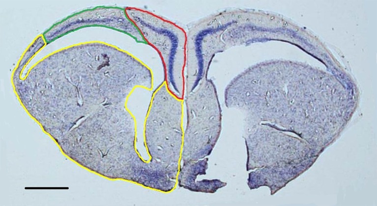
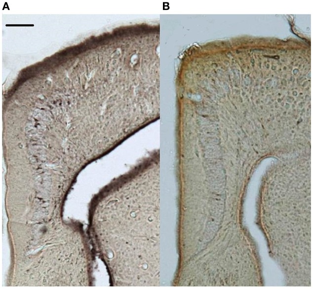
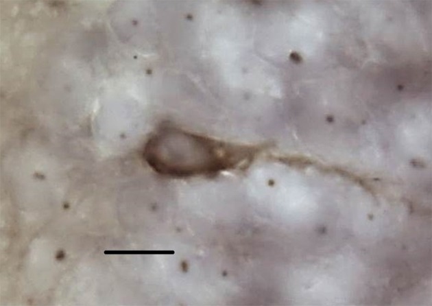
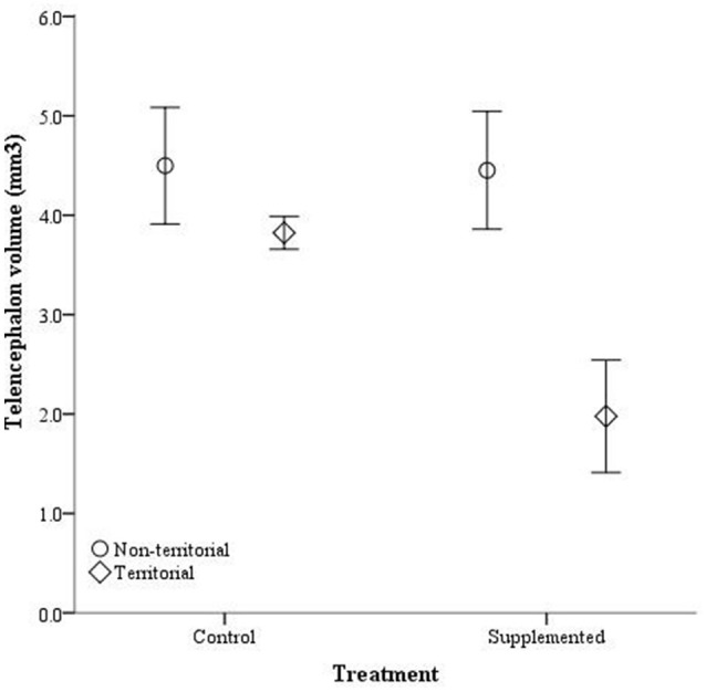
Variation in an animal's spatial environment can induce variation in the hippocampus, an area of the brain involved in spatial cognitive processing. Specifically, increased spatial area use is correlated with increased hippocampal attributes, such as volume and neurogenesis. In the side-blotched lizard (Uta stansburiana), males demonstrate alternative reproductive tactics and are either territorial-defending large, clearly defined spatial boundaries-or non-territorial-traversing home ranges that are smaller than the territorial males' territories. Our previous work demonstrated cortical volume (reptilian hippocampal homolog) correlates with these spatial niches. We found that territorial holders have larger medial cortices than non-territory holders, yet these differences in the neural architecture demonstrated some degree of plasticity as well. Although we have demonstrated a link among territoriality, spatial use, and brain plasticity, the mechanisms that underlie this relationship are unclear. Previous studies found that higher testosterone levels can induce increased use of the spatial area and can cause an upregulation in hippocampal attributes. Thus, testosterone may be the mechanistic link between spatial area use and the brain. What remains unclear, however, is if testosterone can affect the cortices independent of spatial experiences and whether testosterone differentially interacts with territorial status to produce the resultant cortical phenotype. In this study, we compared neurogenesis as measured by the total number of doublecortin-positive cells and cortical volume between territorial and non-territorial males supplemented with testosterone. We found no significant differences in the number of doublecortin-positive cells or cortical volume among control territorial, control non-territorial, and testosterone-supplemented non-territorial males, while testosterone-supplemented territorial males had smaller medial cortices containing fewer doublecortin-positive cells. These results demonstrate that testosterone can modulate medial cortical attributes outside of differential spatial processing experiences but that territorial males appear to be more sensitive to alterations in testosterone levels compared with non-territorial males.

Keywords: Uta stansburiana; dorsal cortex; lizards; medial cortex; neurogenesis; testosterone.


Figures

Figure 1
Figure 1
Representative nissl-stained section (1.25x) with the medial cortex (red), dorsal cortex (green), and remainder of the telencephalon (yellow) outlined in the left hemisphere. Both hemispheres were contoured in order to construct volumetric estimations. Scale bar = 500 μm.
Figure 2
Figure 2
Representative doublecortin-stained sections (5x) of the medial cortices from (A) control and (B) supplemented territorial male side-blotched lizards (Uta stansburiana). Scale bar = 100 μm.
Figure 3
Figure 3
Representative doublecortin-positive cell at 100x. Scale bar = 10 μm.
Figure 4
Figure 4
Circulating testosterone levels (ng/mL) ± SEM in non-territorial (open circles) and territorial (diamonds) male side-blotched lizards (Uta stansburiana). Individuals were either subjected to testosterone implants (supplemented) or an empty vehicle (control) for 2 months. Implants were sufficient to raise testosterone levels similarly in non-territorial and territorial males. Different letters indicate statistically significant differences (p ≤ 0.05).
Figure 5
Figure 5
Absolute telencephalon volume (mm3) ± SEM in non-territorial (open circles) and territorial (diamonds) male side-blotched lizards (Uta stansburiana). Individuals were either subjected to testosterone implants (supplemented) or an empty vehicle (control) for 2 months. Supplemented territorial males had smaller telencephalons than any other group (p = 0.013).
Figure 6
Figure 6
Absolute medial cortex volume (mm3) ± SEM in non-territorial (open circles) and territorial (diamonds) male side-blotched lizards (Uta stansburiana). Individuals were either subjected to testosterone implants (supplemented) or an empty vehicle (control) for 2 months. Supplemented territorial males had smaller medial cortices than any other group (p = 0.014).
Figure 7
Figure 7
The total number of doublecortin-positive cells in the medial cortex ± SEM in non-territorial (open circles) and territorial (diamonds) male side-blotched lizards (Uta stansburiana). Individuals were either subjected to testosterone implants (supplemented) or an empty vehicle (control) for 2 months. Supplemented territorial males had fewer doublecortin-positive cells in the medial cortex compared with the other groups (p = 0.05).

Similar articles

Cited by

References

    1. Absil P., Pinxten R., Balthazart J., Eens M. (2003). Effect of age and testosterone on autumnal neurogenesis in male European starlings (Sturnus vulgaris). Behav. Brain Res. 143, 15–30. 10.1016/S0166-4328(03)00006-8 - DOI - PubMed
    1. Allen K. M., Fung S. J., Rothmond D. A., Noble P. L., Weickert C. S. (2014). Gonadectomy increases neurogenesis in the male adolescent rhesus macaque hippocampus. Hippocampus 24, 225–238. 10.1002/hipo.22217 - DOI - PubMed
    1. Alvarez-Buylla A., Kirn J. R. (1997). Birth, migration, incorporation, and death of vocal control neurons in adult songbirds. J. Neurobiol. 33, 585–601. - PubMed
    1. Amrein I., Slomianka L., Poletaeva I. I., Bologova N. V., Lipp H.-P. (2004). Marked species and age-dependent differences in cell proliferation and neurogenesis in the hippocampus of wild-living rodents. Hippocampus 14, 1000–1010. 10.1002/hipo.20018 - DOI - PubMed
    1. Aubin-Horth N., Landry C. R., Letcher B. H., Hofmann H. A. (2005). Alternative life histories shape brain gene expression profiles in males of the same population. Proc. R. Soc. Lond. B Biol. Sci. 272, 1655–1662. 10.1098/rspb.2005.3125 - DOI - PMC - PubMed

LinkOut - more resources