Review
. 2017 Mar 1:10:54.
doi: 10.3389/fnmol.2017.00054. eCollection 2017.

Functional Roles of the Interaction of APP and Lipoprotein Receptors

Affiliations
Review

Functional Roles of the Interaction of APP and Lipoprotein Receptors

Theresa Pohlkamp et al. Front Mol Neurosci. .

Abstract

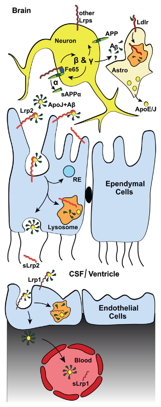
The biological fates of the key initiator of Alzheimer's disease (AD), the amyloid precursor protein (APP), and a family of lipoprotein receptors, the low-density lipoprotein (LDL) receptor-related proteins (LRPs) and their molecular roles in the neurodegenerative disease process are inseparably interwoven. Not only does APP bind tightly to the extracellular domains (ECDs) of several members of the LRP group, their intracellular portions are also connected through scaffolds like the one established by FE65 proteins and through interactions with adaptor proteins such as X11/Mint and Dab1. Moreover, the ECDs of APP and LRPs share common ligands, most notably Reelin, a regulator of neuronal migration during embryonic development and modulator of synaptic transmission in the adult brain, and Agrin, another signaling protein which is essential for the formation and maintenance of the neuromuscular junction (NMJ) and which likely also has critical, though at this time less well defined, roles for the regulation of central synapses. Furthermore, the major independent risk factors for AD, Apolipoprotein (Apo) E and ApoJ/Clusterin, are lipoprotein ligands for LRPs. Receptors and ligands mutually influence their intracellular trafficking and thereby the functions and abilities of neurons and the blood-brain-barrier to turn over and remove the pathological product of APP, the amyloid-β peptide. This article will review and summarize the molecular mechanisms that are shared by APP and LRPs and discuss their relative contributions to AD.

Keywords: APOE; LDL receptor gene family; LRP; amyloid beta; glutamate receptors; neuromuscular junction; synapse; trafficking.


Figures

Figure 1
Figure 1
The low-density lipoprotein (LDL) receptor family. Schematic diagram depicting the domain structure of the LDL receptor family members classified as (from left to right): core, distant and the far side. The seven core members (left) are LDL receptor (Ldlr), very-LDL receptor (Vldlr), Apolipoprotein E (ApoE) receptor 2 (Apoer2/Lrp8), LDL receptor related protein (Lrp)-4 (Lrp4), Lrp1, Lrp1b and Lrp2. These members are classified as core members by the presence of at least one NPxY-motif (asterisk) and a combination of two classical LDL receptor domains: (1) N-terminal ligand binding domain composed of cysteine-rich ligand binding-type repeats (blue); and (2) epidermal growth factor (EGF)-precursor homology domain (orange) composed of EGF-repeats and YWTD/β-propeller domain. Ldlr, Vldlr and Apoer2 express an additional extracellular O-linked sugar (OLS) domain adjacent to the transmembrane (TM) segment. The more distant members (middle) are the NPxY-lacking Lrp5/Lrp6 and hybrid SorLA with additional Fibronectin repeats (pink) and importantly the VPS10p-sorting motif (green). Four very distant “far side” proteins (right, Lrp3, Lrp10, Lrp12, and Lrad3) only encode ligand binding-type repeats. Lrp3, Lrp10 and Lrp12 also contain atypical CUB-domain (binds Complement, Uegf and Bmp1). In addition to the OLS domains of Apoer2 and Vldlr, alternative splicing of Apoer2 produces splice variants lacking N-terminal ligand binding type repeats (repeats 4–6; Brandes et al., ; gray).
Figure 2
Figure 2
Lipoprotein receptors modulate amyloid precursor protein (APP) trafficking and processing in neurons. Neurons are the major source of Aβ (depicted as green droplets) in the brain. APP (green), all core LDL receptor family members as well as the more distant member SorLA contains at least one NPxY-motif, which acts as a docking site for PTB-domains of intracellular adaptor/scaffold proteins. Both Fe65 and Dab1 bind APP, as well as a number of LDL receptor family members (red and orange), via their PTB-domains. The simultaneous binding of these intracellular adaptor/scaffolding proteins to the NPxY motifs of APP and LDL receptors coordinate their intracellular trafficking, thus regulating APP localization and processing. The adapter/scaffold proteins control the speed of endocytosis of the receptors in that Fe65 and Dab1 binding to APP masks the endocytosis signal of APP, resulting in the surface retention of APP. This increases the exposure of APP to α-secretase (α), which cleaves APP inside the Aβ region (dark green) to release a soluble APPα (sAPPα) fragment and ultimately preventing the production of Aβ. Importantly, Lrp1 and Lrp1b (both orange in the diagram) have drastically different rates of endocytosis, with the internalization rate of Lrp1 exceeding that of Lrp1b by many-fold. Both bind Fe65, connecting them in a complex APP, and have opposite effects on APP processing. The fast endocytosis rate of Lrp1 increases the exposure of APP to the endosomal β- (BACE1, β) and γ-secretase (γ), producing Aβ (green tears) and soluble APPβ (sAPPβ) fragment. Another intraendosomal sorting receptor of the LDL receptor family, SorLA, can bind and reroute receptors from the endosome back to the trans-Golgi network (TGN), where it is either sequestered, sorted back to the cell surface, or sent to the lysosome for degradation. Apoer2, which also recycles slowly, binds Fe65 via its NPxY-motif, promoting APP surface stability and decrease amyloidogenic processing. Additionally, simultaneous binding of the secreted, extracellular ligand, F-spondin, to the ECDs of APP and Apoer2 also promotes APP stability at the surface.
Figure 3
Figure 3
Lrp2 mediates Aβ-clearance via the blood cerebrospinal fluid (CSF) barrier (BCSFB). Diagram depicting the Lrp2-mediated clearance of interstitial Aβ through the cerebral spinal fluid (CSF) into the blood. In addition to direct astrocytic Lrp2 clearance of Aβ, Lrp2 expressed in the ependymal cells of the choroid plexus also facilitate Aβ removal. The choroid plexus functions to produce and filter CSF. This filtration removes metabolic waste, excess neurotransmitters and foreign/toxic particles, such as Aβ, which is mainly produced by neurons (see Figure 2). Apolipoproteins, such as ApoE and ApoJ/Clusterin (yellow dots), mainly secreted from astrocytes (“Astro”), bind circulating interstitial Aβ. These Aβ-laden apolipoproteins then bind lipoprotein receptors (red) and mediate their cellular uptake. ApoJ/Clusterin is eliminated rapidly across the BCSFB by ependymal Lrp2 (light red), facilitating the clearance of Aβ via lysosomal degradation in ependymal cells and subsequent exocytosis into the CSF, where soluble Lrp2 (sLrp2) has been detected (Spuch et al., 2015). BACE1 is the enzyme that processes Lrp2 and Lrp1 to release sLrp2 and sLrp1, respectively. BACE1 is also found in the choroid plexus (Crossgrove et al., ; Liu et al., 2013). Other lipoprotein receptors (dark red, most notably Lrp1) then transport Aβ and the apolipoproteins across the endothelial cells from the CSF to the blood vessels of the choroid plexus. sLrp1 can also be detected in plasma, albeit its origin there is mainly peripheral.
Figure 4
Figure 4
Lrp4 and APP interaction during neuromuscular junction (NMJ) formation. Illustration depicting the interaction of Lrp4, MuSK, Agrin and APP/APLP1/APLP2 in the formation of the NMJ. Agrin binds Lrp4 resulting in phosphorylation (P) of MuSK, which leads to the recruitment and clustering of acetylcholine receptors (AchRs). The recruitment of AChR to the NMJ depends on all components of the complex. Knockouts of Lrp4, MuSK, Agrin, or APP/APLP1/APLP2 result in defective NMJ formation and perinatal lethality. APP and its family members (APLP1 and APLP2) have redundant functions, allowing them to compensate if one is knocked out. APLP1 is expressed on the presynaptic motor neuron, whereas APLP2 and APP are expressed by both nerve cells and muscle cells. Double knockouts lacking both APP and APLP1 form functional NMJs and are viable, whereas APP−/−/APLP2−/− and APLP1−/−/APLP2−/− mice have severely defective neuromuscular synapses and high postnatal lethality, indicating that APLP2 is an essential component in NMJ formation, but APP and APLP1 together can partially compensate in the absence of APLP2. Agrin is expressed in both neurons and muscle cells, but each express different isoforms. Isoforms expressed by neurons differ from muscular Agrin by the Z+ splice insert (yellow star), required for Lrp4 binding (Zong et al., 2012) and NMJ-formation (Burgess et al., 1999). In addition, besides secreted Agrin, motorneurons express a TM Agrin, which is not required for NMJ-formation. Extracellular cleavage of Agrin (α- and β-sites) can be mediated by Neurotrypsin and other as-yet unidentified proteases (black) expressed at the muscle. While Agrin cleavage is required for proper NMJ maturation, Neurotrypsin-mediated cleavage of Agrin is not—despite the fact that Neurotrypsin overexpression leads to NMJ-failures (Bolliger et al., 2010). The small soluble Z+ containing C-terminal fragment (after β-cleavage) is sufficient to bind Lrp4 and induce AChR-clustering, but it is less efficient compared to full length Agrin or Agrin cleaved at the α-site, only (Zong et al., 2012).

Similar articles

Cited by

References

    1. Aledo R., Alonso R., Mata P., Llorente-Cortés V., Padró T., Badimon L. (2012). LRP1 gene polymorphisms are associated with premature risk of cardiovascular disease in patients with familial hypercholesterolemia. Rev. Esp. Cardiol. (Engl. Ed.) 65, 807–812. 10.1016/j.rec.2012.03.012 - DOI - PubMed
    1. Alvira-Botero X., Carro E. M. (2010). Clearance of amyloid-β peptide across the choroid plexus in Alzheimer’s disease. Curr. Aging Sci. 3, 219–229. 10.2174/1874609811003030219 - DOI - PubMed
    1. Alvira-Botero X., Pérez-Gonzalez R., Spuch C., Vargas T., Antequera D., Garzón M., et al. . (2010). Megalin interacts with APP and the intracellular adapter protein FE65 in neurons. Mol. Cell. Neurosci. 45, 306–315. 10.1016/j.mcn.2010.07.005 - DOI - PubMed
    1. Andersen O. M., Dagil R., Kragelund B. B. (2013). New horizons for lipoprotein receptors: communication by β-propellers. J. Lipid Res. 54, 2763–2774. 10.1194/jlr.m039545 - DOI - PMC - PubMed
    1. Andersen O. M., Reiche J., Schmidt V., Gotthardt M., Spoelgen R., Behlke J., et al. . (2005). Neuronal sorting protein-related receptor sorLA/LR11 regulates processing of the amyloid precursor protein. Proc. Natl. Acad. Sci. U S A 102, 13461–13466. 10.1073/pnas.0503689102 - DOI - PMC - PubMed

LinkOut - more resources