. 2017 Mar 1:11:9.
doi: 10.3389/fncir.2017.00009. eCollection 2017.

Mapping and Analysis of the Connectome of Sympathetic Premotor Neurons in the Rostral Ventrolateral Medulla of the Rat Using a Volumetric Brain Atlas

Affiliations

Mapping and Analysis of the Connectome of Sympathetic Premotor Neurons in the Rostral Ventrolateral Medulla of the Rat Using a Volumetric Brain Atlas

Bowen Dempsey et al. Front Neural Circuits. .

Abstract

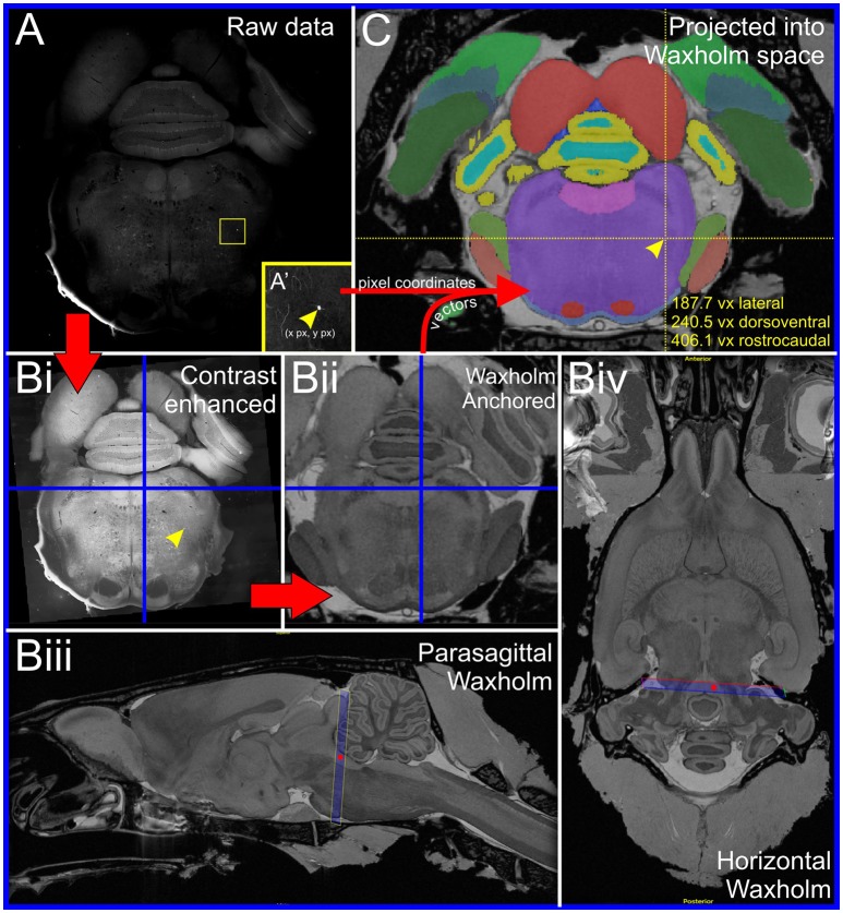
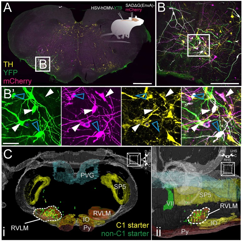
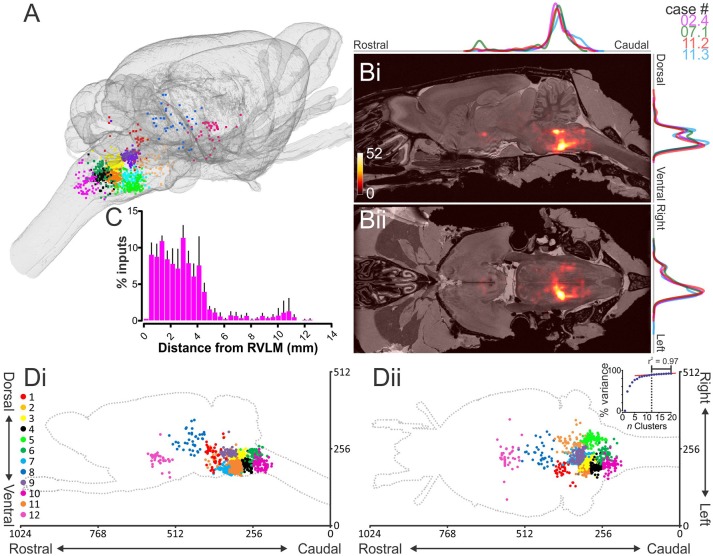
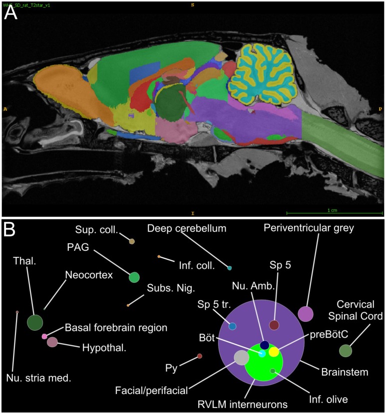
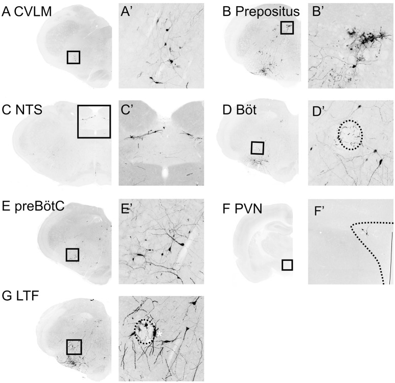
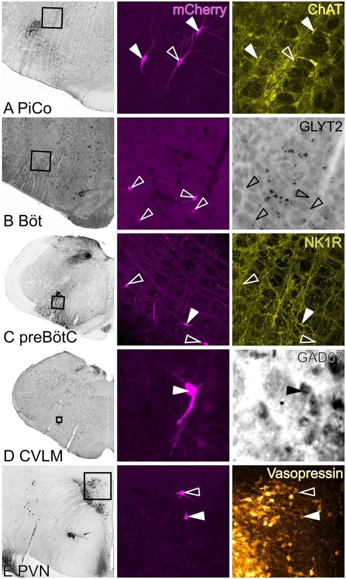
Spinally projecting neurons in the rostral ventrolateral medulla (RVLM) play a critical role in the generation of vasomotor sympathetic tone and are thought to receive convergent input from neurons at every level of the neuraxis; the factors that determine their ongoing activity remain unresolved. In this study we use a genetically restricted viral tracing strategy to definitively map their spatially diffuse connectome. We infected bulbospinal RVLM neurons with a recombinant rabies variant that drives reporter expression in monosynaptically connected input neurons and mapped their distribution using an MRI-based volumetric atlas and a novel image alignment and visualization tool that efficiently translates the positions of neurons captured in conventional photomicrographs to Cartesian coordinates. We identified prominent inputs from well-established neurohumoral and viscero-sympathetic sensory actuators, medullary autonomic and respiratory subnuclei, and supramedullary autonomic nuclei. The majority of inputs lay within the brainstem (88-94%), and included putative respiratory neurons in the pre-Bötzinger Complex and post-inspiratory complex that are therefore likely to underlie respiratory-sympathetic coupling. We also discovered a substantial and previously unrecognized input from the region immediately ventral to nucleus prepositus hypoglossi. In contrast, RVLM sympathetic premotor neurons were only sparsely innervated by suprapontine structures including the paraventricular nucleus, lateral hypothalamus, periaqueductal gray, and superior colliculus, and we found almost no evidence of direct inputs from the cortex or amygdala. Our approach can be used to quantify, standardize and share complete neuroanatomical datasets, and therefore provides researchers with a platform for presentation, analysis and independent reanalysis of connectomic data.

Keywords: RVLM; connectome; mesoscale; rabies; respiratory-sympathetic; segmentation; sympathetic; volumetric.


Figures

Figure 1
Figure 1
Anchoring workflow. (A) Original coronal epifluorescence image showing the location of a single rabies-labeled input neuron (yellow box, inset in A'). Following manual annotation, the pixel coordinates of the neuron were exported to a spreadsheet and the image contrast adjusted for optimal visualization of anatomical landmarks (Bi). The image was then aligned to a corresponding section plane through the Waxholm atlas template (Bii) using a beta version of an image alignment/anchoring tool embedded in the Navigator-3 system. The anchoring tool allowed accurate positioning of the image plane in the MRI-derived atlas template. The orientation of the plane of the image is close to coronal, as shown in the blue frames in (Biii) and (Biv). The parasagittal plane through the atlas template shown in (Biii) corresponds to the vertical blue line in (Bi) and (Bii), whereas the horizontal plane in (Biv) corresponds to the horizontal blue lines in (Bi) and (Bii). The red dots in (Biii) and (Biv) represent the intersections with the horizontal and parasagittal planes, respectively. Anchoring vectors generated by Navigator-3 were then used to translate the pixel co-ordinates of annotated neurons into xyz Waxholm coordinates and integrated into the Waxholm segmentation model (C).
Figure 2
Figure 2
Experimental strategy (inset in A): HSV-hCMV-YTB was injected at the T2 IML, retrogradely driving the expression of TVA, rabies glycoprotein, and YFP in spinally projecting neurons. SADΔG(EnvA)-mCherry was subsequently injected into the RVLM, exclusively infecting neurons that express TVA, and seeding trans-synaptic infection of monosynaptically connected input neurons. (A) Coronal brainstem section at the level of the rostral ventrolateral medulla (RVLM) processed for immunoreactivity to TH and YFP; starter neurons are defined by the expression of both YFP and mCherry. (B) Confocal detail with magnified (B') examples of C1 (solid arrowheads) and non-C1 (cyan arrowheads) starter neurons (individual channels and merged image). Blue asterisk indicates a C1 input neuron. (C) Distribution of C1 (yellow) and non-C1 (green) starter neurons from four experiments plotted in Waxholm space, shown from coronal (Ci) and parasagittal (Cii) perspectives. Anatomical landmarks are Waxholm-segmented boundaries of the inferior olive (IO), spinal trigeminal nucleus (SP5), RVLM, pyramidal tract (Py), facial nerve (VII), and periventricular gray (PVG). Scale bars 1000 μm (A), 250 μm (B), and 100 μm (B').
Figure 3
Figure 3
Distribution of monosynaptic input neurons. (A) Input neurons to putative sympathetic premotor neurons in the rostral ventrolateral medulla (RVLM) segregated into 12 distinct clusters and plotted in the Waxholm volumetric rat atlas. (B) Heat maps showing input neuron density overlaid on the Waxholm MRI dataset in the parasagittal (i) and horizontal (ii) planes. Scale indicates number of neurons per 10 voxels. Normalized plots of input densities for individual experiments are plotted alongside heat maps. (C) Absolute distance of input neurons from RVLM epicenter (n = 4 rats). (D) Cluster plots presented in the sagittal (i) and horizontal (ii) planes. Inset in (Dii) shows the proportion of total variance accounted for by incremental increases in cluster number; the turning point of the curve, after which slope becomes linear, occurs at 12 clusters.
Figure 4
Figure 4
RVLM input neurons organized by Waxholm segment. (A) Parasagittal view through the Waxholm volumetric atlas for reference; different colors indicate different brain segments. (B) Proportions of input neurons per Waxholm segment (summed from 4 experiments) colored and positioned according to their corresponding Waxholm regions. The diameter of each circle corresponds to the number of inputs therein. See Table S2 for source data and abbreviations.
Figure 5
Figure 5
Medullary inputs to RVLM sympathetic premotor neurons. Inverted epifluorescence micrographs with high power insets (A'–F') of boxed regions illustrating monosynaptic inputs from the caudal ventrolateral medulla (CVLM: A), nucleus prepositus (B), nucleus of the solitary tract (NTS: C), Bötzinger (Böt: D), pre-Bötzinger Complex (preBötC: E), paraventricular nucleus of the hypothalamus (PVN: F) and lateral tegmental field (LTF: G). Nucleus ambiguus is indicated by the hatched ovals in (D,G). *in Panel (G) indicates a TH-positive starter neuron.
Figure 6
Figure 6
Neurochemical phenotypes of input neurons. Left hand panels are low power images of in situ hybridization/immunohistochemistry indicating region shown in high power images. Middle panel shows rabies-labeled input neurons, right panels show in situ hybridization/immunofluorescence. Closed arrowheads indicate double-labeled neurons; open arrowheads indicate the positions of rabies labeled input neurons. (A) ChAT-immunoreactive input neurons in the region of the lateral reticular formation that corresponds to the mouse PiCo. (B) Bötzinger input neurons were abundant but none were identified as GlyT2-positive. (C) NK1R-positive and -negative pre-Bötzinger Complex inputs. (D) GAD67-positive input neurons in the CVLM. (E) Vasopressin-positive and -negative PVN inputs. PiCo: post-inspiratory complex, Böt: Bötzinger, preBötC: pre-Bötzinger Complex, CVLM: caudal ventrolateral medulla, PVN: paraventricular nucleus.

Similar articles

Cited by

References

    1. Allen A. M. (2002). Inhibition of the hypothalamic paraventricular nucleus in spontaneously hypertensive rats dramatically reduces sympathetic vasomotor tone. Hypertension 39, 275–280. 10.1161/hy0202.104272 - DOI - PubMed
    1. Anderson T. M., Garcia A. J., Baertsch N. A., Pollak J., Bloom J. C., Wei A. D., et al. . (2016). A novel excitatory network for the control of breathing. Nature 536, 76–80. 10.1038/nature18944 - DOI - PMC - PubMed
    1. Bou Farah L., Bowman B. R., Bokiniec P., Karim S., Le S., Goodchild A. K., et al. . (2016). Somatostatin in the rat rostral ventrolateral medulla: origins and mechanism of action. J. Comp. Neurol. 524, 323–342. 10.1002/cne.23846 - DOI - PubMed
    1. Bowman B. R., Kumar N. N., Hassan S. F., McMullan S., Goodchild A. K. (2013). Brain sources of inhibitory input to the rat rostral ventrolateral medulla. J. Comp. Neurol. 521, 213–232. 10.1002/cne.23175 - DOI - PubMed
    1. Brennand K. J., Simone A., Jou J., Gelboin-Burkhart C., Tran N., Sangar S., et al. . (2011). Modelling schizophrenia using human induced pluripotent stem cells. Nature 473, 221–225. 10.1038/nature09915 - DOI - PMC - PubMed

Publication types