White Matter Deterioration May Foreshadow Impairment of Emotional Valence Determination in Early-Stage Dementia of the Alzheimer Type
- PMID: 28298891
- PMCID: PMC5331035
- DOI: 10.3389/fnagi.2017.00037
White Matter Deterioration May Foreshadow Impairment of Emotional Valence Determination in Early-Stage Dementia of the Alzheimer Type
Abstract
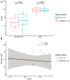
In Alzheimer Disease (AD), non-verbal skills often remain intact for far longer than verbally mediated processes. Four (1 female, 3 males) participants with early-stage Clinically Diagnosed Dementia of the Alzheimer Type (CDDAT) and eight neurotypicals (NTs; 4 females, 4 males) completed the emotional valence determination test (EVDT) while undergoing BOLD functional magnetic resonance imaging (fMRI). We expected CDDAT participants to perform just as well as NTs on the EVDT, and to display increased activity within the bilateral amygdala and right anterior cingulate cortex (r-ACC). We hypothesized that such activity would reflect an increased reliance on these structures to compensate for on-going neuronal loss in frontoparietal regions due to the disease. We used diffusion tensor imaging (DTI) to determine if white matter (WM) damage had occurred in frontoparietal regions as well. CDDAT participants had similar behavioral performance and no differences were observed in brain activity or connectivity patterns within the amygdalae or r-ACC. Decreased fractional anisotropy (FA) values were noted, however, for the bilateral superior longitudinal fasciculi and posterior cingulate cortex (PCC). We interpret these findings to suggest that emotional valence determination and non-verbal skill sets are largely intact at this stage of the disease, but signs foreshadowing future decline were revealed by possible WM deterioration. Understanding how non-verbal skill sets are altered, while remaining largely intact, offers new insights into how non-verbal communication may be more successfully implemented in the care of AD patients and highlights the potential role of DTI as a presymptomatic biomarker.
Keywords: Alzheimer; DTI; brain networks; chimeric faces; emotional valence; fMRI.
Figures


Similar articles
-
Lower Activation in Frontal Cortex and Posterior Cingulate Cortex Observed during Sex Determination Test in Early-Stage Dementia of the Alzheimer Type.Front Aging Neurosci. 2017 May 22;9:156. doi: 10.3389/fnagi.2017.00156. eCollection 2017. Front Aging Neurosci. 2017. PMID: 28588478 Free PMC article.
-
The role of diffusion tensor imaging and fractional anisotropy in the evaluation of patients with idiopathic normal pressure hydrocephalus: a literature review.Neurosurg Focus. 2016 Sep;41(3):E12. doi: 10.3171/2016.6.FOCUS16192. Neurosurg Focus. 2016. PMID: 27581308 Review.
-
Multiple DTI index analysis in normal aging, amnestic MCI and AD. Relationship with neuropsychological performance.Neurobiol Aging. 2012 Jan;33(1):61-74. doi: 10.1016/j.neurobiolaging.2010.02.004. Epub 2010 Apr 3. Neurobiol Aging. 2012. PMID: 20371138
-
Is there an association between peripheral immune markers and structural/functional neuroimaging findings?Prog Neuropsychopharmacol Biol Psychiatry. 2014 Jan 3;48:295-303. doi: 10.1016/j.pnpbp.2012.12.013. Epub 2013 Jan 11. Prog Neuropsychopharmacol Biol Psychiatry. 2014. PMID: 23313563 Review.
-
Changes in anatomical and functional connectivity of Parkinson's disease patients according to cognitive status.Eur J Radiol. 2015 Jul;84(7):1318-24. doi: 10.1016/j.ejrad.2015.04.014. Epub 2015 Apr 27. Eur J Radiol. 2015. PMID: 25963506
Cited by
-
Altered Functional Connectivity of Basal Ganglia in Mild Cognitive Impairment and Alzheimer's Disease.Brain Sci. 2022 Nov 15;12(11):1555. doi: 10.3390/brainsci12111555. Brain Sci. 2022. PMID: 36421879 Free PMC article.
-
What We Know About the Brain Structure-Function Relationship.Behav Sci (Basel). 2018 Apr 18;8(4):39. doi: 10.3390/bs8040039. Behav Sci (Basel). 2018. PMID: 29670045 Free PMC article. Review.
-
Hearing impairment and the risk of neurodegenerative dementia: A longitudinal follow-up study using a national sample cohort.Sci Rep. 2018 Oct 15;8(1):15266. doi: 10.1038/s41598-018-33325-x. Sci Rep. 2018. PMID: 30323320 Free PMC article.
References
-
- Albert M. S., DeKosky S. T., Dickson D., Dubois B., Feldman H. H., Fox N. C., et al. . (2011). The diagnosis of mild cognitive impairment due to Alzheimer disease: recommendations from the National institute on aging-Alzheimer’s association workgroups on diagnostic guidelines for Alzheimer disease. Alzheimers Dement. 7, 270–279. 10.1016/j.jalz.2011.03.008 - DOI - PMC - PubMed
-
- Arnold S. E., Hyman B. T., Flory J., Damasio A. R., Van Hoesen G. W. (1991). The topographical and neuroanatomical distribution of neurofibrillary tangles and neuritic plaques in the cerebral cortex of patients with Alzheimer’s disease. Cereb. Cortex 1, 103–116. 10.1093/cercor/1.1.103 - DOI - PubMed
Grants and funding
LinkOut - more resources
Full Text Sources
Other Literature Sources

