. 2017 Mar 1:8:86.
doi: 10.3389/fphys.2017.00086. eCollection 2017.

Two Phase Modulation of [Formula: see text] Entry and Cl-/[Formula: see text] Exchanger in Submandibular Glands Cells by Dexmedetomidine

Affiliations

Two Phase Modulation of [Formula: see text] Entry and Cl-/[Formula: see text] Exchanger in Submandibular Glands Cells by Dexmedetomidine

Minjeong Ji et al. Front Physiol. .

Abstract

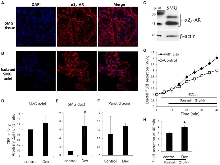
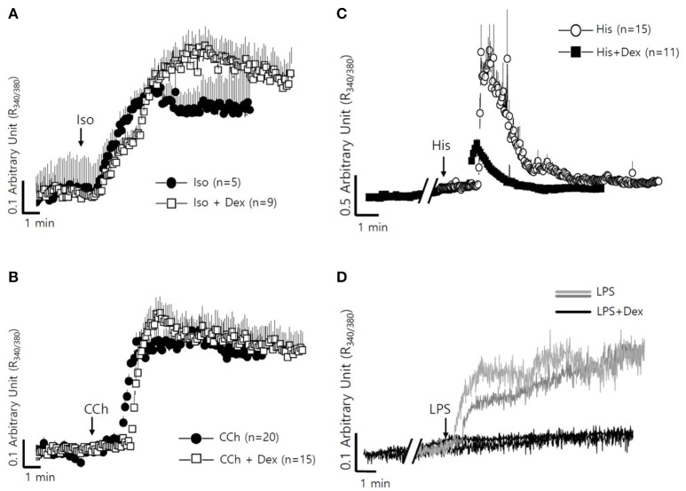
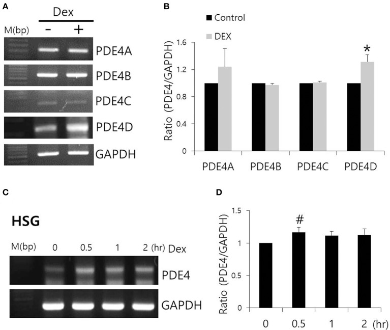
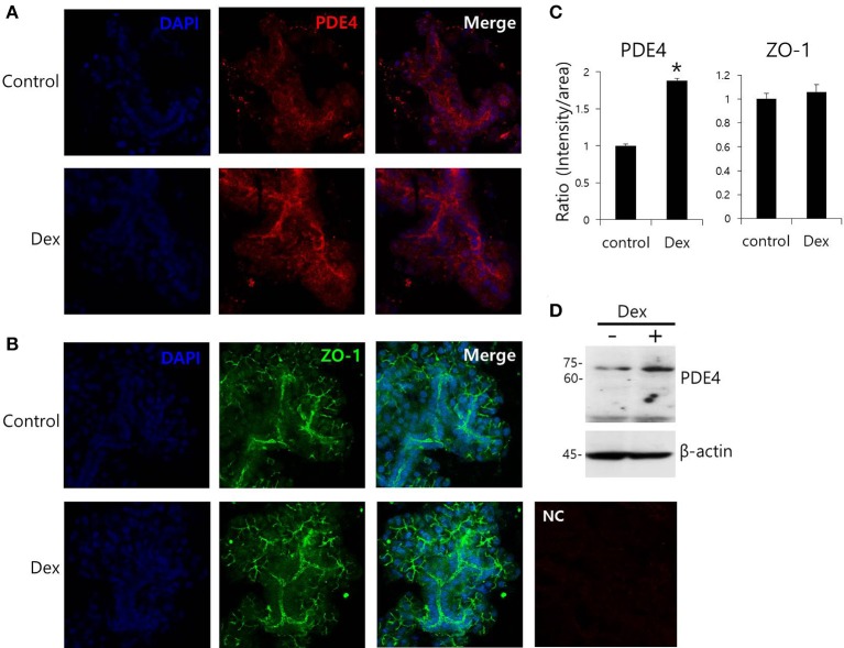
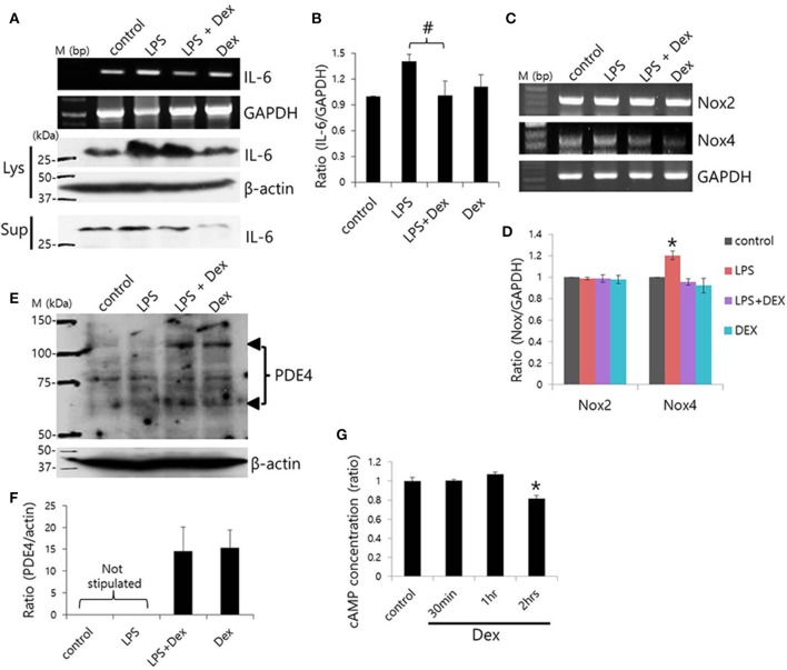
Dexmedetomidine (Dex), a highly selective α2-adrenoceptor agonist, attenuates inflammatory responses induced by lipopolysaccharide (LPS) and induces sedative and analgesic effects. Administration of Dex also reduces salivary secretion in human subjects and inhibits osmotic water permeability in rat cortical collecting ducts. However, little is known about the mechanisms underlying the effects of Dex on salivary glands fluid secretion. We demonstrated the α2-adrenoceptor expression in the basolateral membrane of mouse submandibular glands (SMG). To investigate fluid secretion upon treatment with Dex, we studied the effects of Dex on the activity of Na+-K+-2Cl- cotransporter1 (NKCC1) and Cl-/[Formula: see text] exchange (CBE), and on downstream pro-inflammatory cytokine expression in isolated primary mouse SMG cells. Dex acutely increased CBE activity and NKCC1-mediated and independent [Formula: see text] entry in SMG duct cells, and enhanced ductal fluid secretion in a sealed duct system. Dex showed differential effects on cholinergic/adrenergic stimulations and inflammatory mediators, histamine, and LPS, stimulations-induced Ca2+ in mouse SMG cells. Both, histamine- and LPS-induced intracellular Ca2+ increases were inhibited by Dex, whereas carbachol-stimulated Ca2+ signals were not. Long-lasting (2 h) treatment with Dex reduced CBE activity in SMG and in human submandibular glands (HSG) cells. Moreover, when isolated SMG cells were stimulated with Dex for 2 h, phosphodiesterase 4D (PDE4D) expression was enhanced. These results confirm the anti-inflammatory properties of Dex on LPS-mediated signaling. Further, Dex also inhibited mRNA expression of interleukin-6 and NADPH oxidase 4. The present study also showed that α2-adrenoceptor activation by Dex reduces salivary glands fluid secretion by increasing PDE4D expression, and subsequently reducing the concentration of cAMP. These findings reveal an interaction between the α2-adrenoceptor and PDE4D, which should be considered when using α2-adrenoceptor agonists as sedative or analgesics.

Keywords: dexmedetomidine; ion transporters; phosphodiesterase 4; secretion; submandibular gland.


Figures

Figure 1
Figure 1
Effect of Dex on CBE activity in isolated mouse SMG cells and fluid secretion in sealed parotid ducts. (A,B) Localization of α2A-adrenergic receptor (AR) in SMG tissue and isolated SMG acini. (C) Protein expression α2A-AR in SMG. CBE activity was determined by measuring changes in pHi in SMG acini (D), SMG ducts (E), and parotid acini (F) with and without 100 ng/ml Dex. The slope of pHi measured CBE activity in the absence of Cl at the beginning of time course (30–45 s), and height to reach the point of maximum pHi from the minimum point. Bars represent the mean ± SEM (n = 4, *p < 0.01, #p < 0.05). (G) Sealed parotid ducts were isolated and used to measure fluid secretion in response to stimulation with 5 μM forskolin in the absence (control, open square) and presence of 100 ng/ml Dex (closed rhombus) (n = 4, *p < 0.01). (H) The mean ± SEM at the 40 min secretion time point shows an increase in basal secretion upon Dex treatment.
Figure 2
Figure 2
Effect of Dex on NH4+ influx in mouse isolated SMG and HSG cells. NH4+ influx was assessed by measuring changes in pHi in SMG acini with and without 100 ng/ml Dex. The rate of NH4+ influx (gray dotted line) was determined from the pHi recovery rate in the second phase after 20 mM NH4Cl pulse. Bars represent the mean ± SEM (n = 5). (A) Traces of the NH4Cl pulses used to measure NH4+ influx with 100 ng/ml Dex or 10 μM YOH in isolated SMG acini. (B) Analysis of NH4+ influx in isolated SMG acini. Bars represent the mean ± SEM (n = 4, *p < 0.01). Analysis of NH4+ influx in HSG cells (C,D) and SMG acini (E) in the presence of the indicated components including 100 μM Bumetanide (Bumeta). Bars represent the mean ± SEM (n = 4, *p < 0.01).
Figure 3
Figure 3
Long-lasting Dex treatment on CBE activity in in mouse isolated SMG and HSG cells. SMG and HSG cells were treated with Dex for 2 h. CBE activity was assessed by measuring changes in pHi in AE2-transfected cells (A), in SLC26A6-transfected cells (B), in SMG acini (C), and in HSG cells (D) with and without 100 ng/ml Dex. Bars show the mean ± SEM (n = 4, *p < 0.01).
Figure 4
Figure 4
Differential effects of Dex on neurotransmitter inputs and inflammatory mediator-induced intracellular calcium signaling in mouse SMG cells. (A) Changes in [Ca2+]i induced by 100 μM isoproterenol without (closed circle) and with pre-treatment with 100 ng/ml Dex (open square). All of the traces were averaged. (B) Changes in [Ca2+]i induced by 10 μM carbachol without (closed circle) and with pre-treatment with 100 ng/ml Dex (open square). All of the traces were averaged. (C) Changes in [Ca2+]i induced by 100 nM histamine without (closed circle) and with pre-treatment with 100 ng/ml Dex (closed square). All of the traces were averaged. (D) Changes in [Ca2+]i induced by 20 μg/ml LPS in ducts and in acini without (gray line for ducts and light gray line for acini) and with (black line) pre-treatment with 100 ng/ml Dex. A single trace is shown. Arrows indicate when stimuli were applied to the cells.
Figure 5
Figure 5
Effect of Dex on PDE4 mRNA expression. (A) The mRNA expression of PDE4A–D subfamily members with and without 100 ng/ml Dex in SMG cells. (B) Results are expressed as fold expression relative to the control and mean ± SEM (n = 3, *p < 0.01). (C,D) Time-dependent PDE4 mRNA expression with and without 100 ng/ml Dex in HSG cells (n = 3, #p < 0.05).
Figure 6
Figure 6
Effect of Dex on PDE4 expression. (A,B) Immunofluorescence staining of PDE4 (red) and ZO-1 (green) in isolated SMG cells. (C) Relative intensities of PDE4 and ZO-1 staining divided by area. Bars represent the mean ± SEM (n = 3, *p < 0.01). NC, negative control. (D) PDE4 and β-actin protein expressions with and without 100 ng/ml Dex in SMG cells.
Figure 7
Figure 7
Dex-induced inhibition of LPS-induced inflammatory cytokine expression and cAMP concentration. (A) SMG cells were stimulated with LPS, treated with 100 ng/ml Dex, and then IL-6 mRNA and protein expression were measured in cell lysates (Lys) and cell supernatants (Sup). (B) Bars represent the mean ± SEM (n = 4, #p < 0.05). (C) LPS-induced Nox2 and Nox4 mRNA expression in the presence of 100 ng/ml Dex. (D) Analysis of Nox2 and Nox4 mRNA expression in the presence of the indicated components (n = 5, *p < 0.01). (E) PDE4 protein expression in LPS-stimulated SMG cells. The arrowheads indicate PDE4A and PDE4D bands at 65 and 102 kDa, respectively. (F) Analysis of PDE4 expression. Bars show the mean ± SEM (n = 4). (G) cAMP ratios in Dex-treated whole SMG cells (n = 4, *p < 0.01).
Figure 8
Figure 8
Schematic model of Dex-modulated signaling. Dex, dexmedetomidine; α2A-AR, α2A-adrenoceptor; NKCC1, Na+-K+-2Cl cotransporter1; AE2, anion exchanger2; SLC26A6, solute carrier 26 family A6; Nox4, NADPH oxidase 4; LPS, lipopolysaccharide; TLR4, toll-like receptor 4; PDE, phosphodiesterase; YOH, yohimbine. Acute (black) and long-lasting (red) treatment with Dex.

Similar articles

Cited by

References

    1. Ahuja M., Jha A., Maleth J., Park S., Muallem S. (2014). cAMP and Ca2+ signaling in secretory epithelia: crosstalk and synergism. Cell Calcium 55, 385–393. 10.1016/j.ceca.2014.01.006 - DOI - PMC - PubMed
    1. Bischoff P., Kochs E. (1993). [Alpha 2-agonists in anesthesia and intensive medicine]. Anasthesiol. Intensivmed. Notfallmed. Schmerzther. 28, 2–12. 10.1055/s-2007-998867 - DOI - PubMed
    1. Blanco V. M., Márquez M. S., Alvarez-Leefmans F. J. (2013). Parallel changes in intracellular water volume and pH induced by NH3/NH4+ exposure in single neuroblastoma cells. Cell. Physiol. Biochem. 32, 57–76. 10.1159/000356624 - DOI - PubMed
    1. Dauphinee S. M., Karsan A. (2006). Lipopolysaccharide signaling in endothelial cells. Lab. Invest. 86, 9–22. 10.1038/labinvest.3700366 - DOI - PubMed
    1. Engelhard K., Werner C., Kaspar S., Möllenberg O., Blobner M., Bachl M., et al. . (2002). Effect of the α2-agonist dexmedetomidine on cerebral neurotransmitter concentrations during cerebral ischemia in rats. Anesthesiology 96, 450–457. 10.1097/00000542-200202000-00034 - DOI - PubMed

LinkOut - more resources