

Cellular Preoxygenation Partially Attenuates the Antitumoral Effect of Cisplatin despite Highly Protective Effects on Renal Epithelial Cells
- PMID: 28298953
- PMCID: PMC5337362
- DOI: 10.1155/2017/7203758
Cellular Preoxygenation Partially Attenuates the Antitumoral Effect of Cisplatin despite Highly Protective Effects on Renal Epithelial Cells
Abstract
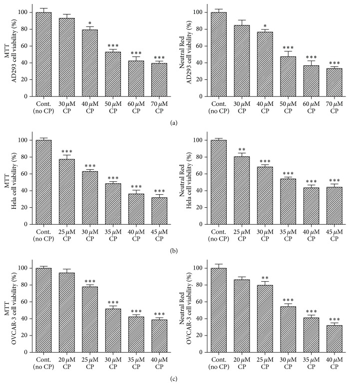
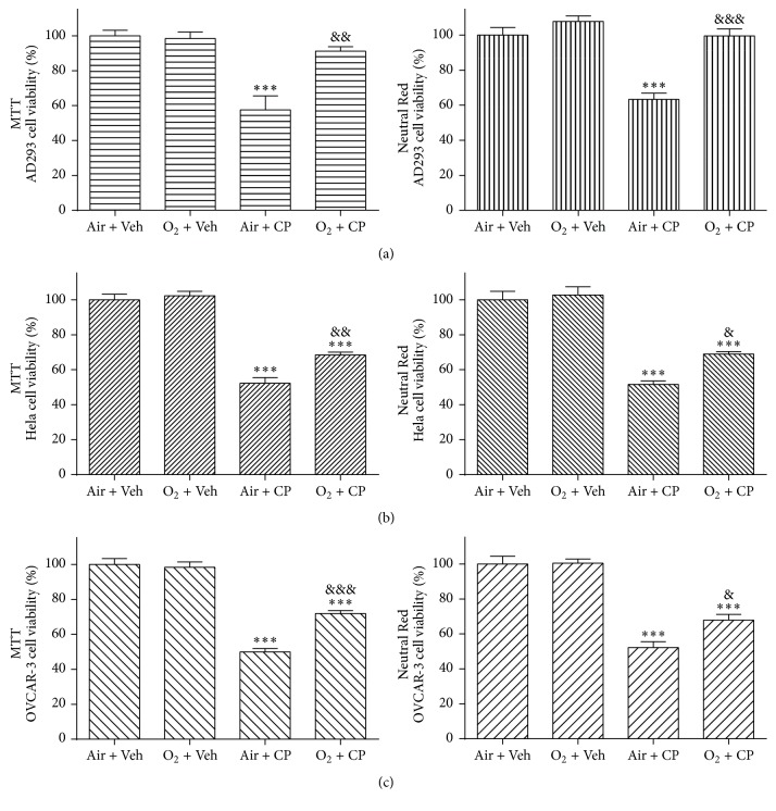
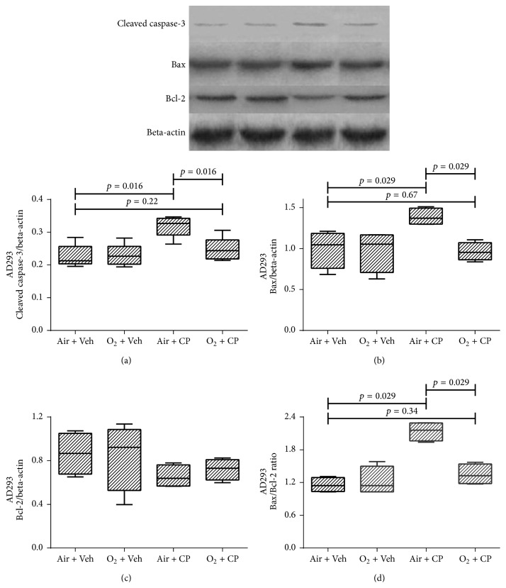
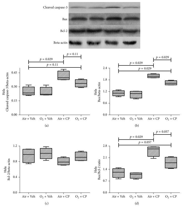
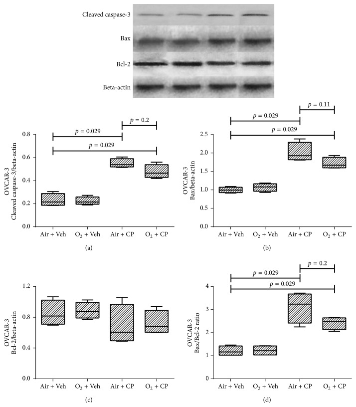
Our previous in vitro studies demonstrated that oxygen pretreatment significantly protects human embryonic renal tubular cell against acute cisplatin- (CP-) induced cytotoxicity. The present study was designed to investigate whether this protective effect is associated with decreasing therapeutic effects of cisplatin on malignant cells. For this purpose, cultured human embryonic kidney epithelial-like (AD293), cervical carcinoma epithelial-like (Hela), and ovarian adenocarcinoma epithelial-like (OVCAR-3) cells were subjected to either 2-hour pretreatment with oxygen (≥90%) or normal air and then to a previously determined 50% lethal dose of cisplatin for 24 hours. Cellular viability was evaluated via MTT and Neutral Red assays. Also, activated caspase-3 and Bax/Bcl-2 ratio, as the biochemical markers of cell apoptosis, were determined using immunoblotting. The hyperoxic preexposure protocol significantly protects renal AD293 cells against cisplatin-induced toxicity. Oxygen pretreatment also partially attenuated the cisplatin-induced cytotoxic effects on Hela and OVCAR-3 cells. However, it did not completely protect these cells against the therapeutic cytotoxic effects of cisplatin. In summary, the protective methods for reducing cisplatin nephrotoxic side effects like oxygen pretreatment might be associated with concurrent reduction of the therapeutic cytotoxic effects of cisplatin on malignant cells like cervical carcinoma (Hela) and ovarian adenocarcinoma (OVCAR-3) cells.
Conflict of interest statement
The authors declare that they have no competing interests regarding the publication of this paper.
Figures
Similar articles
-
Cisplatin toxicity reduced in human cultured renal tubular cells by oxygen pretreatment.Ren Fail. 2013;35(10):1382-6. doi: 10.3109/0886022X.2013.829406. Epub 2013 Sep 4. Ren Fail. 2013. PMID: 24001324
-
Penta-O-galloyl-β-D-glucose attenuates cisplatin-induced nephrotoxicity via reactive oxygen species reduction in renal epithelial cells and enhances antitumor activity in Caki-2 renal cancer cells.Toxicol In Vitro. 2012 Mar;26(2):206-14. doi: 10.1016/j.tiv.2011.11.012. Epub 2011 Dec 7. Toxicol In Vitro. 2012. PMID: 22172427
-
Antitumor activity of a new platinum(II) complex with low nephrotoxicity and genotoxicity.Chem Biol Interact. 2004 Jun 30;148(1-2):37-48. doi: 10.1016/j.cbi.2004.04.002. Chem Biol Interact. 2004. PMID: 15223355
-
Effects of pretreatment with single-dose or intermittent oxygen on Cisplatin-induced nephrotoxicity in rats.Nephrourol Mon. 2014 Sep 5;6(5):e19680. doi: 10.5812/numonthly.19680. eCollection 2014 Sep. Nephrourol Mon. 2014. PMID: 25695032 Free PMC article.
-
Autophagy delays apoptosis in renal tubular epithelial cells in cisplatin cytotoxicity.Autophagy. 2008 Jul;4(5):710-2. doi: 10.4161/auto.6309. Epub 2008 May 20. Autophagy. 2008. PMID: 18497570 Review.
Cited by
-
Silver Nanoparticles Potentiates Cytotoxicity and Apoptotic Potential of Camptothecin in Human Cervical Cancer Cells.Oxid Med Cell Longev. 2018 Dec 12;2018:6121328. doi: 10.1155/2018/6121328. eCollection 2018. Oxid Med Cell Longev. 2018. PMID: 30647812 Free PMC article.
-
Flurbiprofen axetil attenuates cerebral ischemia/reperfusion injury by reducing inflammation in a rat model of transient global cerebral ischemia/reperfusion.Biosci Rep. 2018 Jul 6;38(4):BSR20171562. doi: 10.1042/BSR20171562. Print 2018 Aug 31. Biosci Rep. 2018. PMID: 29540536 Free PMC article.
-
Hypoxia signaling in cancer: Implications for therapeutic interventions.MedComm (2020). 2023 Jan 23;4(1):e203. doi: 10.1002/mco2.203. eCollection 2023 Feb. MedComm (2020). 2023. PMID: 36703877 Free PMC article. Review.
-
The therapeutic approaches of renal recovery after relief of the unilateral ureteral obstruction: A comprehensive review.Iran J Basic Med Sci. 2020 Nov;23(11):1367-1373. doi: 10.22038/ijbms.2020.41984.9926. Iran J Basic Med Sci. 2020. PMID: 33235692 Free PMC article. Review.
-
Anticancer and cytotoxic effects of troxerutin on HeLa cell line: an in-vitro model of cervical cancer.Mol Biol Rep. 2020 Aug;47(8):6135-6142. doi: 10.1007/s11033-020-05694-y. Epub 2020 Aug 1. Mol Biol Rep. 2020. PMID: 32740797
References
-
- Lieberthal W., Triaca V., Levine J. Mechanisms of death induced by cisplatin in proximal tubular epithelial cells: apoptosis vs. necrosis. American Journal of Physiology. 1996;270(4):F700–F708. - PubMed
-
- Meyer K. B., Madias N. E. Cisplatin nephrotoxicity. Mineral and Electrolyte Metabolism. 1994;20(4):201–213. - PubMed
MeSH terms
Substances
LinkOut - more resources
Full Text Sources
Other Literature Sources
Research Materials
Miscellaneous
