

Magnetic resonance imaging in the assessment of brain involvement in alcoholic and nonalcoholic Wernicke's encephalopathy
- PMID: 28298967
- PMCID: PMC5334504
- DOI: 10.4329/wjr.v9.i2.72
Magnetic resonance imaging in the assessment of brain involvement in alcoholic and nonalcoholic Wernicke's encephalopathy
Abstract
Aim: To present the typical and atypical magnetic resonance (MR) imaging findings of alcoholic and non-alcoholic Wernicke's encephalopathy.
Methods: This study included 7 patients with Wernicke's encephalopathy (2 men, 5 women; mean age, 52.3 years) that underwent brain MR examination between January 2012 and March 2016 in a single institution. Three patients were alcoholics and 4 patients were non-alcoholics. MR protocol included a T2-weighted sequence, a fluid attenuation inversion recovery (FLAIR) sequence, a diffusion-weighted sequence (b = 0 and 1000 s/mm2), and a contrast-enhanced MR sequence. All MR images were retrospectively reviewed at baseline and follow-up by two radiologists.
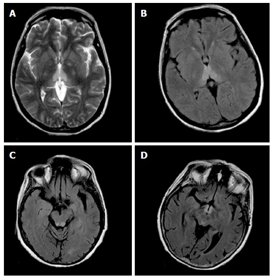
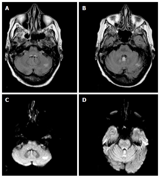
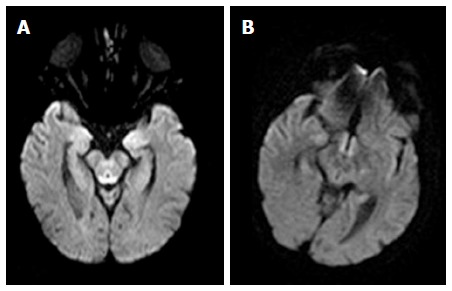
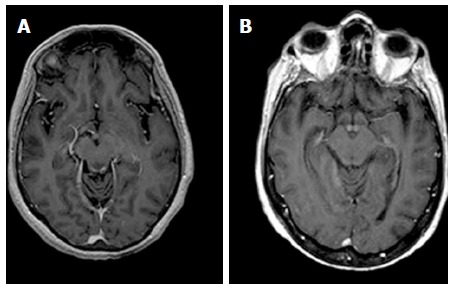
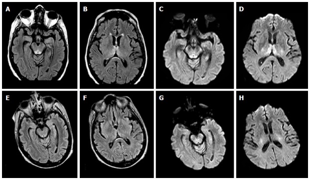
Results: All patients with Wernicke's encephalopathy had bilateral areas showing high signal intensity on both T2-weighted and FLAIR MR images in the typical sites (i.e., the periaqueductal region and the tectal plate). Signal intensity abnormalities in the atypical sites (i.e., the cerebellum and the cerebellar vermis) were seen in 4 patients, all of which had no history of alcohol abuse. Six patients had areas with restricted diffusion in the typical and atypical sites. Four patients had areas showing contrast-enhancement in the typical and atypical sites. Follow-up MR imaging within 6 mo after therapy (intravenous administration of thiamine) was performed in 4 patients, and demonstrated a complete resolution of all the signal intensities abnormalities previously seen in all patients.
Conclusion: MR imaging is valuable in the diagnosis of Wernicke's encephalopathy particularly in patients presenting with atypical clinical symptoms, or with no history of alcohol abuse.
Keywords: Brain; Magnetic resonance imaging; Neurodegenerative disorder; Wernicke’s encephalopathy.
Conflict of interest statement
Conflict-of-interest statement: All authors have no conflict-of-interest.
Figures
References
-
- Gui QP, Zhao WQ, Wang LN. Wernicke’s encephalopathy in nonalcoholic patients: clinical and pathologic features of three cases and literature reviewed. Neuropathology. 2006;26:231–235. - PubMed
-
- Thomson AD, Marshall EJ. The natural history and pathophysiology of Wernicke’s Encephalopathy and Korsakoff’s Psychosis. Alcohol Alcohol. 2006;41:151–158. - PubMed
-
- Nolli M, Barbieri A, Pinna C, Pasetto A, Nicosia F. Wernicke’s encephalopathy in a malnourished surgical patient: clinical features and magnetic resonance imaging. Acta Anaesthesiol Scand. 2005;49:1566–1570. - PubMed
LinkOut - more resources
Full Text Sources
Other Literature Sources
