Review
doi: 10.4103/2008-322X.200162.
Optical Coherence Tomography Angiography of the Optic Disc; an Overview
Affiliations
- PMID: 28299012
- PMCID: PMC5340069
- DOI: 10.4103/2008-322X.200162
Item in Clipboard
Review
Optical Coherence Tomography Angiography of the Optic Disc; an Overview
J Ophthalmic Vis Res.
2017 Jan-Mar.
Abstract
Different diseases of the optic disc may be caused by or lead to abnormal vasculature at the optic nerve head. Optical coherence tomography angiography (OCTA) is a novel technology that provides high resolution mapping of the retinal and optic disc vessels. Recent studies have shown the ability of OCTA to visualize vascular abnormalities in different optic neuropathies. In addition, quantified OCTA measurements were found promising for differentiating optic neuropathies from healthy eyes.
Keywords: Glaucoma; Optic Disc; Optic Nerve Head; Optic Neuropathy; Optical Coherence Tomography Angiography.
Conflict of interest statement
There are no conflicts of interest.
Figures
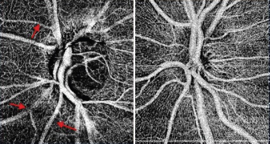
Optical coherence angiography of the optic nerve head of a glaucomatous disc (left) and a healthy disc (right). In addition to the general reduction in the visibility of the disc and peripapillary microvasculature in the glaucomatous disc, focal areas of vascular attenuation are visible (arrows).
References
-
- Regillo CD. The present role of indocyanine green angiography in ophthalmology. Curr Opin Ophthalmol. 1999;10:189–196. - PubMed
-
- Keane PA, Sadda SR. Retinal imaging in the twenty- first century: State of the art and future directions. Ophthalmology. 2014;121:2489–2500. - PubMed
-
- Stein MR, Parker CW. Reactions following intravenous fluorescein. Am J Ophthalmol. 1971;72:861–868. - PubMed
-
- Avila CP, Jr, Bartsch DU, Bitner DG, Cheng L, Mueller AJ, Karavellas MP, et al. Retinal blood flow measurements in branch retinal vein occlusion using scanning laser Doppler flowmetry. Am J Ophthalmol. 1998;126:683–690. - PubMed
Publication types
LinkOut - more resources
Full Text Sources
Other Literature Sources