Tolosa-Hunt Syndrome Demonstrated by Constructive Interference Steady State Magnetic Resonance Imaging
- PMID: 28299013
- PMCID: PMC5340048
- DOI: 10.4103/2008-322X.200171
Tolosa-Hunt Syndrome Demonstrated by Constructive Interference Steady State Magnetic Resonance Imaging
Abstract
Purpose: To highlight the role of constructive interference steady state (CISS) magnetic resonance imaging (MRI) in the diagnosis of Tolosa-Hunt Syndrome (THS).
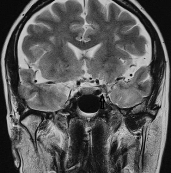
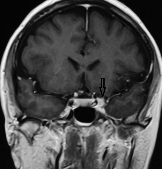
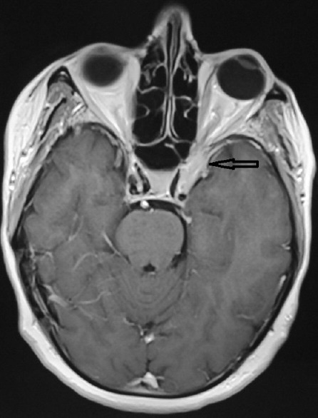
Case report: We describe a case of THS in a 55-year-old woman presenting with left painful opthalmoplegia that was diagnosed by CISS MRI. Patient responded to steroid treatment and the lesion resolved.
Conclusion: Imaging with MRI can help in making the diagnosis of THS by demonstrating an enhancing soft tissue lesion in the cavernous sinus and orbital apex resolving with steroids. CISS MRI is a sensitive sequence for diagnosis and follow-up imaging in THS.
Keywords: Cavernous Sinus; Constructive Interference Steady State Magnetic Resonance Imaging; Painful Ophthalmoplegia; Tolosa-Hunt Syndrome.
Conflict of interest statement
There are no conflicts of interest.
Figures

References
-
- Zhang X, Zhou Z, Steiner TJ, Zhang W, Liu R, Dong Z, et al. Validation of ICHD-3 beta diagnostic criteria for 13.7 Tolosa-Hunt syndrome: Analysis of 77 cases of painful ophthalmoplegia. Cephalalgia. 2014;34:624–632. - PubMed
-
- Okawa S, Hanazono A, Sugawara M, Takahashi S, Otani T, Hanyu N, et al. Contrast-enhanced 3D FIESTA imaging in Tolosa-Hunt syndrome. Headache. 2012;52:822–824. - PubMed
-
- Jain R, Sawhney S, Koul RL, Chand P. Tolosa-Hunt syndrome: MRI appearances. J Med Imaging Radiat Oncol. 2008;52:447–451. - PubMed
-
- Kwan ES, Wolpert SM, Hedges TR, 3rd, Laucella M. Tolosa-Hunt syndrome revisited: Not necessarily a diagnosis of exclusion. AJNR Am J Neuroradiol. 1987;8:1067–1072. - PubMed
Publication types
LinkOut - more resources
Full Text Sources
Other Literature Sources
Miscellaneous