. 2017 May;112(3):23.
doi: 10.1007/s00395-017-0612-7. Epub 2017 Mar 15.

Cardiomyocyte Ogt limits ventricular dysfunction in mice following pressure overload without affecting hypertrophy

Affiliations

Cardiomyocyte Ogt limits ventricular dysfunction in mice following pressure overload without affecting hypertrophy

Sujith Dassanayaka et al. Basic Res Cardiol. 2017 May.

Abstract

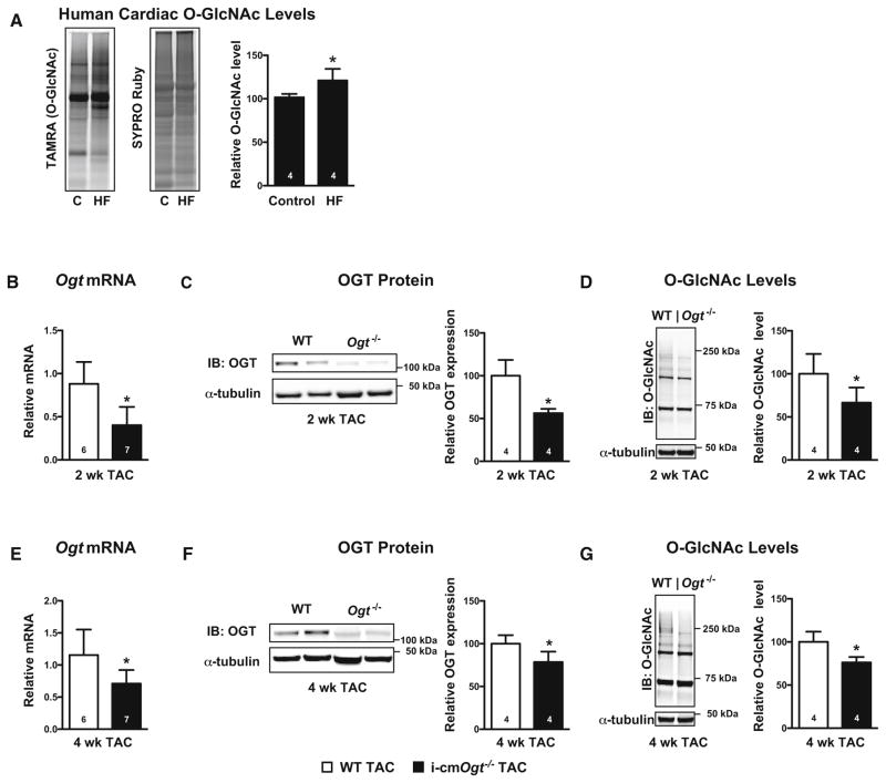
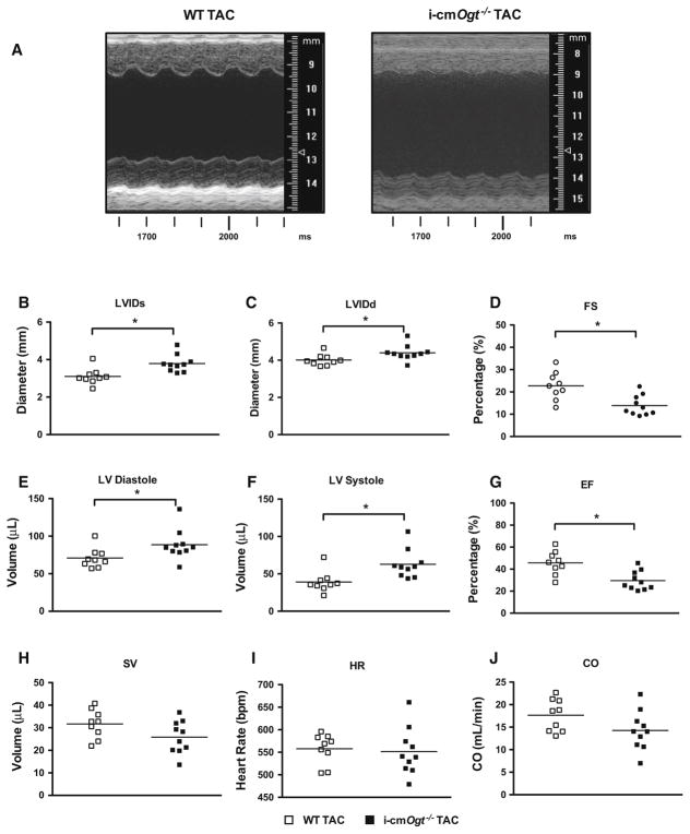
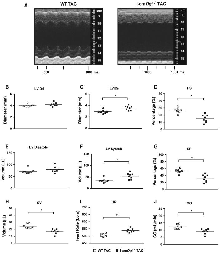
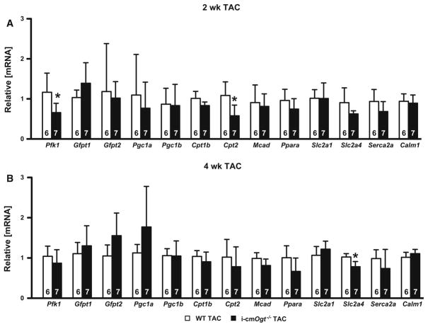
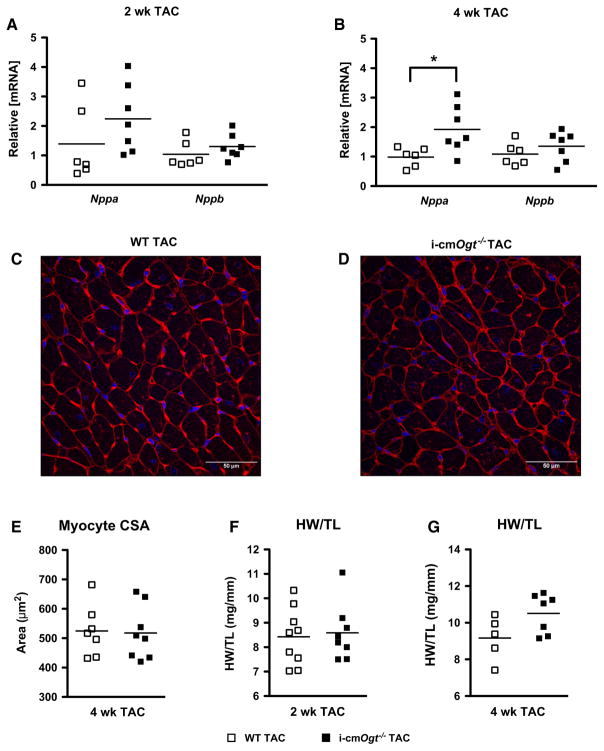
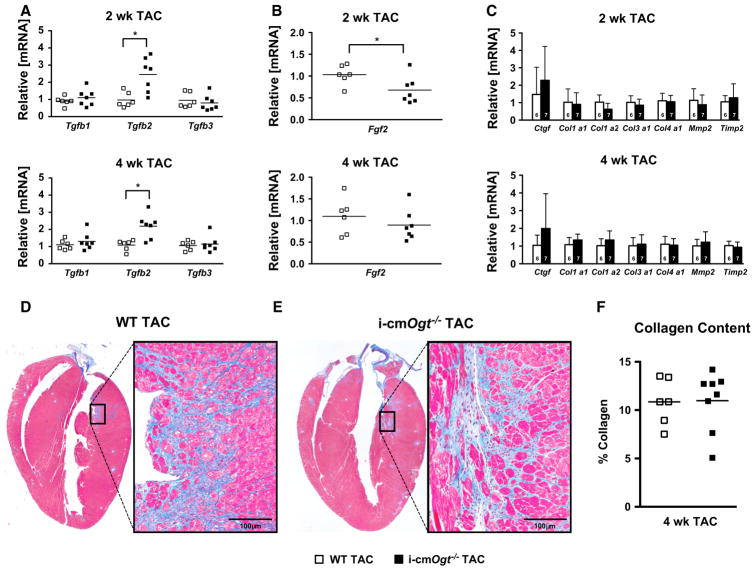
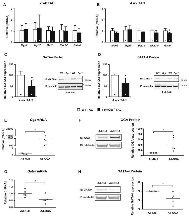
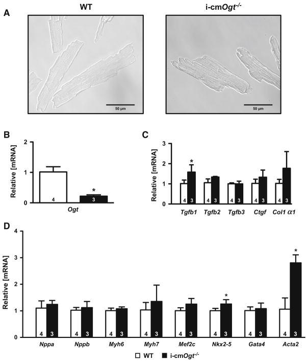
The myocardial response to pressure overload involves coordination of multiple transcriptional, posttranscriptional, and metabolic cues. The previous studies show that one such metabolic cue, O-GlcNAc, is elevated in the pressure-overloaded heart, and the increase in O-GlcNAcylation is required for cardiomyocyte hypertrophy in vitro. Yet, it is not clear whether and how O-GlcNAcylation participates in the hypertrophic response in vivo. Here, we addressed this question using patient samples and a preclinical model of heart failure. Protein O-GlcNAcylation levels were increased in myocardial tissue from heart failure patients compared with normal patients. To test the role of OGT in the heart, we subjected cardiomyocyte-specific, inducibly deficient Ogt (i-cmOgt -/-) mice and Ogt competent littermate wild-type (WT) mice to transverse aortic constriction. Deletion of cardiomyocyte Ogt significantly decreased O-GlcNAcylation and exacerbated ventricular dysfunction, without producing widespread changes in metabolic transcripts. Although some changes in hypertrophic and fibrotic signaling were noted, there were no histological differences in hypertrophy or fibrosis. We next determined whether significant differences were present in i-cmOgt -/- cardiomyocytes from surgically naïve mice. Interestingly, markers of cardiomyocyte dedifferentiation were elevated in Ogt-deficient cardiomyocytes. Although no significant differences in cardiac dysfunction were apparent after recombination, it is possible that such changes in dedifferentiation markers could reflect a larger phenotypic shift within the Ogt-deficient cardiomyocytes. We conclude that cardiomyocyte Ogt is not required for cardiomyocyte hypertrophy in vivo; however, loss of Ogt may exert subtle phenotypic differences in cardiomyocytes that sensitize the heart to pressure overload-induced ventricular dysfunction.

Keywords: Glycosylation; Heart failure; Hexosamine biosynthetic pathway; Metabolism.


Figures

Fig. 1
Fig. 1
Ogt ablation depresses protein O-GlcNAcylation following pressure overload. Human O-GlcNAcylation (a) was measured from cardiac biopsies of disease free patients (control; C) and heart failure (HF) patients. TAMRA-positive bands indicate the presence of O-GlcNAc. SYPRO Ruby stain was used to indicate total protein content and served as a loading control. Cardiac OGT mRNA and protein levels were measured following TAC in mice with cardiac-specific deletion of Ogt. Ogt mRNA, OGT protein, and O-GlcNAc levels were depressed in i-cmOgt−/− mice at both 2 weeks (bd) and 4 weeks (eg) following pressure overload. *p < 0.05 vs. WT TAC (or vs. control human heart group, as in a)
Fig. 2
Fig. 2
Ogt ablation depresses cardiac function following 2 weeks of pressure overload. OGT ablation depresses cardiac function. Following cardiac-specific ablation of Ogt, mice were subjected to pressure overload. Echocardiography was taken at 2 weeks with representative M modes of WT and i-cmOgt−/− TAC (a). Cardiac ablation of Ogt prior to TAC demonstrated significant increases in diastolic (b) and systolic (c) diameters and depressed fractional shortening (d) at 2 weeks. Cardiac left ventricular diastolic (e) and systolic (f) volumes were also increased with a concomitant reduction in ejection fraction (g). Stroke volume (h), heart rate (i), and cardiac output (j) were not disturbed after 2 weeks of pressure overload. *p < 0.05 vs. WT TAC
Fig. 3
Fig. 3
Ogt ablation exacerbates cardiac dysfunction following 4 weeks of pressure overload. Ogt ablation exacerbates pressure overload-induced heart failure. Cardiac function was assessed with echocardiography after 4 weeks of TAC. Representative M modes for WT and i-cmOgt−/− are shown (a). At 4 weeks, diastolic diameter was not changed (b), but systolic diameter (c) was increased in i-cmOgt−/−. Ogt ablation exacerbated fractional shortening (d). Left ventricular diastolic volume (e) was unaltered; however, systolic volume (f) was significantly increased. Ejection fraction worsened in i-cmOgt−/− mice (g). Stroke volume was depressed (h), heart rate increased (i), and cardiac output (j) was exacerbated in i-cmOgt−/− after 4 weeks of TAC. *p < 0.05 vs. WT TAC
Fig. 4
Fig. 4
Metabolic transcripts are not significantly different without Ogt during pressure overload. Cardiac mRNA metabolic profiles of WT TAC and i-cmOgt−/− TAC were analyzed at 2 (a) and 4 weeks (b). Ogt ablation in cardiomyocytes demonstrated decreased Pfk1 and Cpt2 expression 2-week post-TAC, indicating alterations in glycolytic and fatty acid metabolism, respectively. Furthermore, Slc2a4 mRNA was depressed in O-GlcNAc-attenuated cohort indicating a reduction in insulin-mediated glucose cell entry. *p < 0.05 vs. WT TAC
Fig. 5
Fig. 5
Ogt is not required for cardiomyocyte hypertrophy during pressure overload. Mediators of cardiac hypertrophy, Nppa and Nppb, were measured following 2 and 4 weeks of pressure overload (a, b). Ogt deletion did not alter Nppa and Nppb expression at 2 weeks. Nppa expression increased at 4-week post-TAC (b). Hearts from 4 weeks WT TAC (c) and i-cmOgt−/− TAC (d) were stained with wheat germ agglutinin and DAPI. Myocyte cross-sectional area (CSA; e) remained unchanged between WT TAC and i-cmOgt−/− TAC. Heart weight to tibia length ratio was measured at 2 (f) and 4 (g)-week post-TAC. *p < 0.05 vs. WT TAC
Fig. 6
Fig. 6
Cardiac-specific deletion of Ogt enhances TGFβ2 mRNA at 2 and 4 weeks following pressure overload. Markers of cardiac fibrosis were assessed via RT-PCR in WT and i-cmOgt−/− cohorts following 2 and 4 weeks of TAC. Gene expression of TGFβ, major regulator of cardiac fibrosis, demonstrated significant upregulation of Tgfb2 isoform at both 2 and 4 weeks (a). Coincidentally, a known repressor of TGFβ1 mediated signaling, FGF2, demonstrated depressed mRNA expression at 2 weeks. We subsequently measured collagen mRNA expression at 2- and 4-week post-TAC (b). Col1α1, Col1α2, Col3α1, and Col4α2 mRNA expression remained unaltered between groups (c). Following 4 weeks of pressure overload, whole hearts from WT TAC (d) and i-cmOgt−/− TAC (e) mice were excised, sectioned, and stained with Masson’s trichrome to measure cardiac fibrosis. i-cmOgt−/− TAC hearts exhibited no difference in cardiac collagen content from WT TAC hearts. *p < 0.05 vs. WT TAC. *p < 0.05 vs. WT TAC
Fig. 7
Fig. 7
Cardiomyocyte deletion of Ogt suppresses GATA-4 expression during pressure overload. Markers of differentiation and lineage were detected via RT-PCR in WT and i-cmOgt−/− groups following 2 and 4 weeks of TAC. Gene expression of adult cardiomyocyte markers (Myh6, Myh7) remained unchanged (a, b) in both 2- and 4-week TAC groups. Markers of lineage commitment (Mef2c, Nxk2-5, and Gata4) remained unchanged at 2-week post-TAC (a). At 4-week post-TAC, Gata4 mRNA expression was significantly down-regulated in the i-cmOgt−/− group (b). GATA-4 protein expression was assessed at both 2 and 4 weeks (c, d). GATA-4 protein was significantly depressed at both 2 and 4 weeks in the i-cmOgt−/− groups. In additional experiments, NRCMs were transduced with adenovirus to overexpress OGA (which decreases O-GlcNAc levels). OGA overexpression was verified by qRT-PCR (e) and western blotting (f). In these same NRCMs, Gata4 mRNA (g) and GATA-4 protein expression (h) were measured. *p < 0.05 vs. WT TAC
Fig. 8
Fig. 8
Ogt deletion in surgically naïve cardiomyocytes enhances genes indicative of dedifferentiation. Cardiomyocytes were isolated from surgically naïve WT and i-cmOgt−/− mice 5-day post-tamoxifen treatment (a). Ogt mRNA suppression was verified in i-cmOgt−/− cardiomyocytes (b). Markers of fibrosis were largely unchanged except for an increase in Tgfb1 mRNA in i-cmOgt−/− cardiomyocytes (c). Markers of remodeling and lineage commitment were assessed in WT and i-cmOgt−/− cardiomyocytes (d). Nkx2-5 and Acata2 mRNA were upregulated in i-cmOgt−/− cardiomyocytes (d). *p < 0.05 vs. WT

Similar articles

Cited by

References

    1. Akao M, Ohler A, O’Rourke B, Marban E. Mitochondrial ATP-sensitive potassium channels inhibit apoptosis induced by oxidative stress in cardiac cells. Circ Res. 2001;88:1267–1275. doi: 10.1161/hh1201.092094. - DOI - PubMed
    1. Bisping E, Ikeda S, Kong SW, Tarnavski O, Bodyak N, McMullen JR, Rajagopal S, Son JK, Ma Q, Springer Z, Kang PM, Izumo S, Pu WT. Gata4 is required for maintenance of postnatal cardiac function and protection from pressure overload-induced heart failure. Proc Natl Acad Sci USA. 2006;103:14471–14476. doi: 10.1073/pnas.0602543103. - DOI - PMC - PubMed
    1. Condorelli G, Roncarati R, Ross J, Jr, Pisani A, Stassi G, Todaro M, Trocha S, Drusco A, Gu Y, Russo MA, Frati G, Jones SP, Lefer DJ, Napoli C, Croce CM. Heart-targeted overexpression of caspase3 in mice increases infarct size and depresses cardiac function. Proc Natl Acad Sci USA. 2001;98:9977–9982. doi: 10.1073/pnas.161120198. - DOI - PMC - PubMed
    1. Cushing MC, Mariner PD, Liao JT, Sims EA, Anseth KS. Fibroblast growth factor represses Smad-mediated myofibroblast activation in aortic valvular interstitial cells. FASEB J. 2008;22:1769–1777. doi: 10.1096/fj.07-087627. - DOI - PMC - PubMed
    1. Dassanayaka S, Readnower RD, Salabei JK, Long BW, Aird AL, Zheng YT, Muthusamy S, Facundo HT, Hill BG, Jones SP. High glucose induces mitochondrial dysfunction independently of protein O-GlcNAcylation. Biochem J. 2015;467:115–126. doi: 10.1042/BJ20141018. - DOI - PMC - PubMed

Publication types

Substances