

The Role of Runx1 in Embryonic Blood Cell Formation
- PMID: 28299650
- PMCID: PMC5813695
- DOI: 10.1007/978-981-10-3233-2_4
The Role of Runx1 in Embryonic Blood Cell Formation
Abstract
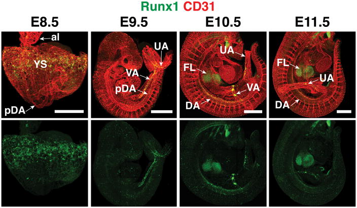
The de novo generation of hematopoietic stem and progenitor cells (HSPC) occurs solely during embryogenesis from a population of epithelial cells called hemogenic endothelium (HE). During midgestation HE cells in multiple intra- and extraembryonic vascular beds leave the vessel wall as they transition into HSPCs in a process termed the endothelial to hematopoietic transition (EHT). Runx1 expression in HE cells orchestrates the transcriptional switch necessary for the transdifferentiation of endothelial cells into functional HSPCs. Runx1 is widely considered the master regulator of developmental hematopoiesis because it plays an essential function during specification of the hematopoietic lineage during embryogenesis. Here we review the role of Runx1 in embryonic HSPC formation, with a particular focus on its role in hemogenic endothelium.
Keywords: Hematopoiesis; Hematopoietic stem cell; Hemogenic endothelium; Runx1.
Figures
References
-
- BEE T, ASHLEY EL, BICKLEY SR, JARRATT A, LI PS, SLOANE-STANLEY J, GOTTGENS B, DE BRUIJN MF. The mouse Runx1 +23 hematopoietic stem cell enhancer confers hematopoietic specificity to both Runx1 promoters. Blood. 2009a;113:5121–4. - PubMed
-
- BEE T, LIDDIARD K, SWIERS G, BICKLEY SR, VINK CS, JARRATT A, HUGHES JR, MEDVINSKY A, DE BRUIJN MF. Alternative Runx1 promoter usage in mouse developmental hematopoiesis. Blood Cells Mol Dis. 2009b;43:35–42. - PubMed
-
- BEE T, SWIERS G, MUROI S, POZNER A, NOTTINGHAM W, SANTOS AC, LI PS, TANIUCHI I, DE BRUIJN MF. Nonredundant roles for Runx1 alternative promoters reflect their activity at discrete stages of developmental hematopoiesis. Blood. 2010;115:3042–50. - PubMed
Publication types
MeSH terms
Substances
Grants and funding
LinkOut - more resources
Full Text Sources
Other Literature Sources
