Review
. 2017 Apr;14(2):358-371.
doi: 10.1007/s13311-017-0520-4.

Convection-Enhanced Delivery

Affiliations
Review

Convection-Enhanced Delivery

A M Mehta et al. Neurotherapeutics. 2017 Apr.

Abstract

Convection-enhanced delivery (CED) is a promising technique that generates a pressure gradient at the tip of an infusion catheter to deliver therapeutics directly through the interstitial spaces of the central nervous system. It addresses and offers solutions to many limitations of conventional techniques, allowing for delivery past the blood-brain barrier in a targeted and safe manner that can achieve therapeutic drug concentrations. CED is a broadly applicable technique that can be used to deliver a variety of therapeutic compounds for a diversity of diseases, including malignant gliomas, Parkinson's disease, and Alzheimer's disease. While a number of technological advances have been made since its development in the early 1990s, clinical trials with CED have been largely unsuccessful, and have illuminated a number of parameters that still need to be addressed for successful clinical application. This review addresses the physical principles behind CED, limitations in the technique, as well as means to overcome these limitations, clinical trials that have been performed, and future developments.

Keywords: Blood–brain barrier; Central nervous system; Convection-enhanced delivery; Drug delivery; Malignant gliomas; Technique.


Figures

Fig. 1
Fig. 1
The surgeon inserts 1 or more catheters through bur holes into the interstitial spaces of the brain. A pressure gradient is generated using an infusion pump, and the agent displaces extracellular fluid after being directly infused into the extracellular space. The tumor is often highly vascularized, and this, as well as a number of factors, can affect the delivery of the infusate (white matter vs gray matter, backflow, etc.) Figure printed with permission from the Journal of Clinical Oncology [35] CED = convection-enhanced delivery; GBM = glioblastoma multiforme
Fig. 2
Fig. 2
Reflux at high infusion rates. This 1.5-T magnetic resonance T1-weighted spoiled gradient image shows (A) little reflux at an infusion rate of 5 μl/min but (B) significant reflux (marked by the arrowheads) along the cannula at 8 μl/min. The infusion was done using a reflux-resistant catheter, on a spontaneous canine piriform lobe tumor, and the infusate being delivered was Gadoteridol-labeled neutral nanoliposomes. Figure printed with permission from Neurotherapeutics [38]
Fig. 3
Fig. 3
Relationship between volume of infusion (Vi) and volume of distribution (Vd), both in the absence and presence of reflux. A normal pattern is shown in Region A, in which the Vd increases linearly with Vd. Point C shows the point at which reflux begins, and in Region B, despite an increase in Vi, Vd does not change. Figure printed with permission from Neurotherapeutics [38]
Fig. 4
Fig. 4
The different designs of catheters used in convection-enhanced delivery. From left to right they are end port cannula, multiport cannula, porous-tipped catheter, balloon-tipped catheter, and stepped-profile catheter. Figure reprinted from Lewis et al. [113]
Fig. 5
Fig. 5
Illustration of the advantage conferred by prolonged infusions of topotecan-gadolinium using a subcutaneous pump. The graph compares the changes in relative volume in 10-day infusions to 3-day infusions. In both scenarios, a peak is observed approximately 2 to 3 days after infusion, after which the 3-day infusion begins to trend downwards and the 10-day infusion hovered around its peak value. Figure reprinted with permission from Oxford University Press [119]
Fig. 6
Fig. 6
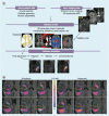
Schematic summarizing the modeling process described in the review. (A) The software requires protocol, and imaging data generates a simulation of fluid and molecular transport. The simulation also allows for interstitial expansion and compromised blood–brain barrier. (B) The simulated and measured distributions of Magnevist™ are compared across six axial slices, with each slice being taken 5 mm apart. Units for concentration color mmol of agent/l of solution. Figure reprinted with permission from Therapeutic Delivery [33]

References

    1. Kanu OO, Mehta A, Di C, et al. Glioblastoma multiforme: a review of therapeutic targets. Expert Opin Ther Targets. 2009;13:701–718. doi: 10.1517/14728220902942348. - DOI - PubMed
    1. Ostrom QT, Gittleman H, Farah P, et al. CBTRUS statistical report: Primary brain and central nervous system tumors diagnosed in the United States in 2006–2010. Neuro Oncol 2013;15(Suppl. 2):ii1-56. - PMC - PubMed
    1. Stupp R, Mason WP, van den Bent MJ, et al. Radiotherapy plus concomitant and adjuvant temozolomide for glioblastoma. N Engl J Med. 2005;352:987–996. doi: 10.1056/NEJMoa043330. - DOI - PubMed
    1. Stupp R, Hegi ME, Mason WP, et al. Effects of radiotherapy with concomitant and adjuvant temozolomide versus radiotherapy alone on survival in glioblastoma in a randomised phase III study: 5-year analysis of the EORTC-NCIC trial. Lancet Oncol. 2009;10:459–466. doi: 10.1016/S1470-2045(09)70025-7. - DOI - PubMed
    1. Blasberg RG, Patlak C, Fenstermacher JD. Intrathecal chemotherapy: brain tissue profiles after ventriculocisternal perfusion. J Pharmacol Exp Ther. 1975;195:73–83. - PubMed

Publication types