. 2017 May;35(5):1303-1315.
doi: 10.1002/stem.2613.

Electroacupuncture Promotes Central Nervous System-Dependent Release of Mesenchymal Stem Cells

Affiliations

Electroacupuncture Promotes Central Nervous System-Dependent Release of Mesenchymal Stem Cells

Tatiana E Salazar et al. Stem Cells. 2017 May.

Abstract

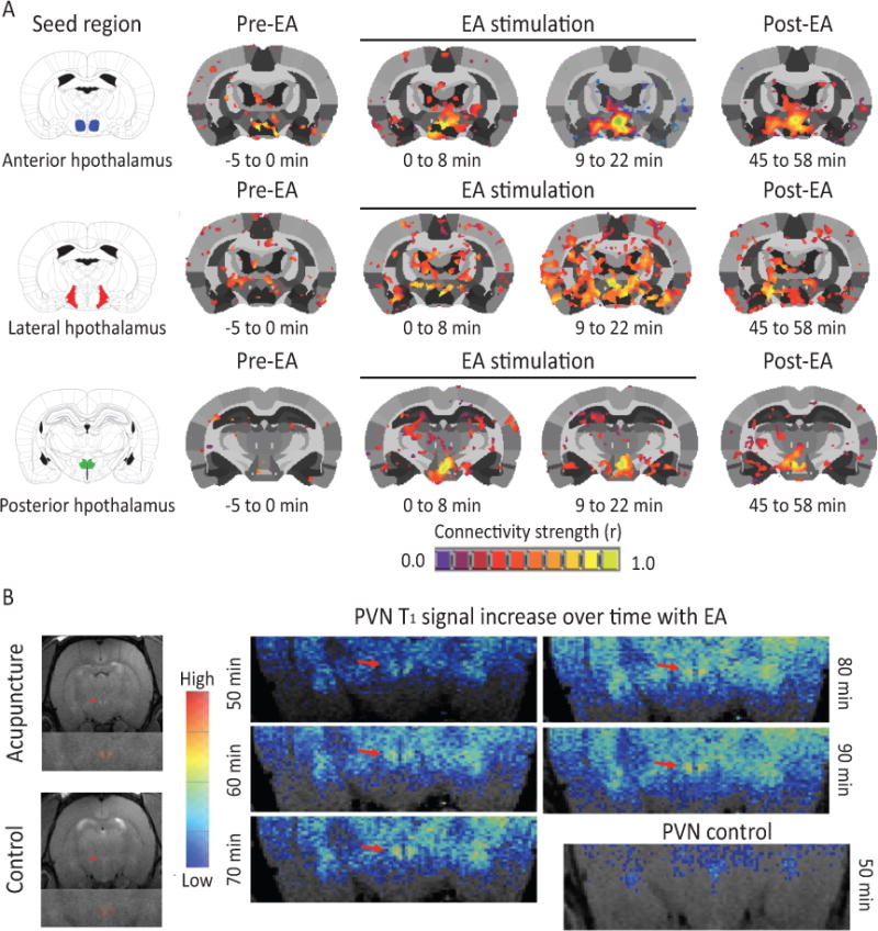
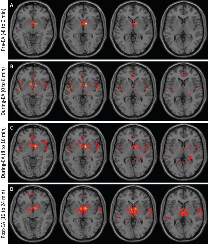
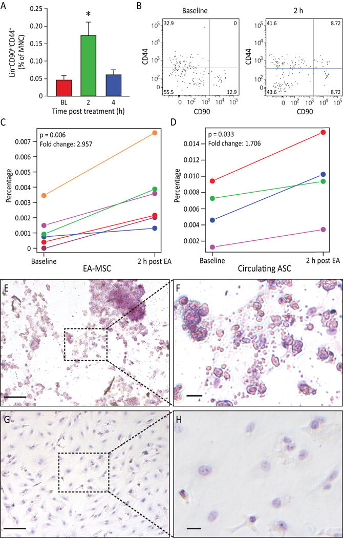
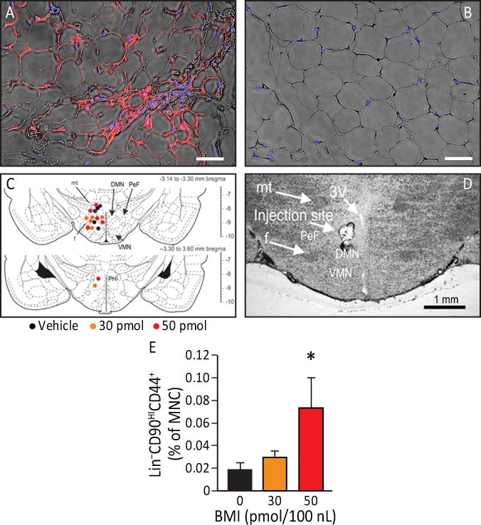
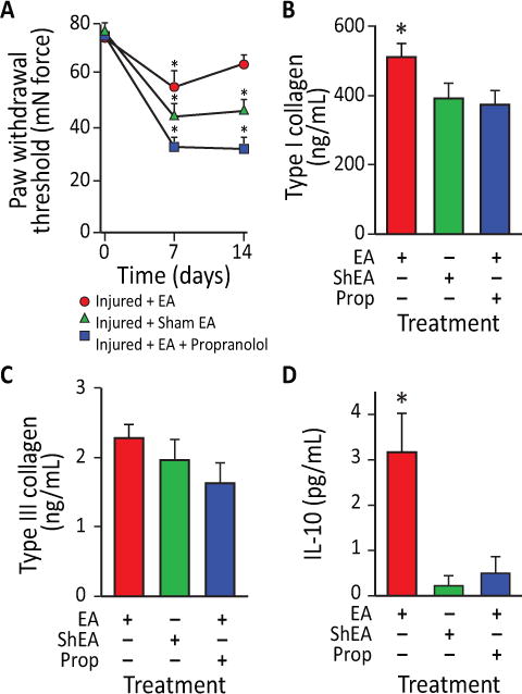
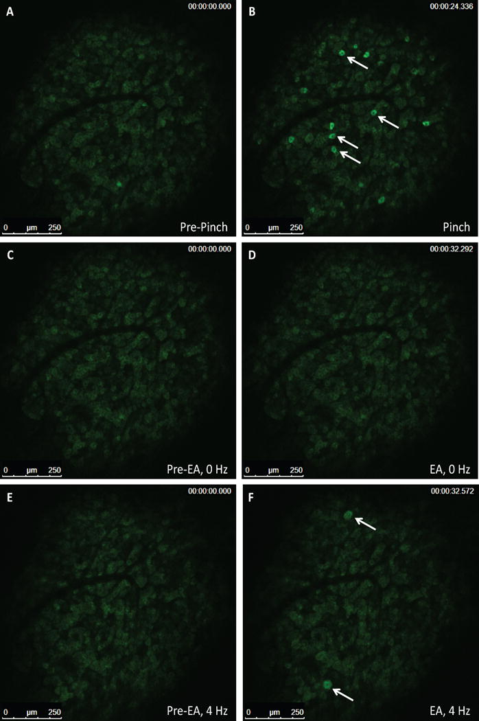
Electroacupuncture (EA) performed in rats and humans using limb acupuncture sites, LI-4 and LI-11, and GV-14 and GV-20 (humans) and Bai-hui (rats) increased functional connectivity between the anterior hypothalamus and the amygdala and mobilized mesenchymal stem cells (MSCs) into the systemic circulation. In human subjects, the source of the MSC was found to be primarily adipose tissue, whereas in rodents the tissue sources were considered more heterogeneous. Pharmacological disinhibition of rat hypothalamus enhanced sympathetic nervous system (SNS) activation and similarly resulted in a release of MSC into the circulation. EA-mediated SNS activation was further supported by browning of white adipose tissue in rats. EA treatment of rats undergoing partial rupture of the Achilles tendon resulted in reduced mechanical hyperalgesia, increased serum interleukin-10 levels and tendon remodeling, effects blocked in propranolol-treated rodents. To distinguish the afferent role of the peripheral nervous system, phosphoinositide-interacting regulator of transient receptor potential channels (Pirt)-GCaMP3 (genetically encoded calcium sensor) mice were treated with EA acupuncture points, ST-36 and LIV-3, and GV-14 and Bai-hui and resulted in a rapid activation of primary sensory neurons. EA activated sensory ganglia and SNS centers to mediate the release of MSC that can enhance tissue repair, increase anti-inflammatory cytokine production and provide pronounced analgesic relief. Stem Cells 2017;35:1303-1315.

Keywords: Adult stem cells; Mesenchymal stem cells; Nervous system; Neurones.


Figures

Figure 1
Figure 1. EA stimulation increases hypothalamic functional connectivity in rats
(A) Rat brains were monitored through fMRI during administration of EA. Functional connectivity increased within the hypothalamus and between the hypothalamus and adjacent brain region with progression of treatment (n=6). (B) A representative example of the increase in signal (over time) in the PVN of a rat receiving acupuncture (n=1).
Figure 2
Figure 2. EA stimulation increases hypothalamic functional connectivity in humans
Brains from normal subjects were monitored using fMRI before, during and after EA. Functional connectivity increased within the hypothalamus and between the hypothalamus and adjacent brain regions with progression of treatment (n=6).
Figure 3
Figure 3. EA stimulation induced MSC stem cell mobilization
A) Rat peripheral blood MSC were increased (p=0.0063) after EA. Circulating MSC were defined as Lin(CD45CD31erythroidCD11b) cells that were positive for CD44 and CD90. Gated cells increased post treatment (n=11 for baseline and 4 hours, n=9 for 2 hours). B) Representative flow charts for rat Lin cells are shown at baseline and 2 h samples. C) The percentage of human peripheral blood MSC increased in post EA-treatment (p=0.006, n=6). D) The percentage of circulating MSCs from adipose tissue (AD-MSC) are significantly elevated in 2 h post EA-treatment (p=0.033, n=4). E, F) EA-mobilized MSC were expanded in vitro. After undergoing adipogenesis differentiation, EA-mobilized MSC developed fat deposits as seen by Oil Red staining, which were not seen in the undifferentiated control cells (G, H). Magnification bars: E,G: 100 μm; F,H: 50 μm.
Figure 4
Figure 4. EA increases sympathetic activation leading to browning of WAT and the effects of EA can be duplicated by pharmacological disinhibition of hypothalamus (sympathetic activation)
A, B) UCP1 immunofluorescence (red) detectable in inguinal subcutaneous adipose tissue (blue: adipocytes nuclei) from animals that underwent EA treatment (A) but not in control (B). EA resulted in an increase in beige adipocytes (n=4). Magnification bars: 50 μm. C) The effects of EA can be duplicated by pharmacological disinhibition of hypothalamus. Rats underwent injection of either vehicle, 30 pmol or 50 pmol/100 nl of the GABAA receptor antagonist bicuculline methiodide (BMI). Sites of injections are represented on a coronal section from the tuberal hypothalamus from a Standard Steroetaxic Atlas of the Rat brain [11]. Colored circles indicate injection sites (black, orange and red represent vehicle, 30 pmol and 50 pmol respectively). D) Representative photomicrograph showing an injection site from one rat. Abbreviations: 3V, 3rd ventricle; DMN, dorsomedial hypothalamic nucleus; f, fornix; mt, mammillothalamic tract; PeF, perifornical hypothalamus; PH, posterior hypothalamic nucleus; ventromedial hypothalamic nucleus. Magnification bar: 1 mm. E) There was a significant increase (p=0.027) in LinCD90HICD44+ cells in the plasma 4 h post an injection (n=6). Data presented as means ± SEM.
Figure 5
Figure 5. EA-treated rodents exhibit reduced mechanical hyperalgesia, enhanced tissue remodeling and increased serum IL-10 levels following partial Achilles tendon rupture
A) Effects of EA application on mechanical allodynia in rats at 7 and 14 days after partial tendon rupture in the right hind leg. Mechanical hypersensitivity was determined by measuring the change in weight-bearing forces on the affected limb. Behavioral changes in the hind paw tactile threshold (in millinewtons, mN) were observed in the hindpaw ipsilateral to the tendon injury 18 hours after EA treatment, EA sham treatment or EA treatment with propranolol. (*p<0.01 versus baseline. EA: n=9; EA: sham n=7; EA+propranolol: n=10). B) EA increased type I collagen content in injured tendons in rats at 14 days after unilateral Achilles tendon partial tenotomy. In EA treated animals (n=6), type-I collagen content was 24% greater in injured tendons than EA sham treated tendons (n=7; p<0.05) and 28% greater in injured tendons than EA+propranolol treated tendons (n=8; p<0.02). C) In contrast, there was no difference in type-III collagen content between injured EA and EA sham or EA+propranolol treated tendons (p=0.67). D) IL-10 serum levels were also elevated in EA-treated rats compared to EA sham and EA+propanolol animals (p=0.0041). Data presented as means ± SEM.
Figure 6
Figure 6. Small diameter primary afferent sensory neurons show sensitivity to pinch stimulus and medium-large diameter neurons show sensitivity to EA in Pirt-GCaMP3 mice with intact dorsal root ganglia (DRG)
Representative fluorescent GCaMP3 neuronal imaging response by lumbar DRG in vivo before (A) and after a mechanical press of the hindpaw using a 100 g force (B). The mechanical force evokes a robust fluorescent GCaMP3 Ca2+ response in numerous small sensory neurons (B; white arrows). Acupuncture needles alone failed to elicit neuronal changes in the same lumbar DRG of the Pirt-GCamp3 mouse (C, D). EA-stimulation of the ST-36 and Liv-3 accupoints produced rapid activation of medium-larger diameter of the lumbar DRG (E, F; white arrows)(n=4, p<0.0001).
Figure 7
Figure 7. EA-mediated sympathetic stimulation induces MSC release into the circulation
A) EA mobilized cells are highly proliferative and potentiate vasculogenesis. Equine peripheral blood MNC showed an increased colony-forming ability 2 h (p<0.05) post administration of EA at immune points (n=7), while cells obtained from the same horses when they underwent mock treatment or treatment at metabolic points did not. B) The EA-mobilized cells demonstrated high proliferative capacity, when plated in a single-cell assay, with over 50% proliferating into large colonies (p<0.001 vs. all groups). C) Equine peripheral blood mononuclear cells were cultured to the third passage and then differentiated into key mesenchymal lineages. a) Following culture with osteogenic induction media, the mobilized equine cells showed strong osteogenic potency, demonstrated by Alizarin Red staining of calcium deposits (Red: Alizarin Red). b) Human mesenchymal cells responded in a similar fashion when cultured under identical conditions. Equine cells when cultured under control media (1:1 Ham’s F12 and Low Glucose DMEM, 15%FBS)(e) and human cells under control conditions (f) did not show Alizarin Red staining. c) The EA-mobilized equine cells showed a weak adipogenic response, demonstrated by Oil Red O staining of lipid deposits (Red: Oil Red O) when cultured under adipogenic conditions; d) human MSC showed a much stronger response than the equine cells when cultured under identical adipogenic conditions. Under control conditions, neither equine MSC (g) or human MSC (h) showed Oil Red O staining. i, j) When cells were cultured under chondrogenesis differentiation medium they were able to differentiate into chondrogenic lineages, demonstrated by Alcian Blue staining of proteoglycans in the cell masses. Magnification bars: 50 μm. D) In vivo angiogenesis assay. a) When equine cells were incorporated into a 3D type I pig skin collagen plug and placed under the skin of NOD/SCID mice no capillaries were formed. b) Human ECFC were inserted into the porcine collagen plugs together with equine MSC, and implanted into the flank of NOD/SCID mice. c) A higher magnification of the boxed area in b), showing a bona fide blood vessel. Magnification bars: a, b: 50 μm; c: 10 μm. E) When quantified, the hECFC-MSC had a significant increase of arteriogenesis compared to hECFC alone (p=0.02, n=5 for ECFCs alone, n=7 for combined hECFC-MSC group). F) After 48 h in vitro, cells were isolated, total mRNA was extracted and Hey2 expression levels were quantified by qRT-PCR. Hey2 was elevated in the mixed cell treatment when compared to ECFC alone (p=0.006, n=4). All data presented as means ± SEM.

References

    1. Zhao ZQ. Neural mechanism underlying acupuncture analgesia. Prog Neurobiol. 2008;85:355–375. - PubMed
    1. Urano K, Ogasawara S. A fundamental study on acupuncture points phenomena of dog body. Kitasato Arch Exp Med. 1978;51:95–109. - PubMed
    1. Gunn CC, Ditchburn FG, King MH, et al. Acupuncture loci: a proposal for their classification according to their relationship to known neural structures. Am J Chin Med (Gard City N Y) 1976;4:183–195. - PubMed
    1. Goldman N, Chen M, Fujita T, et al. Adenosine A1 receptors mediate local anti-nociceptive effects of acupuncture. Nat Neurosci. 2010;13:883–888. - PMC - PubMed
    1. Kim SK, Bae H. Acupuncture and immune modulation. Auton Neurosci. 2010;157:38–41. - PubMed

MeSH terms