

The value of combining conventional, diffusion-weighted and dynamic contrast-enhanced MR imaging for the diagnosis of parotid gland tumours
- PMID: 28299943
- PMCID: PMC5606280
- DOI: 10.1259/dmfr.20160434
The value of combining conventional, diffusion-weighted and dynamic contrast-enhanced MR imaging for the diagnosis of parotid gland tumours
Abstract
Objectives: The aim of this study was to determine the value of combining conventional MRI, diffusion-weighted imaging (DWI) and dynamic contrast-enhanced (DCE)-MRI in diagnosing solid neoplasms in the parotid gland.
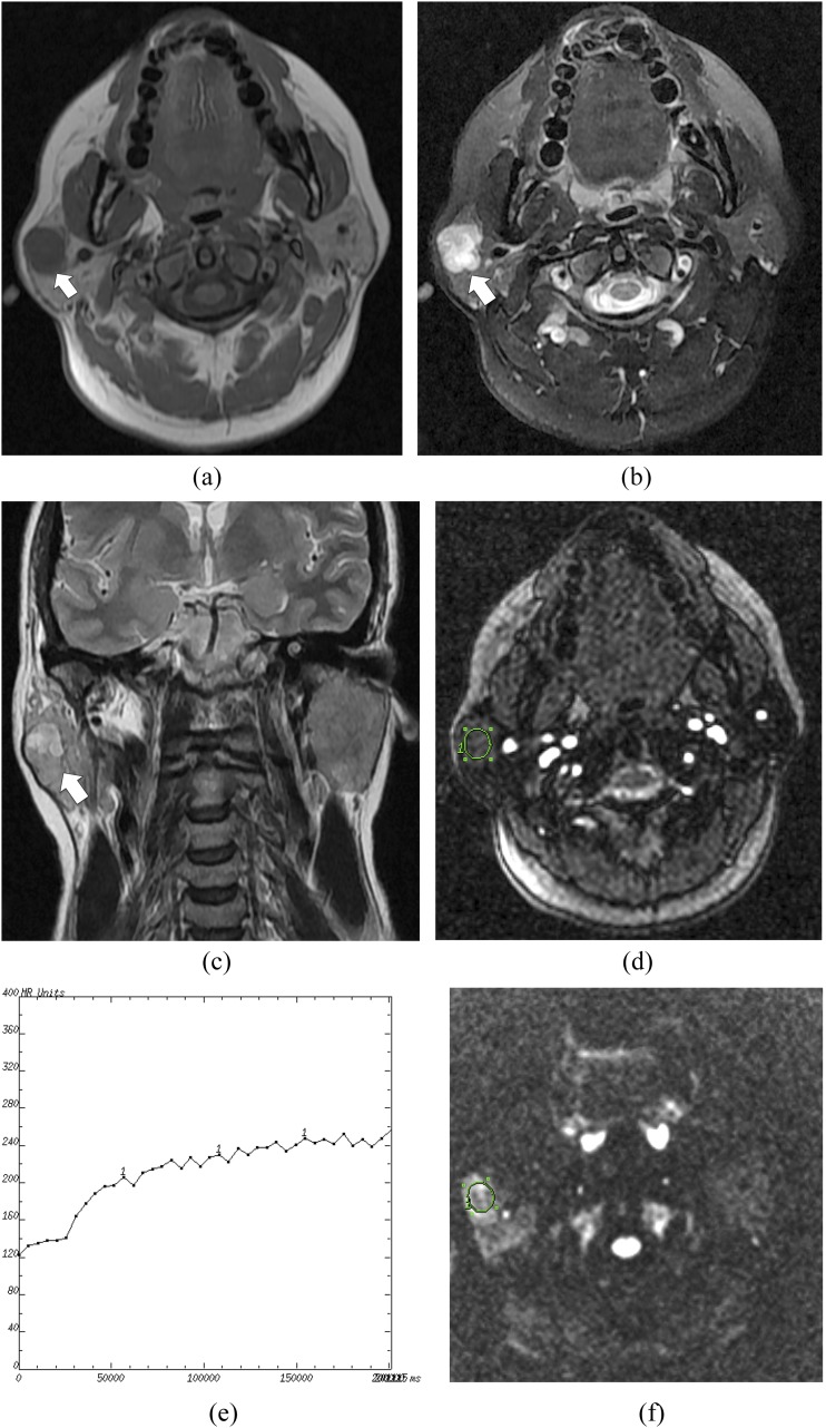
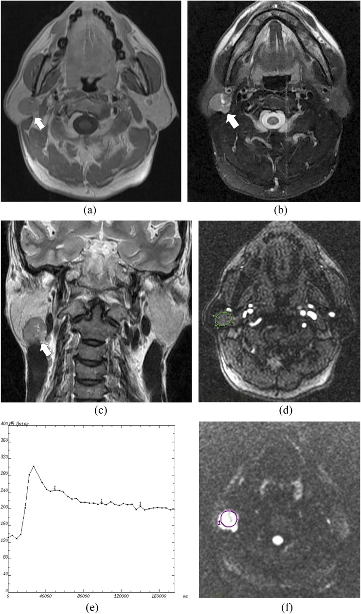
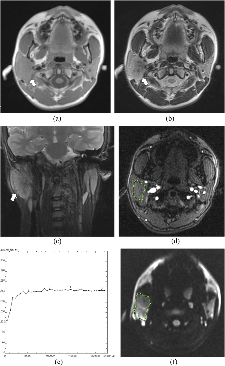
Methods: A total of 148 subjects (101 subjects with benign and 47 subjects with malignant tumours) were evaluated with conventional MRI, DWI and DCE-MRI prior to surgery and pathologic verification. The items observed with conventional MRI included the shape, capsule and signal intensity of parotid masses. The apparent diffusion coefficient (ADC) was calculated from DWI that was obtained with a b-factor of 0 and 1000 s mm-2. A time-intensity curve (TIC) was obtained from DCE-MRI.
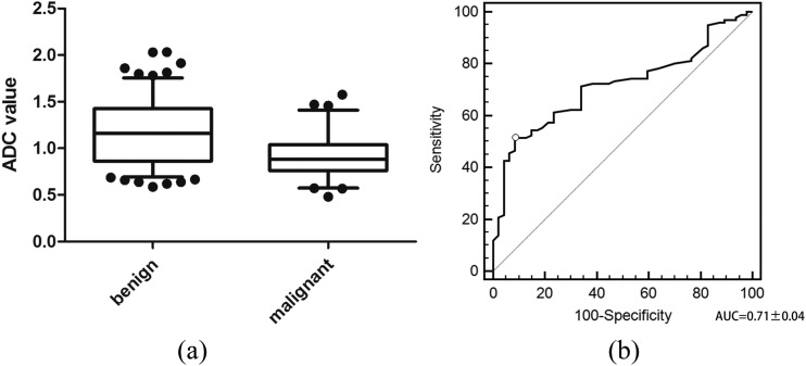
Results: There were significant differences (p < 0.01) in the shape, capsule, ADC and TIC between benign and malignant parotid tumours. Irregular neoplasms without a capsule, ADC <1.12 × 10⁻³ mm2 s-1 and a plateau enhancement pattern were valuable parameters for predicting malignant neoplasms. A combination of all of these parameters yielded sensitivity, specificity, accuracy, positive-predictive value and negative-predictive value of 85.1%, 94.1%, 91.2%, 87.0% and 93.1%, respectively.
Conclusions: A combined analysis using conventional MRI, DWI and DCE-MRI is helpful in distinguishing benign from malignant tumours in the parotid gland.
Keywords: diffusion-weighted MR imaging (DW-MRI); dynamic contrast enhanced MR imaging (DCE-MRI); magnetic resonance imaging (MRI); neoplasm; parotid gland.
Figures
References
-
- Scianna JM, Petruzzelli GJ. Contemporary management of tumors of the salivary glands. Curr Oncol Rep 2007; 9: 134–8. doi: https://doi.org/10.1007/s11912-007-0011-6 - DOI - PubMed
-
- Swoboda H, Franz P. Salivary gland tumors. Clinical aspects and therapy. [In German.] Radiologe 1994; 34: 232–8. - PubMed
-
- Mihashi H, Kawahara A, Kage M, Kojiro M, Nakashima T, Umeno H, et al. Comparison of preoperative fine-needle aspiration cytology diagnosis and histopathological diagnosis of salivary gland tumors. Kurume Med J 2006; 53: 23–7. doi: https://doi.org/10.2739/kurumemedj.53.23 - DOI - PubMed
-
- Das DK, Petkar MA, Al-Mane NM, Sheikh ZA, Mallik MK, Anim JT. Role of fine needle aspiration cytology in the diagnosis of swellings in the salivary gland regions: a study of 712 cases. Med Princ Pract 2004; 13: 95–106. - PubMed
-
- Zbären P, Nuyens M, Loosli H, Stauffer E. Diagnostic accuracy of fine-needle aspiration cytology and frozen section in primary parotid carcinoma. Cancer 2004; 100: 1876–83. - PubMed
MeSH terms
Substances
LinkOut - more resources
Full Text Sources
Other Literature Sources
Medical
