Review
. 2017 Mar 16:3:17011.
doi: 10.1038/nrdp.2017.11.

Alopecia areata

Affiliations
Review

Alopecia areata

C Herbert Pratt et al. Nat Rev Dis Primers. .

Abstract

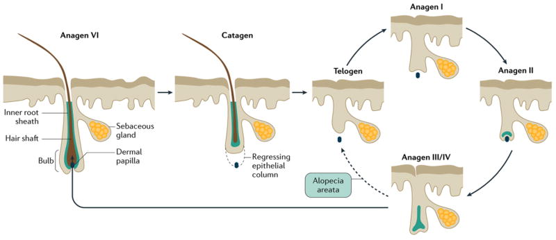
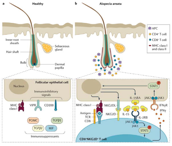
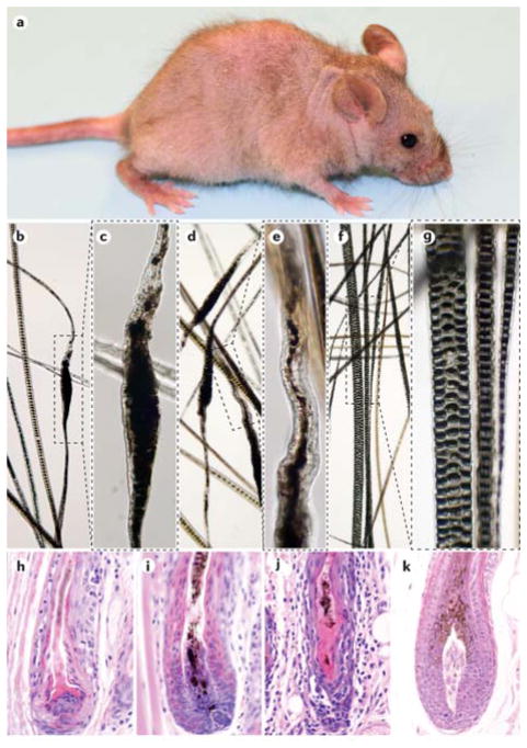
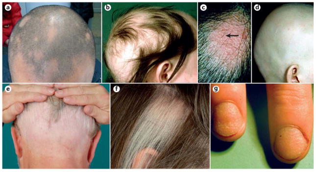
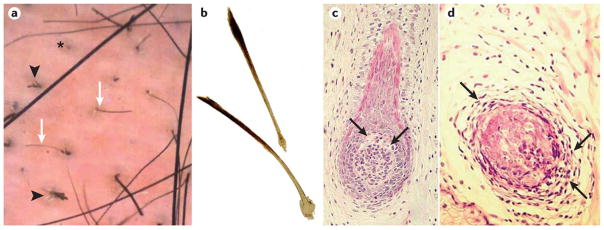
Alopecia areata is an autoimmune disorder characterized by transient, non-scarring hair loss and preservation of the hair follicle. Hair loss can take many forms ranging from loss in well-defined patches to diffuse or total hair loss, which can affect all hair-bearing sites. Patchy alopecia areata affecting the scalp is the most common type. Alopecia areata affects nearly 2% of the general population at some point during their lifetime. Skin biopsies of affected skin show a lymphocytic infiltrate in and around the bulb or the lower part of the hair follicle in the anagen (hair growth) phase. A breakdown of immune privilege of the hair follicle is thought to be an important driver of alopecia areata. Genetic studies in patients and mouse models have shown that alopecia areata is a complex, polygenic disease. Several genetic susceptibility loci were identified to be associated with signalling pathways that are important to hair follicle cycling and development. Alopecia areata is usually diagnosed based on clinical manifestations, but dermoscopy and histopathology can be helpful. Alopecia areata is difficult to manage medically, but recent advances in understanding the molecular mechanisms have revealed new treatments and the possibility of remission in the near future.


Conflict of interest statement

Competing interests

L.E.K. is on the scientific advisory committee for the National Alopecia Areata Foundation (NAAF) and Cicatricial Alopecia Research Foundation (CARF). A.M.C. is on the scientific advisory committee for NAAF and is a consultant for Aclaris Therapeutics, Inc. J.P.S. has or has had sponsored research contract with Biocon LLC and the NAAF for preclinical trials using mouse models for alopecia areata, and is on the scientific advisory committee for NAAF and Chairman for CARF. C.H.P. and A.G.M. have no conflicts to disclose.

Figures

Figure 1
Figure 1. Hair cycle
During the anagen phase (the period when a hair follicle is actively growing), which can vary from a few weeks to several years, epithelial cells in the hair bulb undergo vigorous mitotic activity and differentiate as they move distally to form the hair fiber and its surrounding inner root sheath and inner layer of the outer root sheath. Eventually, epithelial cell division ceases, and the follicle enters the catagen phase, in which the proximal end of the hair shaft keratinizes to form a club-shaped structure, which eventually sheds, and the lower part of the follicle involutes by apoptosis. The telogen phase marks the period between follicular regression and the onset of the next anagen phase (solid arrows). The development of the anagen follicle, which closely replicates embryonic development of the hair follicle, is conventionally divided into six stages (I–VI, with anagen VI representing the fully formed anagen follicle). In most mammals, hair cycles are coordinated in a wave-like fashion across the skin (moult waves), but in humans, follicles cycle independently of their neighbors. In alopecia areata (dashed arrow), anagen VI follicles are precipitated prematurely into telogen phase. Although they are probably able to re-enter anagen phase, the development of the follicle is halted at the anagen III/IV stage,, when they prematurely return to the telogen phase. Truncated cycles may continue if and until disease activity declines, and follicles are able to progress further into the anagen phase.
Figure 2
Figure 2. Breakdown of immune privilege in alopecia areata
a) Immune privilege of the hair follicle can be achieved through several strategies including: down regulation of MHC class I and β2 microglobulin, which normally stimulate natural killer (NK) cells; local production of immunosuppressants; expression of immunoinhibitory signals (for example, CD200; also known as OX2 membrane glycoprotein); and repression of intrafollicular antigen-presenting cell (APC), perifollicular NK cell and mast cell functions owing to increased levels of macrophage migration inhibitory factor (MIF). Vasoactive intestinal peptide (VIP), released by perifollicular sensory nerve fibers, is also believed to be an immunoinhibitory neuropeptide that might have a role in immune privilege. b) Late anagen hair follicles in patients with alopecia areata have perifollicular infiltrations of APCs, CD4+ and CD8+ T cells, and abnormal expression of MHC class I and II molecules. CD8+ T cells also infiltrate into the hair follicle root sheaths. Molecules involved in the lymphocyte co-stimulatory cascade are involved in the pathogenesis of alopecia area and provide targets for therapeutic intervention. No inflammatory cells are found in the surrounding of nomal follicles in late anagen phase. INFγ, interferon gamma; IFNγR, interferon gamma receptor; IL2RB, IL2 receptor subunit beta; IL15RA, IL15 receptor subunit alpha; JAK, Janus kinase; NKG2D, NK cell receptor D; NKG2DL, NKG2D ligand; P, phosphorylated; POMC, pro-opiomelanocortin; STAT1, signal transducer and activator of transcription 1; TCR, T cell receptor; TGFB, transforming growth factor beta, VIPR, VIP receptor.
Figure 3
Figure 3. C3H/HeJ mouse model of alopecia areata
a) Alopecia areata in C3H/HeJ mice is shown (part a). Spontaneous alopecia development in C3H/HeJ mice starts with patchy alopecia areata in the axillary and inguinal regions, which spreads across the ventral skin to the dorsal skin and eventually results in diffuse alopecia. Note that the fine hair of the tail and ears are not involved. Analysis of the hair of C3H/HeJ mice with alopecia areata (parts b–e). The regular sepate or seputulate (ladder-like) pattern of the normal hair medulla (part f and part g) is lost and the hair shaft becomes thin and serpentine (parts b–e). These deformities weaken the hair and result in shaft breakage (parts b–e). Normal hair shafts from an unaffected C3H/HeJ mouse reveal the regular septulate (3 rows) and septate (1 row) cellular pattern within the hair shafts (part f and part g). Parts c, e and g are enlargements of the boxes in parts b, d and f, respectively. Histopathological analysis of skin is shown (parts h–k). Mice with alopecia areata have various degrees of inflammation and follicular dystrophy in and around the anagen hair shafts (parts h–j). A healthy, late anagen stage hair follicle is shown in part k.
Figure 4
Figure 4. Clinical manifestations of alopecia areata
a) Limited patchy alopecia areata (<50% scalp involvement). b) Extensive patchy alopecia areata (>50% scalp involvement). c) Active patch of alopecia areata showing exclamation point hairs (arrow) and slight skin erythema. d) Alopecia universalis. e) Ophiasis pattern of alopecia areata. f) Sparing of white hairs in alopecia areata. g) Nail pitting and longitudinal striations (trachyonychia) associated with alopecia areata. Part c and part f are reproduced with permission from REF 208, Wiley. Part e is reproduced with permission from REF 209, Springer. Part c and part f are reproduced with permission from Messenger, A. G., Sinclair, R. D., Farrant, P. & de Berker, D. A. R. in Rook’s Textbook of Dermatology 9th edn (eds Griffiths, C., Barker, J., Bleiker, T., Chalmers, R. & Creamer, D.) 89 (Wiley-Blackwell, 2016), Wiley. Part e is reproduced with permission from Bohm, M. in European Handbook of Dermatological Treatments (eds Katsambas, A. D., Lotti, T. M., Dessinioti, C, & D’Erme, A. M.) 45-53 (Springer Berlin Heidelberg, 2015), Springer
Figure 5
Figure 5. Diagnostic tools to validate and differentiate alopecia areata
a) Dermoscopy of the skin of patient with alopecia areata. Black arrowheads point to dystrophic, cadaverized and twisted hairs. The asterisk denotes a yellow dot with follicular plugging by keratinaceous debris. White arrows point to short, regrowing miniaturized hairs. b) Plucked exclamation point hairs are characterized by proximal hair shaft narrowing and are always broken; they might show reduced pigmentation. c) Histopathology of a vertical section of a skin biopsy from patch of alopecia areata with very early regrowth of hypopigmented hair. An early anagen hair bulb with vacuolar degeneration (arrows) of epithelial cells around the upper pole of the dermal papilla. d) A horizontal section from a similar area showing lymphocytic infiltrate (arrows) surrounding and infiltrating an anagen hair bulb. Part b is reproduced with permission from REF 208, Wiley. Part c is adopted with permission from REF, Wiley. Part b is reproduced with permission from Messenger, A. G., Sinclair, R. D., Farrant, P. & de Berker, D. A. R. in Rook’s Textbook of Dermatology 9th edn (eds Griffiths, C., Barker, J., Bleiker, T., Chalmers, R. & Creamer, D.) 89 (Wiley-Blackwell, 2016), Wiley. Part c is adapted with permission from Messenger, A. G., Slater, D. N. & Bleehen, S. S. Alopecia areata: alterations in the hair growth cycle and correlation with the follicular pathology. Br. J. Dermatol. 114, 337-347 (1986), Wiley

Similar articles

Cited by

References

    1. McElwee KJ, et al. Comparison of alopecia areata in human and nonhuman mammalian species. Pathobiology : journal of immunopathology, molecular and cellular biology. 1998;66:90–107. - PubMed
    1. Xing L, et al. Alopecia areata is driven by cytotoxic T lymphocytes and is reversed by JAK inhibition. Nat Med. 2014;20:1043–1049. doi: 10.1038/nm.3645. - DOI - PMC - PubMed
    1. Duvic M, et al. The national alopecia areata registry-update. J Invest Dermat Symp Proc. 2013;16:S53. - PMC - PubMed
    1. Petukhova L, et al. Genome-wide association study in alopecia areata implicates both innate and adaptive immunity. Nature. 2010;466:113–117. - PMC - PubMed
    1. Shi Q, et al. Health-Related Quality of Life (HRQoL) in alopecia areata patients-a secondary analysis of the National Alopecia Areata Registry Data. J Invest Dermat Symp Proc. 2013;16:S49–50. - PubMed