. 2017 Mar 16:7:44420.
doi: 10.1038/srep44420.

Brucella spp. of amphibians comprise genomically diverse motile strains competent for replication in macrophages and survival in mammalian hosts

Affiliations

Brucella spp. of amphibians comprise genomically diverse motile strains competent for replication in macrophages and survival in mammalian hosts

Sascha Al Dahouk et al. Sci Rep. .

Abstract

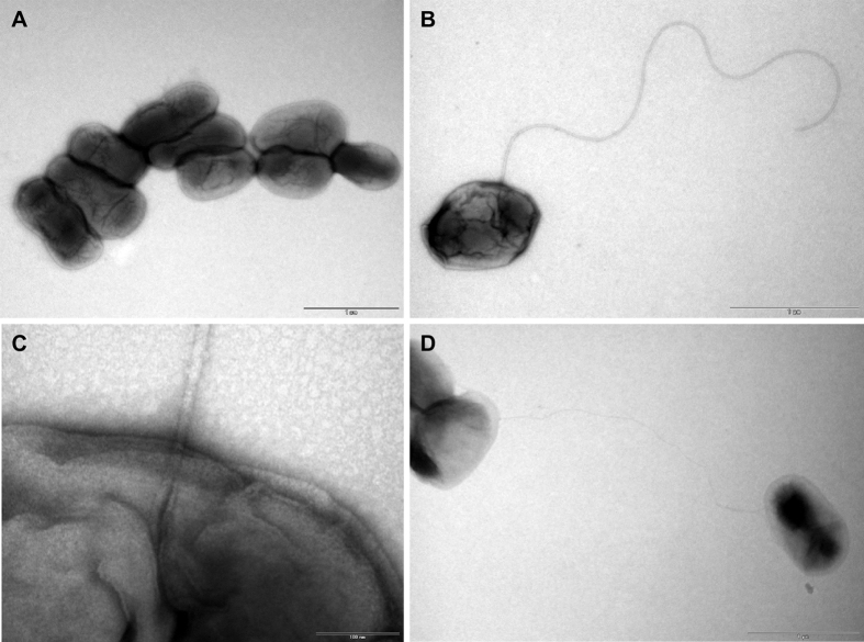
Twenty-one small Gram-negative motile coccobacilli were isolated from 15 systemically diseased African bullfrogs (Pyxicephalus edulis), and were initially identified as Ochrobactrum anthropi by standard microbiological identification systems. Phylogenetic reconstructions using combined molecular analyses and comparative whole genome analysis of the most diverse of the bullfrog strains verified affiliation with the genus Brucella and placed the isolates in a cluster containing B. inopinata and the other non-classical Brucella species but also revealed significant genetic differences within the group. Four representative but molecularly and phenotypically diverse strains were used for in vitro and in vivo infection experiments. All readily multiplied in macrophage-like murine J774-cells, and their overall intramacrophagic growth rate was comparable to that of B. inopinata BO1 and slightly higher than that of B. microti CCM 4915. In the BALB/c murine model of infection these strains replicated in both spleen and liver, but were less efficient than B. suis 1330. Some strains survived in the mammalian host for up to 12 weeks. The heterogeneity of these novel strains hampers a single species description but their phenotypic and genetic features suggest that they represent an evolutionary link between a soil-associated ancestor and the mammalian host-adapted pathogenic Brucella species.


Conflict of interest statement

The authors declare no competing financial interests.

Figures

Figure 1
Figure 1. Electron micrographs of flagellated Brucella sp. isolated from African bullfrogs.
Electron micrographs showing individual cells or clusters of strain no. 10RB9206 (A). Some bacteria were flagellated with a polar flagellum (B,C) and presented pili-like structures (D).
Figure 2
Figure 2. Phylogenetic analysis of the bullfrog isolates in comparison to Brucella spp. inferred by MLSA.
Phylogenetic relationship of amphibian isolates with other Brucella species based on eight-locus MLSA. Numbers at nodes correspond to proportions of 500 resamplings that support the topology shown with only values >80% indicated. The bar indicates the number of substitutions per nucleotide position. UK8/14 represents a previously described amphibian isolate from the United Kingdom.
Figure 3
Figure 3. Phylogenetic placement of the African bullfrog strains based on whole genome sequencing data.
Trees were constructed using maximum parsimony with nodal support for bootstrapping shown only for the branch that had less than 90% support within the basal clade containing the atypical Brucella strains. Strains NF2653 and 83-13 were isolated from Australian rodents.
Figure 4
Figure 4. Electrophoretic lipopolysaccharide (LPS) profiles produced by the amphibian Brucella sp. strains.
Silver staining (A) and Western blot (B) profiles with mAb A68/10A06/B11 (anti-R-LPS) after SDS-PAGE of proteinase K-digested S-LPS preparations of B. melitensis 16 M (M-dominant reference strain) (lanes 1), B. suis 1330 (A-dominant reference strain) (lanes 2), B. microti CCM 4915 (lanes 3), Brucella sp. strain 83/13 (wild rodent isolate from Australia) (lanes 4), B. inopinata BO1 (lanes 5), Brucella sp. strain BO2 (lanes 6), African bullfrog strains 09RB8910 (lanes 7), 10RB9207 (lanes 8), 10RB9215 (lanes 9), 10RB9213 (lanes 10), 09RB8915 (lanes 11).
Figure 5
Figure 5. Variable organization of the wbk region in the Brucella sp. strains isolated from African bullfrogs.
Comparison of the wbk region, involved in the synthesis of LPS in the classical Brucella strains, across B. microti, B. inopinata (BO1), the BO2 strain, B13-0095 isolated from a Pacman frog and five African bullfrog strains. Genes that are shared across different species have similar color and border patterns, and are also indicated by background shading. Genes with sequence similarity above 91% have solid borders and shading, but those with similarity between 70–90% are indicated by dashed borders. Coding sequences appear as arrows, mobile element proteins as grey triangles and tRNAs as solid boxes. Genes united on the same contig or chromosome are found within the same rectangle. The classically known wbk region can be seen in B. microti and B. inopinata. All genes, their product description, the assigned protein family, and the gene order in the newly annotated genomes are identified by a peg identifier (Supplementary Table S3).
Figure 6
Figure 6. Schematic representation of flagellar gene expression.
Regulators of flagellar gene expression and proteins participating in assembly of the flagellum. Those genes that appear to be functional across all of the compared strains (see Supplementary Table S4) are marked in red.
Figure 7
Figure 7. Intracellular replication of the amphibian Brucella sp. strains in macrophages.
Intracellular multiplication of (A) the African bullfrog strains 09RB8471 (●), 09RB8910 (▲), 09RB8913 (▼) and 10RB9213 (■) in comparison with (B) B. microti CCM 4915 (●), B. inopinata BO1 (▼), and B. suis 1330 (▲) in murine J774 macrophage-like cells. All experiments were performed three times in triplicate each, and results of one typical experiment are shown, presented as the means ± standard deviation.
Figure 8
Figure 8. Course of amphibian Brucella sp. infection in BALB/c mice.
Bacterial counts and organ weights of spleen and liver. Growth curves of Brucella sp. strains 09RB8471 (○), 09RB8910 (△), 09RB8913 (▽), 10RB9213 (□), and B. suis 1330 (●) in spleens (A) and livers (C) of BALB/c mice after intraperitoneal inoculation of 104 colony-forming units (CFU) of the bacteria. For each infection experiment, five mice were killed per time point after 3, 5, 7, 14, 28, 56, and 84 days and the number of viable bacteria was counted. Spleen (B) and liver weights (D) were also determined. Data are presented as mean values ± standard deviation.

References

    1. Scholz H. C. et al.. Brucella vulpis sp. nov., isolated from mandibular lymph nodes of red foxes (Vulpes vulpes). Int. J. Syst. Evol. Microbiol. 66, 2090–2098 (2016). - PubMed
    1. Pappas G., Papadimitriou P., Akritidis N., Christou L. & Tsianos E. V. The new global map of human brucellosis. Lancet Infect. Dis. 6, 91–99 (2006). - PubMed
    1. Christopher G. W., Agan M. B., Cieslak T. J. & Olson P. E. History of U.S. military contributions to the study of bacterial zoonoses. Mil. Med. 170, 39–48 (2005). - PubMed
    1. Pappas G., Panagopoulou P., Christou L. & Akritidis N. Brucella as a biological weapon. Cell. Mol. Life Sci. 63, 2229–2236 (2006). - PMC - PubMed
    1. Foster G., Osterman B. S., Godfroid J., Jacques I. & Cloeckaert A. Brucella ceti sp. nov. and Brucella pinnipedialis sp. nov. for Brucella strains with cetaceans and seals as their preferred hosts. Int. J. Syst. Evol. Microbiol. 57, 2688–2693 (2007). - PubMed

Publication types

MeSH terms

Substances

LinkOut - more resources