. 2017 Mar 16:7:44582.
doi: 10.1038/srep44582.

Exonuclease processivity of archaeal replicative DNA polymerase in association with PCNA is expedited by mismatches in DNA

Affiliations

Exonuclease processivity of archaeal replicative DNA polymerase in association with PCNA is expedited by mismatches in DNA

Takuya Yoda et al. Sci Rep. .

Abstract

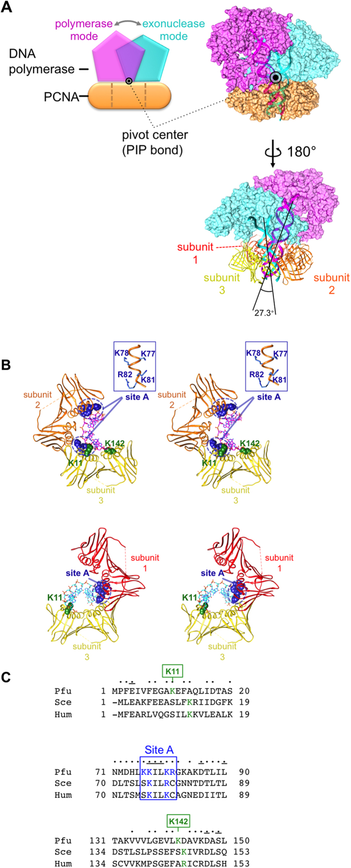
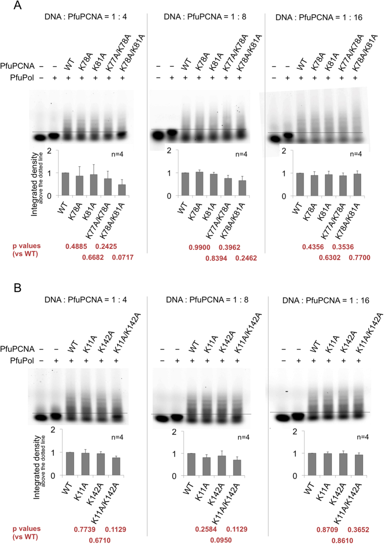
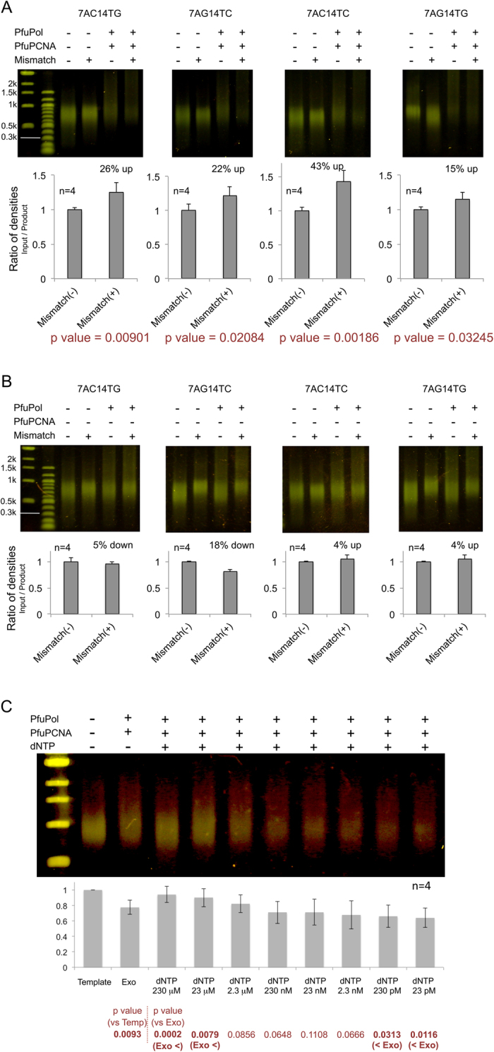
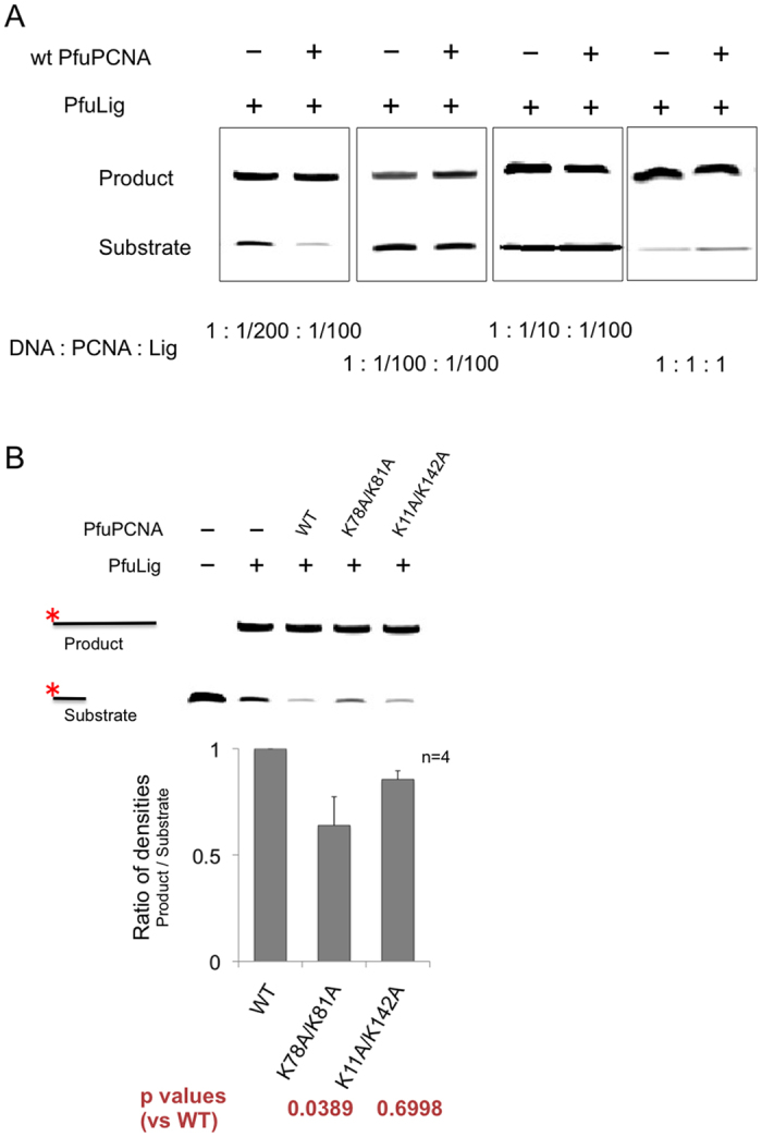
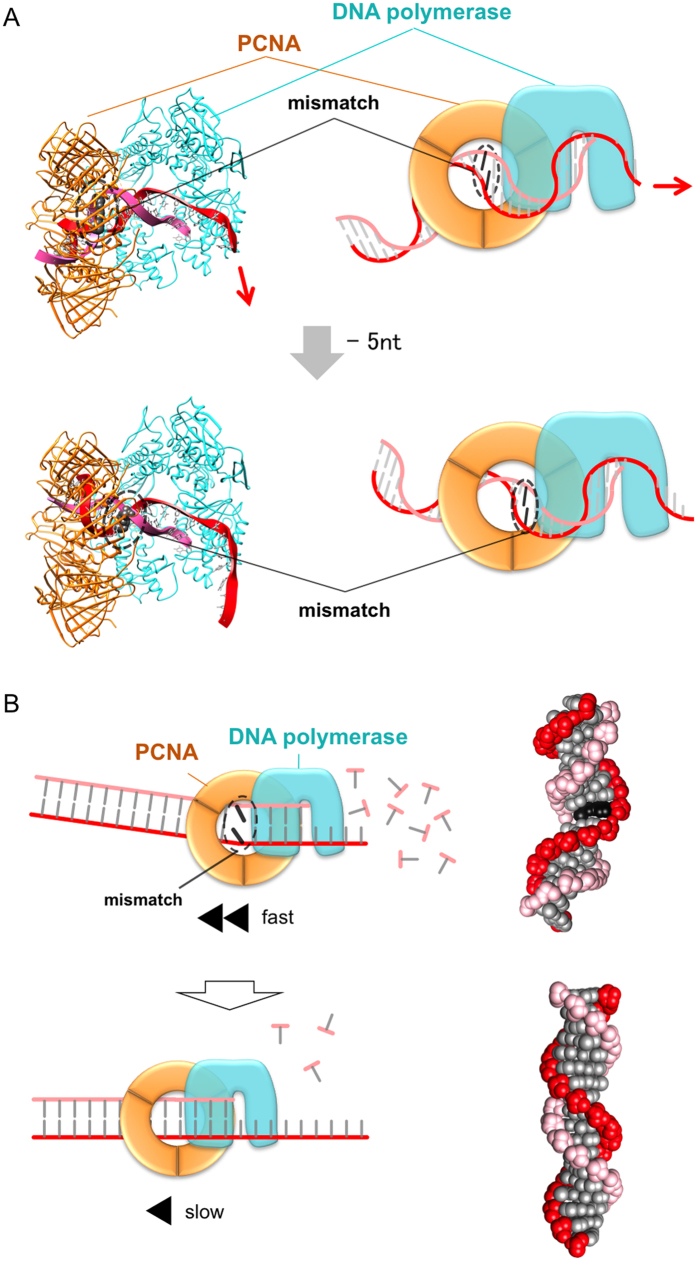
Family B DNA polymerases comprise polymerase and 3' ->5' exonuclease domains, and detect a mismatch in a newly synthesized strand to remove it in cooperation with Proliferating cell nuclear antigen (PCNA), which encircles the DNA to provide a molecular platform for efficient protein-protein and protein-DNA interactions during DNA replication and repair. Once the repair is completed, the enzyme must stop the exonucleolytic process and switch to the polymerase mode. However, the cue to stop the degradation is unclear. We constructed several PCNA mutants and found that the exonuclease reaction was enhanced in the mutants lacking the conserved basic patch, located on the inside surface of PCNA. These mutants may mimic the Pol/PCNA complex processing the mismatched DNA, in which PCNA cannot interact rigidly with the irregularly distributed phosphate groups outside the dsDNA. Indeed, the exonuclease reaction with the wild type PCNA was facilitated by mismatched DNA substrates. PCNA may suppress the exonuclease reaction after the removal of the mismatched nucleotide. PCNA seems to act as a "brake" that stops the exonuclease mode of the DNA polymerase after the removal of a mismatched nucleotide from the substrate DNA, for the prompt switch to the DNA polymerase mode.


Conflict of interest statement

The authors declare no competing financial interests.

Figures

Figure 1
Figure 1. Models of the PfuPolB/PfuPCNA/DNA complex and the DNA interactions in the polymerase and exonuclease modes.
(A) Schematic diagram of the pivot motion of PfuPolB on PfuPCNA between the polymerase-exonuclease modes, with the PIP bond (black circle) as the pivot center. PfuPolB in the polymerase mode is colored magenta, PfuPolB in the exonuclease mode is colored cyan, and PfuPCNA is colored orange. Ribbon representations of the substrate DNA molecules (magenta: polymerase mode, cyan: exonuclease mode) are superimposed on the surface model of the proteins. (B) Stereo views of the substrate DNA interactions with PfuPCNA in each mode. A stick model of the strand in elongation (magenta) is overlaid with the interacting subunits of PfuPCNA in the polymerase mode (top). Two sets of Site A (close-up view in the box) from subunits 2 and 3 interact with the DNA strand, flanked by Lys11 and Lys142. A stick model of the strand to be hydrolyzed (cyan) is superimposed on the relevant subunits of PfuPCNA in the exonuclease mode (bottom). (C) Amino acid alignment of the Pyrococcus furiosus (Pfu), Saccharomyces cerevisiae (Sce), and human (Hum) PCNAs. Amino acids conserved between two of the three organisms are marked by dots, and lines mark those conserved among all three organisms.
Figure 2
Figure 2. In vitro primer extension reactions with/without the wild type and mutant PfuPCNAs.
The gel images displayed here are representative of several trials (n = 4). The bar charts at the bottom of the corresponding gel images show the average ratio of the quantities of the elongated primers to that of the unreacted primers and the non-PCNA-assisted-elongation band, as judged from the lane with PfuPolB alone, in which the ratio of the wild type was normalized to 1.0. The borders between the elongated and the unreacted (including non-PCNA assisted elongation) substrates are depicted as dotted lines. The ratios of the DNA substrates to the proteins (PfuPolB and PfuPCNA) in each panel are (A) 1:4, 1:8, and 1:16 (left to right), using the Site A mutants, and (B) 1:4, 1:8, and 1:16 (left to right), using the non-Site A mutants.
Figure 3
Figure 3. In vitro exonuclease reactions with/without the wild type and mutant PfuPCNAs.
The gel images displayed here are representative of several trials (n = 4). The borders between the degraded and the unreacted (including non-PCNA assisted degradation) substrates are depicted as dotted lines. The ratios of the DNA substrates to the proteins (PfuPolB and PfuPCNA) in each panel are (A) 1:4, 1:8, and 1:16 (left to right), using the Site A mutants, and (B) 1:4, 1:8, and 1:16 (left to right), using the non-Site A mutants.
Figure 4
Figure 4. In vitro exonuclease reactions using the substrate dsDNA with/without several types of mismatches.
The values of the degradation efficiencies using the substrates with (right) or without (left) mismatches are depicted in the bar charts, in which the values with the non-mismatched substrates were set to 1.0. (A) Exonuclease reactions with PCNA using the mismatch-induced substrates, compared to the substrate without a mismatch (left lane in each panel). The samples in the substrate only lanes (-PfuPolB, -PfuPCNA) were incubated in the same manner as those with enzymes. In this figure, the ratios of the densities were produced by dividing the density of the input template by that of the reaction product. (B) The same experiments without PCNA. (C) The addition of dNTPs (final concentrations of 230 μM, 23 μM, 2.3 μM, 230 nM, 23 nM, 2.3 nM, 230 pM, and 23 pM dNTPs) after the exonuclease reaction by PfuPolB + PfuPCNA using the mismatched substrate 4AG14TC, the high concentrations of dNTP (23 μM<) switches the enzyme from the exonuclease mode to the polymerase mode. On the other hand, at the lower concentration range of dNTP (<230 pM), the exonuclease reaction continued despite the existence of dNTP. The ratios of densities in the bar chart were produced by the dividing the density of the reaction product by that of the input substrate. The significant p values (<0.05) are in bold characters.
Figure 5
Figure 5. In vitro nick ligation reactions by PfuLig with/without the wild type and mutant PfuPCNAs.
(A) Search for the conditions in which the addition of PfuPCNA enhances the nick ligation reaction by PfuLig. A large excess of the substrate DNA was required for the PCNA-enhanced nick ligation reaction. (B) In vitro nick ligation reactions of PfuLig with/without the wild type and mutant PfuPCNAs.
Figure 6
Figure 6. The difference in the stability of the interactions between mismatched- and ideal B-type double-stranded DNA.
(A) Dynamic interaction between the substrate with a mismatch and the inside face of PCNA, in accordance with the progression of the exonuclease reaction. This figure was depicted by connecting the CA mismatch dsDNA with a substrate DNA binding to the PfuPolB/PfuPCNA complex in the exonuclease mode. On the one hand, if the location of the CA mismatch is the 11th position from the nucleolytic site, then the left portion of the substrate bends toward the lower side (top). On the other hand, if the location is the 6th position, then the substrate bends toward the upper side (bottom). Since the substrate is bending at the mismatched site, the latter portion of the substrate DNA undergoes a precession movement, and thus the DNA fails to interact stably with PCNA. Therefore, PCNA cannot brake the exonuclease reaction unless the mismatch is removed. (B) The difference in the exonuclease activities between mismatched- and ideal B-type double-stranded DNA. Schematic diagrams representing the difference in the efficacies of exonuclease reactions with a mismatched-dsDNA (top) or a correctly base paired-DNA (bottom), flanked by the tertiary structures of a mismatched-DNA in which the G-T mismatch pair is highlighted in black (top), and an ideal dsDNA (bottom).

References

    1. Tsurimoto T. PCNA, a multifunctional ring on DNA. Biochim. Biophys. Acta 1443, 23–39 (1998). - PubMed
    1. Kelman Z. & Hurwitz J. Protein-PCNA interactions: a DNA scanning mechanism? Trends Biochem. Sci. 23, 236–238 (1998). - PubMed
    1. Maga G. & Hübscher U. Proliferating cell nuclear antigen (PCNA): a dancer with many partners. J. Cell Sci. 116, 3051–3060 (2003). - PubMed
    1. Krishna T. S. R., Kong X.-P., Gary S., Burgers P. M. & Kuriyan J. Crystal structure of the eukaryotic DNA polymerase processivity factor PCNA. Cell 79, 1233–1243 (1994). - PubMed
    1. Gulbis J. M., Kelman Z., Hurwitz J., O’Donnell M. & Kuriyan J. Structure of the C-terminal region of p21WAF1/CIP1 complexed with human PCNA. Cell 87, 297–306 (1996). - PubMed

Publication types

MeSH terms

LinkOut - more resources