Painful After-Sensations in Fibromyalgia are Linked to Catastrophizing and Differences in Brain Response in the Medial Temporal Lobe
- PMID: 28300650
- PMCID: PMC6102715
- DOI: 10.1016/j.jpain.2017.02.437
Painful After-Sensations in Fibromyalgia are Linked to Catastrophizing and Differences in Brain Response in the Medial Temporal Lobe
Abstract
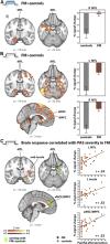
Fibromyalgia (FM) is a complex syndrome characterized by chronic widespread pain, hyperalgesia, and other disabling symptoms. Although the brain response to experimental pain in FM patients has been the object of intense investigation, the biological underpinnings of painful after-sensations (PAS), and their relation to negative affect have received little attention. In this cross-sectional cohort study, subjects with FM (n = 53) and healthy controls (n = 17) were assessed for PAS using exposure to a sustained, moderately painful cuff stimulus to the leg, individually calibrated to a target pain intensity of 40 of 100. Despite requiring lower cuff pressures to achieve the target pain level, FM patients reported more pronounced PAS 15 seconds after the end of cuff stimulation, which correlated positively with clinical pain scores. Functional magnetic resonance imaging revealed reduced deactivation of the medial temporal lobe (MTL; amygdala, hippocampus, parahippocampal gyrus) in FM patients, during pain stimulation, as well as in the ensuing poststimulation period, when PAS are experienced. Moreover, the functional magnetic resonance imaging signal measured during the poststimulation period in the MTL, as well as in the insular and anterior middle cingulate and medial prefrontal cortices, correlated with the severity of reported PAS by FM patients. These results suggest that the MTL plays a role in PAS in FM patients.
Perspective: PAS are more common and severe in FM, and are associated with clinical pain and catastrophizing. PAS severity is also associated with less MTL deactivation, suggesting that the MTL, a core node of the default mode network, may be important in the prolongation of pain sensation in FM.
Keywords: Human; default mode network; neuroimaging; psychophysics; psychosocial; sensitization; temporal summation.
Copyright © 2017 American Pain Society. Published by Elsevier Inc. All rights reserved.
Conflict of interest statement
The authors have no conflicts of interest to declare.
Figures


Similar articles
-
Aberrant Salience? Brain Hyperactivation in Response to Pain Onset and Offset in Fibromyalgia.Arthritis Rheumatol. 2020 Jul;72(7):1203-1213. doi: 10.1002/art.41220. Epub 2020 May 21. Arthritis Rheumatol. 2020. PMID: 32017421 Free PMC article.
-
The somatosensory link in fibromyalgia: functional connectivity of the primary somatosensory cortex is altered by sustained pain and is associated with clinical/autonomic dysfunction.Arthritis Rheumatol. 2015 May;67(5):1395-1405. doi: 10.1002/art.39043. Arthritis Rheumatol. 2015. PMID: 25622796 Free PMC article.
-
Effects of Cognitive-Behavioral Therapy (CBT) on Brain Connectivity Supporting Catastrophizing in Fibromyalgia.Clin J Pain. 2017 Mar;33(3):215-221. doi: 10.1097/AJP.0000000000000422. Clin J Pain. 2017. PMID: 27518491 Free PMC article. Clinical Trial.
-
[Functional brain mapping of pain perception].Med Sci (Paris). 2011 Jan;27(1):82-7. doi: 10.1051/medsci/201127182. Med Sci (Paris). 2011. PMID: 21299967 Review. French.
-
[Functional imaging of pain].Biol Aujourdhui. 2014;208(1):5-12. doi: 10.1051/jbio/2014003. Epub 2014 Jun 23. Biol Aujourdhui. 2014. PMID: 24948014 Review. French.
Cited by
-
Habituation deficit of visual evoked potentials in migraine patients with hypermobile Ehlers-Danlos syndrome.Front Neurol. 2023 Mar 9;14:1072785. doi: 10.3389/fneur.2023.1072785. eCollection 2023. Front Neurol. 2023. PMID: 36970542 Free PMC article.
-
Brief Self-Compassion Training Alters Neural Responses to Evoked Pain for Chronic Low Back Pain: A Pilot Study.Pain Med. 2020 Oct 1;21(10):2172-2185. doi: 10.1093/pm/pnaa178. Pain Med. 2020. PMID: 32783054 Free PMC article.
-
Behavioral, Psychological, Neurophysiological, and Neuroanatomic Determinants of Pain.J Bone Joint Surg Am. 2020 May 20;102 Suppl 1(Suppl 1):21-27. doi: 10.2106/JBJS.20.00082. J Bone Joint Surg Am. 2020. PMID: 32251127 Free PMC article. Review. No abstract available.
-
Aftersensations and Lingering Pain After Examination in Patients with Fibromyalgia Syndrome.Pain Med. 2022 Dec 1;23(12):1928-1938. doi: 10.1093/pm/pnac089. Pain Med. 2022. Retraction in: Pain Med. 2025 Aug 06:pnaf079. doi: 10.1093/pm/pnaf079. PMID: 35652761 Free PMC article. Retracted.
-
Herpes zoster chronification to postherpetic neuralgia induces brain activity and grey matter volume change.Am J Transl Res. 2018 Jan 15;10(1):184-199. eCollection 2018. Am J Transl Res. 2018. PMID: 29423004 Free PMC article.
References
-
- Andrew D, Greenspan JD. Peripheral coding of tonic mechanical cutaneous pain: Comparison of nociceptor activity in rat and human psychophysics. J Neurophysiol. 1999;82:2641–2648. - PubMed
-
- Aoki Y, Inokuchi R, Suwa H. Reduced N-acetylaspartate in the hippocampus in patients with fibromyalgia: A meta-analysis. Psychiatry Res. 2013;213:242–248. - PubMed
-
- Arendt-Nielsen L. Central sensitization in humans: Assessment and pharmacology. Handb Exp Pharmacol. 2015;227:79–102. - PubMed
Publication types
MeSH terms
Grants and funding
LinkOut - more resources
Full Text Sources
Other Literature Sources
Medical

