

Comparison of femtosecond laser-assisted descemetic and predescemetic lamellar keratoplasty for keratoconus
- PMID: 28300735
- PMCID: PMC5369287
- DOI: 10.4103/ijo.IJO_688_16
Comparison of femtosecond laser-assisted descemetic and predescemetic lamellar keratoplasty for keratoconus
Abstract
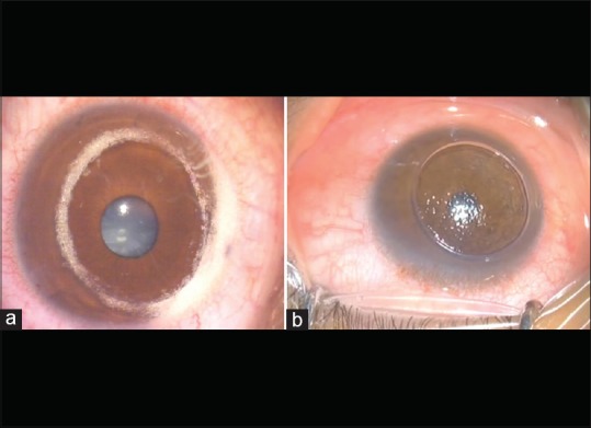
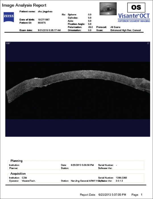
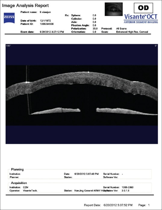
Purpose: The purpose of this study is to compare the outcomes following femtosecond laser-assisted deep anterior lamellar keratoplasty (DALK) with 75% of stromal dissection (predescemetic group) and femtosecond laser-assisted DALK using big-bubble technique with total stromal resection (descemetic group) for the treatment of keratoconus.
Subjects and methods: Twenty eyes of 17 patients with keratoconus were studied. There were 10 eyes of 9 patients in predescemetic group and 10 eyes of 8 patients in descemetic group. The postoperative best-corrected visual acuity (BCVA), manifest refraction, keratometry, endothelial cell density (ECD), and central corneal thickness (CCT) were analyzed.
Results: All surgeries were performed uneventfully. At 1 year after surgery, the BCVA, corneal astigmatism, keratometry, CCT, and ECD between two groups were not statistically significant (all P > 0.05). However, the mean manifest refraction was -9.43 ± 7.44 diopter (D) and -1.03 ± 1.13D in predescemetic and descemetic groups, respectively, which was statistically significant between two groups (P < 0.05).
Conclusions: The results of BCVA and corneal astigmatism, keratometry, ECD, and CCT were comparable between two groups. However, the mean postoperative manifest refraction was lower in descemetic group.
Conflict of interest statement
There are no conflicts of interest.
Figures
References
-
- Tan DT, Dart JK, Holland EJ, Kinoshita S. Corneal transplantation. Lancet. 2012;379:1749–61. - PubMed
-
- Baradaran-Rafii A, Eslani M, Sadoughi MM, Esfandiari H, Karimian F. Anwar versus Melles deep anterior lamellar keratoplasty for keratoconus: A prospective randomized clinical trial. Ophthalmology. 2013;120:252–9. - PubMed
-
- Watson SL, Ramsay A, Dart JK, Bunce C, Craig E. Comparison of deep lamellar keratoplasty and penetrating keratoplasty in patients with keratoconus. Ophthalmology. 2004;111:1676–82. - PubMed
-
- Anwar M, Teichmann KD. Deep lamellar keratoplasty: Surgical techniques for anterior lamellar keratoplasty with and without baring of Descemet's membrane. Cornea. 2002;21:374–83. - PubMed
-
- Fogla R, Padmanabhan P. Results of deep lamellar keratoplasty using the big-bubble technique in patients with keratoconus. Am J Ophthalmol. 2006;141:254–9. - PubMed
Publication types
MeSH terms
LinkOut - more resources
Full Text Sources
Other Literature Sources
