. 2017 Mar 16;8(3):e2668.
doi: 10.1038/cddis.2017.37.

Regulation of RhoA activity by the cellular prion protein

Affiliations

Regulation of RhoA activity by the cellular prion protein

Hee-Jun Kim et al. Cell Death Dis. .

Abstract

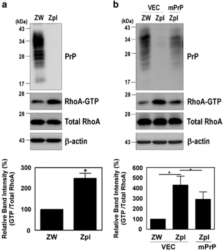
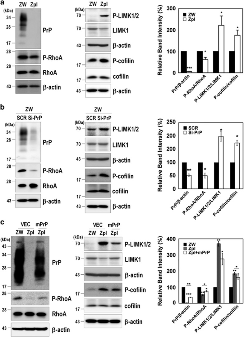
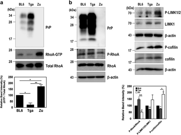
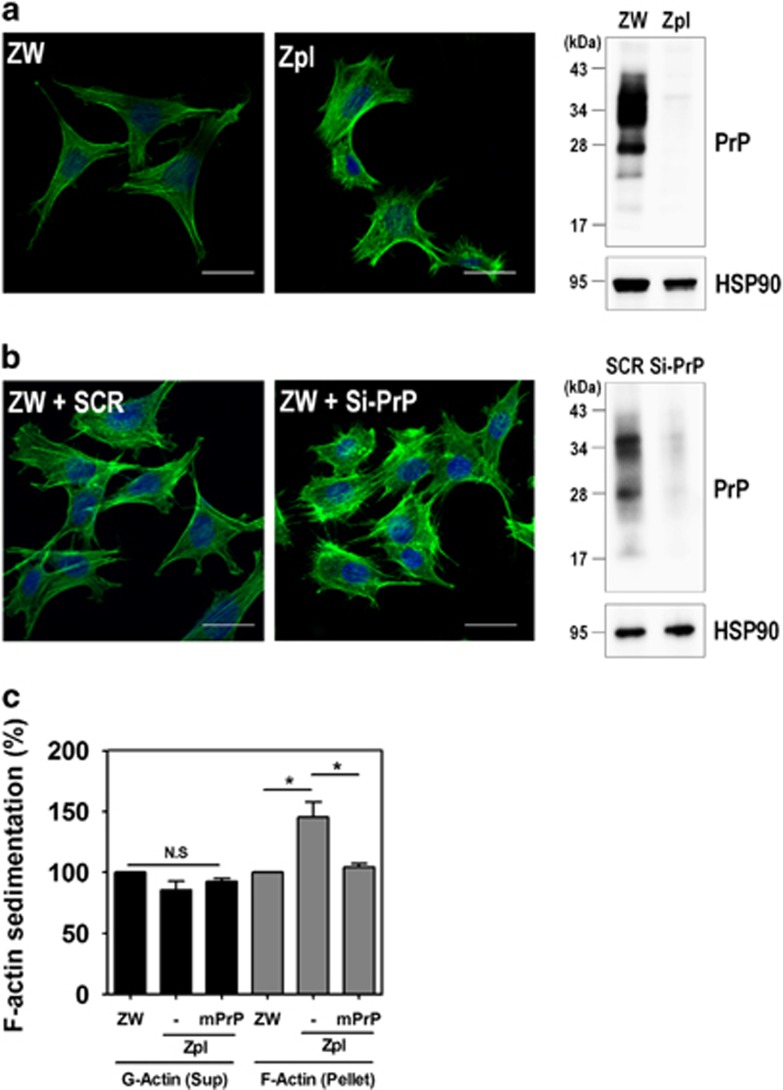
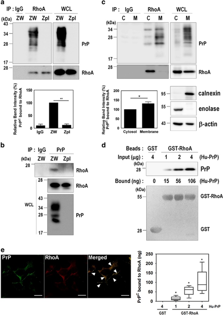
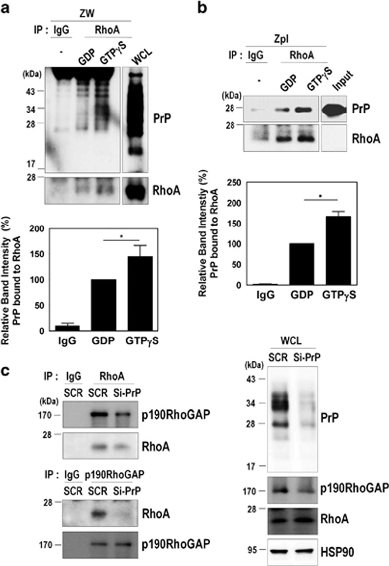
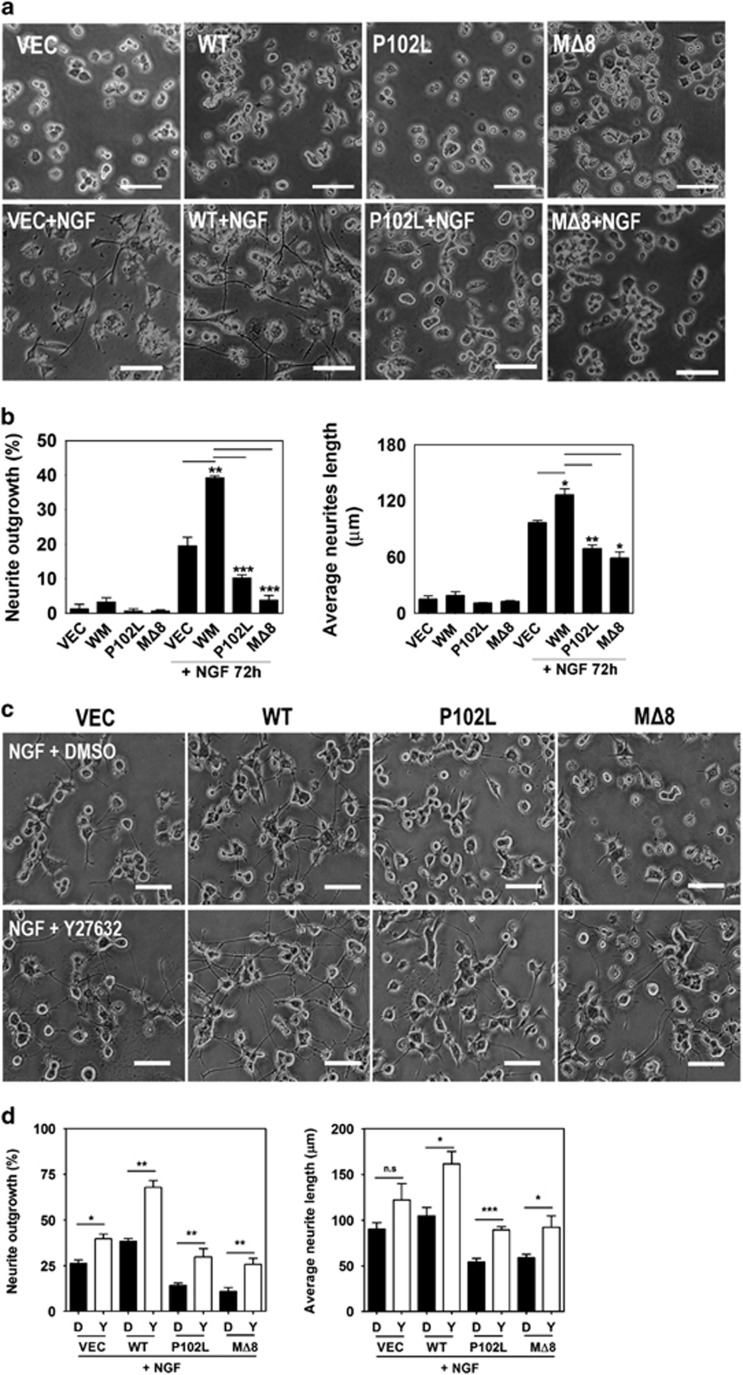
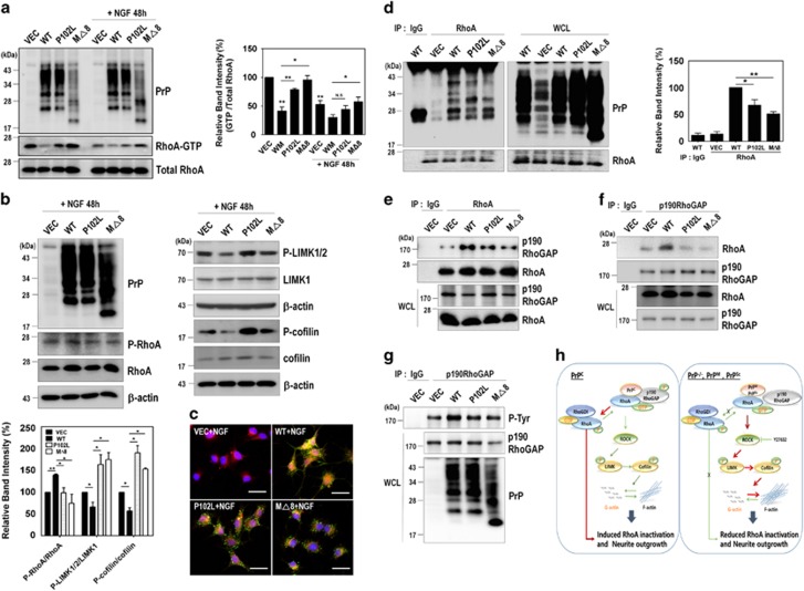
The cellular prion protein (PrPC) is a highly conserved glycosylphosphatidylinositol (GPI)-anchored membrane protein that is involved in the signal transduction during the initial phase of neurite outgrowth. The Ras homolog gene family member A (RhoA) is a small GTPase that is known to have an essential role in regulating the development, differentiation, survival, and death of neurons in the central nervous system. Although recent studies have shown the dysregulation of RhoA in a variety of neurodegenerative diseases, the role of RhoA in prion pathogenesis remains unclear. Here, we investigated the regulation of RhoA-mediated signaling by PrPC using both in vitro and in vivo models and found that overexpression of PrPC significantly induced RhoA inactivation and RhoA phosphorylation in hippocampal neuronal cells and in the brains of transgenic mice. Using siRNA-mediated depletion of endogenous PrPC and overexpression of disease-associated mutants of PrPC, we confirmed that PrPC induced RhoA inactivation, which accompanied RhoA phosphorylation but reduced the phosphorylation levels of LIM kinase (LIMK), leading to cofilin activation. In addition, PrPC colocalized with RhoA, and the overexpression of PrPC significantly increased neurite outgrowth in nerve growth factor-treated PC12 cells through RhoA inactivation. However, the disease-associated mutants of PrPC decreased neurite outgrowth compared with wild-type PrPC. Moreover, inhibition of Rho-associated kinase (ROCK) substantially facilitated neurite outgrowth in NGF-treated PC12 cells, similar to the effect induced by PrPC. Interestingly, we found that the induction of RhoA inactivation occurred through the interaction of PrPC with RhoA and that PrPC enhanced the interaction between RhoA and p190RhoGAP (a GTPase-activating protein). These findings suggest that the interactions of PrPC with RhoA and p190RhoGAP contribute to neurite outgrowth by controlling RhoA inactivation and RhoA-mediated signaling and that disease-associated mutations of PrPC impair RhoA inactivation, which in turn leads to prion-related neurodegeneration.


Conflict of interest statement

The authors declare no conflict of interest.

Figures

Figure 1
Figure 1
PrPC regulates RhoA activation. (a and b) Detection of RhoA-GTP by GST-Rhotekin-RBD pull-down assay in cells expressing PrPC (ZW) and PrP knockout (Zpl) with or without expressing mPrP. The level of RhoA-GTP was determined by western blot with anti-RhoA antibody following a pull-down assay. The data are expressed as the mean±S.E. of three independent experiments (*P<0.05, n=3)
Figure 2
Figure 2
PrPC modulates the RhoA-ROCK-LIMK-cofilin pathway. (a-c) Phosphorylation of RhoA, LIMK1/2, and cofilin in ZW and Zpl cells (a), in ZW cells transfected with scrambled RNA (SCR) or mPrP-targeted siRNA (Si-PrP) (b) and in Zpl cells with or without expressing mPrP (c) was analyzed in triplicate by western blot. The intensities of the bands in each panel were measured and quantified for each group, and the values are expressed as the mean±S.E. of three independent experiments (*P<0.05, **P<0.01, ***P<0.001, n=3)
Figure 3
Figure 3
PrPC is involved in RhoA inactivation in the brains of three different types of mice. (a) Detection of RhoA-GTP levels in the brains of C57BL/6J (BL6, WT), Tga20 (Tga, PrP overexpression) and Zürich I (Zu, PrP-deficient) mice (n=3 per each group). (b) Phosphorylation of RhoA, LIMK, and cofilin was assessed in the whole-brain lysates of C57BL/6J, Tga20, and Zürich I mice. The data are expressed as the mean±S.E. of three independent experiments (*P<0.05, **P<0.01, ***P<0.001, n=3)
Figure 4
Figure 4
Depletion of PrPC increases F-actin formation. (a and b) Immunocytochemical staining for F-actin in ZW and Zpl cells (a), and ZW cells transfected with either scrambled RNA (SCR) or mPrP-targeted siRNA (Si-PrP) (b) using Alexa Fluor 488-phalloidin (green). DAPI (blue) was used to counterstain the nuclei. All pictures are representative of multiple images from three independent experiments (scale bars, 20 μm). The expression of PrPC was determined by western blot with anti-PrP (3F10) antibody and HSP90 was used as a loading control. (c) The expression of F-actin assessed by a sedimentation assay in ZW, Zpl, and Zpl expressing mPrP cells was analyzed by western blot with anti-β-actin, anti-PrP (3F10) and anti-HSP90 antibodies. The intensities of the bands in each panel were measured and quantified for each group, and the values are expressed as the mean±S.E. of three independent experiments (*P<0.05, n=3)
Figure 5
Figure 5
PrPC interacts with RhoA. (a and b) Co-immunoprecipitation of PrP with RhoA using ZW and Zpl cell lysates were performed with either anti-RhoA (a) or anti-PrP (3F10) (b) antibodies, and then analyzed by western blot with anti-PrP and anti-RhoA antibodies, respectively. WCL, whole-cell lysates. (c) The subcellular fractions from ZW cells were used to immunoprecipitate RhoA with anti-RhoA antibody and then analyzed by western blot with anti-PrP (3F10) and anti-RhoA antibodies. Enolase and calnexin were used as makers for the cytosol (C) and membrane (M) fractions, respectively. β-Actin as a loading control. (d) GST and GST-RhoA beads were incubated with human recombinant PrP (Hu-PrP) as indicated, and the level of Hu-PrP bound to GST-RhoA was determined by western blot with anti-PrP (3F4) antibody. The boxplot showing the means±S.E. of abundance of the PrP-RhoA complex, was calculated from the BSA standard curve in three (n=3) independent experiments. The GST and GST-RhoA samples were stained with Ponceau S to confirm the equal loading. (e) Colocalization of PrP with RhoA was assessed by double immunofluorescence staining and confocal microscopy. All above data are expressed as the mean±S.E. of three independent experiments (*P<0.05, **P<0.01, n=3)
Figure 6
Figure 6
PrPC binds to GTP-bound RhoA and p190RhoGAP. (a) ZW cell lysates were preloaded with GDP or GTPγS followed by immunoprecipitation with the anti-RhoA antibody and analyzed by western blot using the anti-PrP (3F10) and anti-RhoA antibodies. WCL, whole-cell lysates. (b) Zpl cell lysates preloaded with GDP or GTPγS were immunoprecipitated with anti-RhoA antibody, incubated with 2 μg of human recombinant PrP (Hu-PrP), and analyzed by western blot with the anti-PrP (3F4) and anti-RhoA antibodies. (c) The co-immunoprecipitation of RhoA or p190RhoGAP using ZW cells transiently transfected with either SCR or Si-PrP was detected by western blot using anti-p190RhoGAP and anti-RhoA antibodies, respectively. The data are expressed as the mean±S.E. of three independent experiments (*P<0.05, n=3)
Figure 7
Figure 7
The disease-associated mutations of PrPC impair neurite outgrowth. (a and b) PC12 cells stably expressing either vector, WT, P102L, or MΔ8 were treated with 50 ng/ml NGF for 72 h. (c and d) The cells expressing either vector, WT, P102L, or MΔ8 were incubated with or without 10 μM Y27632 in the presence of NGF. Changes in the cell morphology, neurite length, and neurite numbers were determined under a microscope. The data are expressed as the mean±S.E. of three independent experiments (*P<0.05, **P<0.01, ***P<0.001, n=3)
Figure 8
Figure 8
The disease-associated mutants of PrPC affect RhoA signaling through the reduced interactions with RhoA and p190RhoGAP. (a and b) The level of RhoA-GTP following a pull-down assay (a) and the phosphorylation of RhoA, LIMK, and cofilin (b) was analyzed by western blot in PC12 cells expressing either vector, WT, P102L, or MΔ8 in response to NGF. (c) Colocalization of PrP with RhoA in the NGF-treated PC12 cells expressing either vector, WT, P102L, or MΔ8 was determined using confocal microscopy (green, PrP; red, RhoA; blue, DAPI). (d) HEK293 cells were transiently transfected with either vector, WT, P102L, or MΔ8. The co-immunoprecipitation of RhoA was detected by western blot using anti-PrP (3F4) and anti-RhoA antibodies. (e-g) PC12 cells transiently transfected WT or disease-associated mutants of PrPC were lysed and immunoprecipitated with anti-RhoA (e) and p190RhoGAP antibodies (f). (g) The p190RhoGAP phosphorylation (p-Tyr) was detected using p-Tyr antibody after p190RhoGAP immunoprecipitation. All above data are expressed as the mean±S.E. of three independent experiments (*P<0.05, **P<0.01, n=3). (h) PrPC–RhoA interaction stimulates RhoA inactivation and neurite outgrowth. In PrPC-expressing cells and mice, PrPC increased the phosphorylation of RhoA and p190RhoGAP, enhancing the interaction between RhoA and p190RhoGAP. This complex led to the inactivation of RhoA and its downstream effectors. Subsequently, RhoA inactivation decreased actin polymerization and enhanced neurite outgrowth. In contrast, depleting PrPC or expressing disease-associated mutants of PrPC prevented RhoA inactivation and neurite outgrowth by interfering with the interaction

References

    1. Bishop AL, Hall A. Rho GTPases and their effector proteins. Biochem J 2000; 348(Pt 2): 241–255. - PMC - PubMed
    1. Burridge K, Wennerberg K. Rho and Rac take center stage. Cell 2004; 116: 167–179. - PubMed
    1. Kim HJ, Kim JG, Moon MY, Park SH, Park JB. IκB kinase gamma/nuclear factor-κB-essential modulator (IKKgamma/NEMO) facilitates RhoA GTPase activation, which, in turn, activates Rho-associated KINASE (ROCK) to phosphorylate IKKbeta in response to transforming growth factor TGF-β1. J Biol Chem 2014; 289: 1429–1440. - PMC - PubMed
    1. Kjoller L, Hall A. Signaling to Rho GTPases. Exp Cell Res 1999; 253: 166–179. - PubMed
    1. Govek EE, Newey SE, Van Aelst L. The role of the Rho GTPases in neuronal development. Genes Dev 2005; 19: 1–49. - PubMed

Publication types

MeSH terms

LinkOut - more resources