

OPHTHALMIC FINDINGS IN LATE STAGE SJOGREN-LARSSON SYNDROME
- PMID: 28301413
- PMCID: PMC5729048
- DOI: 10.1097/ICB.0000000000000583
OPHTHALMIC FINDINGS IN LATE STAGE SJOGREN-LARSSON SYNDROME
Abstract
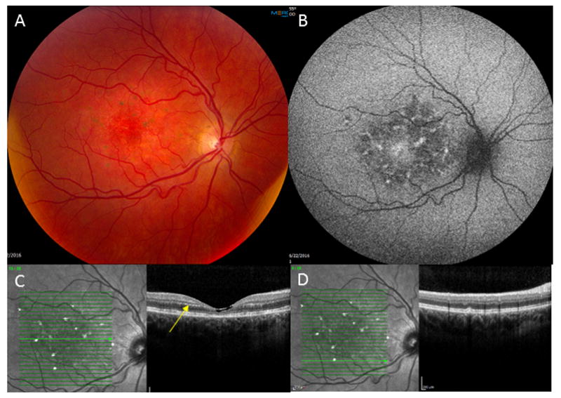
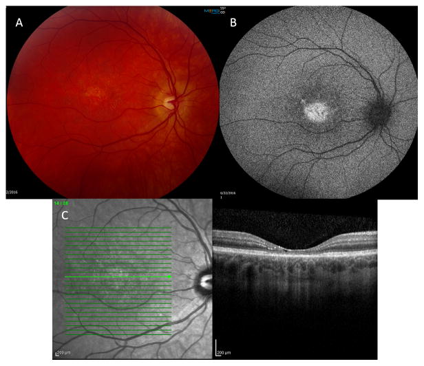
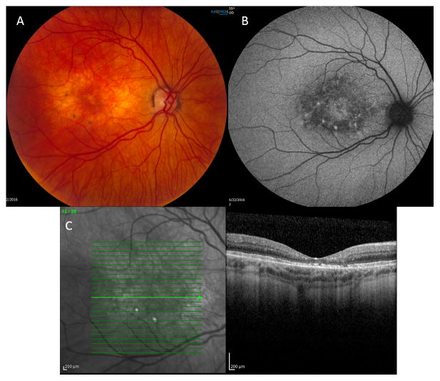
Purpose: To report spectral domain optical coherence tomography and fundus autofluorescence documentation of late stage macular findings associated with Sjogren-Larsson Syndrome in three adult siblings.
Methods: Three adult siblings with Sjogren-Larsson Syndrome underwent ophthalmic examination and imaging.
Results: Crystalline maculopathy and subretinal deposits, presumably lipofuscin accumulation, with macular atrophy were present in varying degrees in all three adult siblings.
Discussion: In adults with Sjogren-Larsson Syndrome, crystalline retinopathy can progress to macular atrophy and the appearance of lipofuscin accumulation.
Figures
References
-
- Sjogren T, Larsson T. Oligophrenia in combination with congenital ichthyosis and spastic disorder; a clinical and genetic study. Acta Psychiatr Neurol Scand Suppl. 1957;113:1–112. - PubMed
-
- Willemsen MAAP, Johannes RMC, Rotteveel JJ, et al. Juvenile macular dystrophy associated with deficient activity of fatty aldehyde dehydrogenase in Sjögren-Larsson syndrome. American Journal of Ophthalmology. 2000;130(6):782–89. - PubMed
-
- De Laurenzi V, Rogers GR, Hamrock DJ, et al. Sjögren-Larsson syndrome is caused by mutations in the fatty aldehyde dehydrogenase gene. Nat Genet. 1996;12:52–7. - PubMed
-
- Newman E, Reichenbach A. The Müller cell: a functional element of the retina. Trends Neurosci. 1996;19:307–12. - PubMed
Publication types
MeSH terms
Substances
Grants and funding
LinkOut - more resources
Full Text Sources
Other Literature Sources
Medical
