. 2017 Mar 16;12(3):e0174077.
doi: 10.1371/journal.pone.0174077. eCollection 2017.

Generation of murine tumor cell lines deficient in MHC molecule surface expression using the CRISPR/Cas9 system

Affiliations

Generation of murine tumor cell lines deficient in MHC molecule surface expression using the CRISPR/Cas9 system

Krishna Das et al. PLoS One. .

Erratum in

Abstract

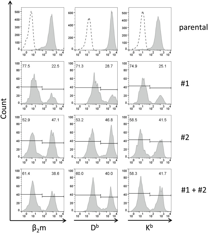
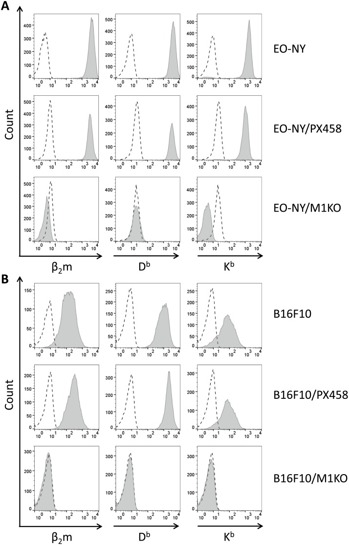
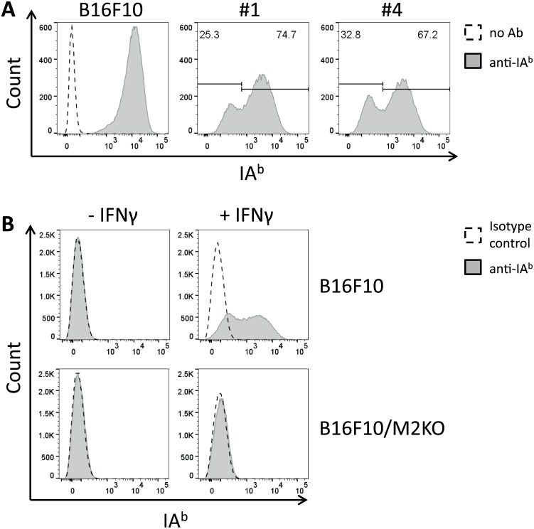
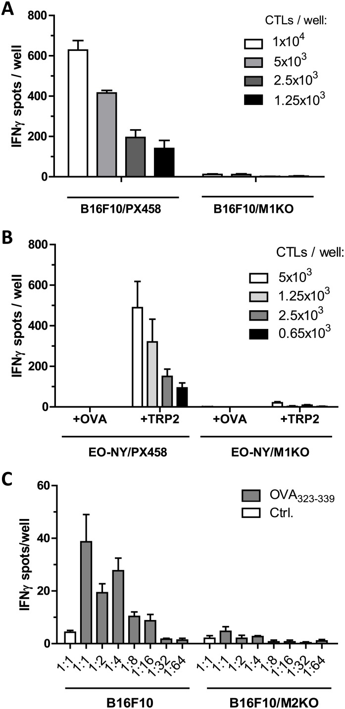
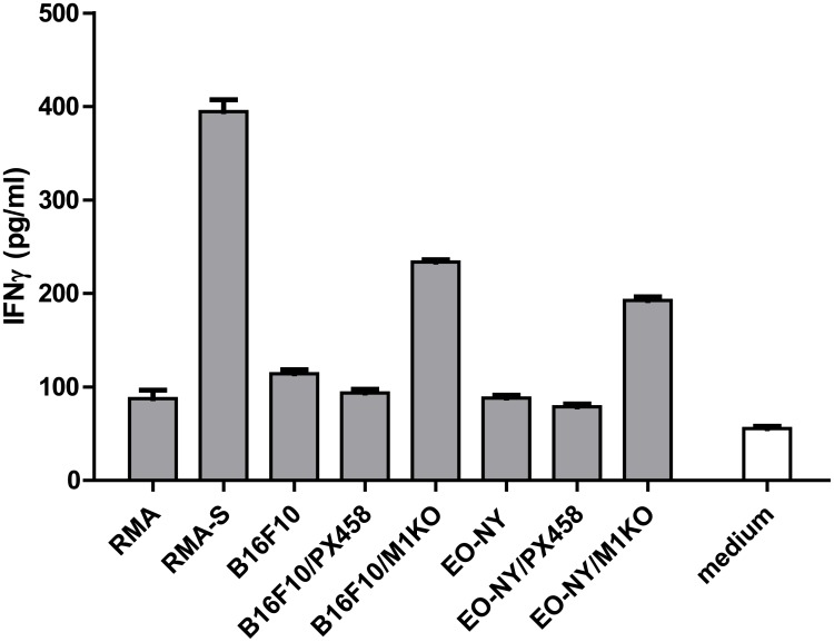
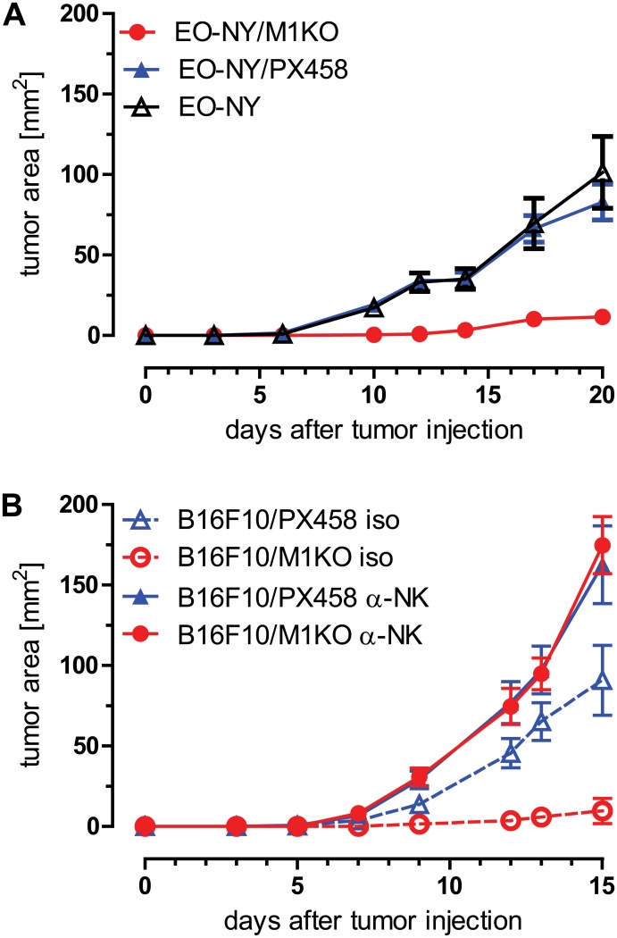
In this study, the CRISPR/Cas9 technology was used to establish murine tumor cell lines, devoid of MHC I or MHC II surface expression, respectively. The melanoma cell line B16F10 and the murine breast cancer cell line EO-771, the latter stably expressing the tumor antigen NY-BR-1 (EO-NY), were transfected with an expression plasmid encoding a β2m-specific single guide (sg)RNA and Cas9. The resulting MHC I negative cells were sorted by flow cytometry to obtain single cell clones, and loss of susceptibility of peptide pulsed MHC I negative clones to peptide-specific CTL recognition was determined by IFNγ ELISpot assay. The β2m knockout (KO) clones did not give rise to tumors in syngeneic mice (C57BL/6N), unless NK cells were depleted, suggesting that outgrowth of the β2m KO cell lines was controlled by NK cells. Using sgRNAs targeting the β-chain encoding locus of the IAb molecule we also generated several B16F10 MHC II KO clones. Peptide loaded B16F10 MHC II KO cells were insusceptible to recognition by OT-II cells and tumor growth was unaltered compared to parental B16F10 cells. Thus, in our hands the CRISPR/Cas9 system has proven to be an efficient straight forward strategy for the generation of MHC knockout cell lines. Such cell lines could serve as parental cells for co-transfection of compatible HLA alleles together with human tumor antigens of interest, thereby facilitating the generation of HLA matched transplantable tumor models, e.g. in HLAtg mouse strains of the newer generation, lacking cell surface expression of endogenous H2 molecules. In addition, our tumor cell lines established might offer a useful tool to investigate tumor reactive T cell responses that function independently from MHC molecule surface expression by the tumor.


Conflict of interest statement

Competing Interests: The authors have declared that no competing interests exist.

Figures

Fig 1
Fig 1. Transfection of EO-NY cells with guide#1 and guide#2 RNA encoding constructs results in outgrowth of β2m negative cell populations.
EO-NY cells transfected with guide#1 or guide #2 constructs, respectively (middle panels), or with pooled guide#1 and guide#2 constructs (lower panel) were stained with monoclonal antibodies specific for β2m (left), H2-Db (center) or H2-Kb molecules (right) and analyzed by FACS seven days after transfection. When gated on EGFP expressing transfectants, β2m negative subpopulations showed up in transfected bulk cultures, but not among parental EO-NY cells (upper panel). Note that the proportion β2m negative cells was equal to the proportion of transfectants lacking MHC I molecule expression.
Fig 2
Fig 2. Phenotype of stable β2m KO clones derived from various tumor entities upon transfection with guide#1 constructs.
EO-NY cells (A) or B16F10 cells (B) transfected with guide #1 constructs (A, B lower panels) or empty vector PX458 as control (A, B, middle panels) were sorted by FACS two days after transfection and cloned by limiting dilution or FACS guided single cell sorting 14 days later. Selected clones were stained with monoclonal antibodies specific for β2m (left), H2-Db (center) or H2-Kb molecules (right) and analyzed by FACS. Living gate was set on 7-AAD negative cells. Guide#1 derived transfectant clones of both tumor entities showed complete loss of β2m as well as MHC I molecule expression (A, B, lower panel), when compared to parental cell lines (A, B, upper panel) or control transfectant clones (A, B, middle panels).
Fig 3
Fig 3. Transfection of B16F10 cells with guide #4 encoding constructs results in generation of a stable B16F10/IAb KO clone.
Parental B16F10 cells and B16F10 cells transfected with guide#1 or guide#4, respectively, were treated with IFNγ (20 U/ml) 9 days post transfection, followed by surface staining with IAb specifc monoclonal antibody. FACS analysis performed on 7-AAD negative cells revealed a higher proportion of IAb negative cells upon transfection with guide#4 (A, right histogram) compared to transfection with guide#1 (A, center). Immunofluorescence staining confirmed complete loss of IAb surface expression on the selected B16F10 KO clone, even when treated with IFNγ (B, lower panel).
Fig 4
Fig 4. Stable β2m KO and IAb KO clones lose susceptibility to cognate T cell recognition.
B16F10/PX458 control transfectants or B16F10/M1KO cells (5 x 10⁴) were incubated overnight with graded numbers of TRP-2-specific CTLs (A). Secretion of IFNγ in response to target cell recognition was retained with B16F10/PX458 control cells but was lost with the B16F10/M1KO clone as measured by ELISpot analysis. Similarly, recognition of peptide incubated EO-NY/PX458 control cells but not of EO-NY/M1KO cells was observed upon incubation with the OVA-specific CTL line (B). Peptide loaded B16F10 cells but not B16F10/M2KO were recognized by OVA-specific OT-II cells. Target cells were treated with IFNγ (20 U/ml) prior to the assay to upregulate IAb expression. Empty bars (Ctrl.), recognition of target cells loaded with IAb restricted HBV core antigen control peptide 128–140 (TPPAYRPPNAPIL). Error bars represent SEM of technical triplicates (C).
Fig 5
Fig 5. Stable β2m KO clones show enhanced susceptibility to NK cell recognition.
Target cells (2.5 x 10⁵) were incubated with (5 x 10⁴) IL2-activated (1,700 IU/ml IL2, 7 d) NK cells for 8 h and IFNγ released by the NK cells was quantified by ELISA. B16F10/M1KO and EO-NY/M1KO clones induce enhanced IFNγ release compared to parental cells or control transfectant clones, respectively. Error bars represent SEM of technical triplicates.
Fig 6
Fig 6. Tumor outgrowth of stable β2m KO clones is controlled by NK cells.
C57BL/6 mice (n = 10) were injected (s.c.) with 2 x 10⁵ parental EO-NY cells or with the transfectant clones EO-NY/PX458 and EO-NY/M1KO derived thereof, showing suppressed outgrowth of EO-NY/M1KO cells (A). Similarly, 2 x 10⁵ B16F10 cells or B16F10 derived transfectant clones were injected (s.c.) into C57BL/6 mice (n = 10). NK cells were depleted by i.p. injections of monoclonal ab PK136 or isotype control on days -2, 0, 7, 13 after tumor cell application, resulting in restored tumor outgrowth (B). Error bars represent SEM within each animal collective.

References

    1. Horvath P, Barrangou R. CRISPR/Cas, the Immune System of Bacteria and Archaea. Science. 2010;327(5962):167–70. 10.1126/science.1179555 - DOI - PubMed
    1. Wiedenheft B, Sternberg SH, Doudna JA. RNA-guided genetic silencing systems in bacteria and archaea. Nature. 2012;482(7385):331–8. 10.1038/nature10886 - DOI - PubMed
    1. Jinek M, Chylinski K, Fonfara I, Hauer M, Doudna JA, Charpentier E. A Programmable Dual-RNA–Guided DNA Endonuclease in Adaptive Bacterial Immunity. Science. 2012;337(6096):816–21. 10.1126/science.1225829 - DOI - PMC - PubMed
    1. Jiang W, Bikard D, Cox D, Zhang F, Marraffini LA. RNA-guided editing of bacterial genomes using CRISPR-Cas systems. Nat Biotech. 2013;31(3):233–9. - PMC - PubMed
    1. Cong L, Ran FA, Cox D, Lin S, Barretto R, Habib N, et al. Multiplex Genome Engineering Using CRISPR/Cas Systems. Science. 2013;339(6121):819–23. 10.1126/science.1231143 - DOI - PMC - PubMed

Substances