Review
. 2017 Jun 20:86:225-244.
doi: 10.1146/annurev-biochem-061516-044820. Epub 2017 Mar 15.

Mechanisms of Autophagy Initiation

Affiliations
Review

Mechanisms of Autophagy Initiation

James H Hurley et al. Annu Rev Biochem. .

Abstract

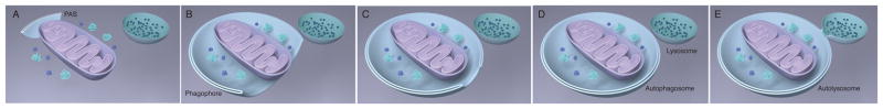
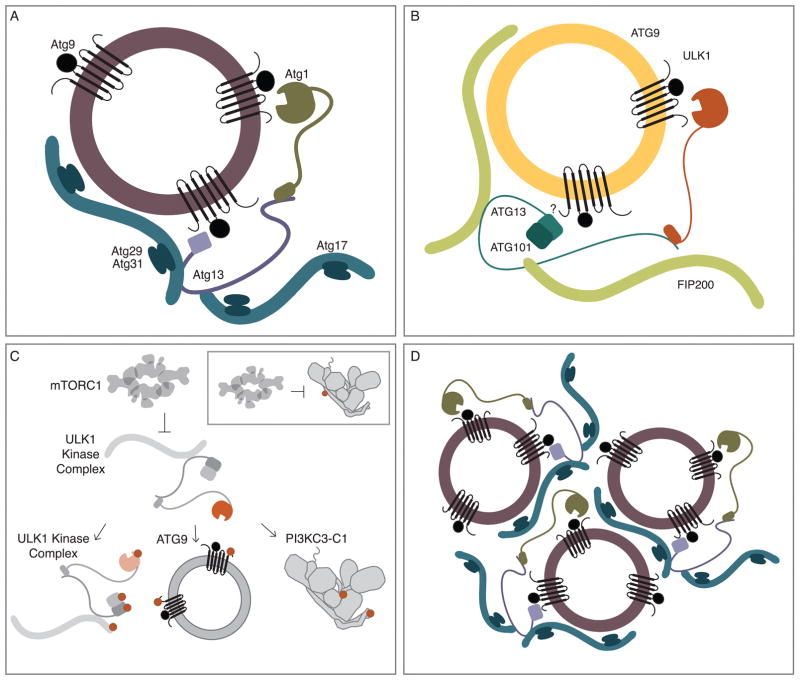
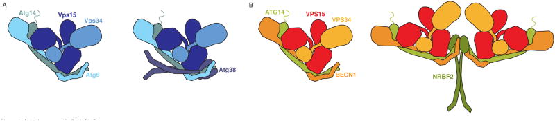
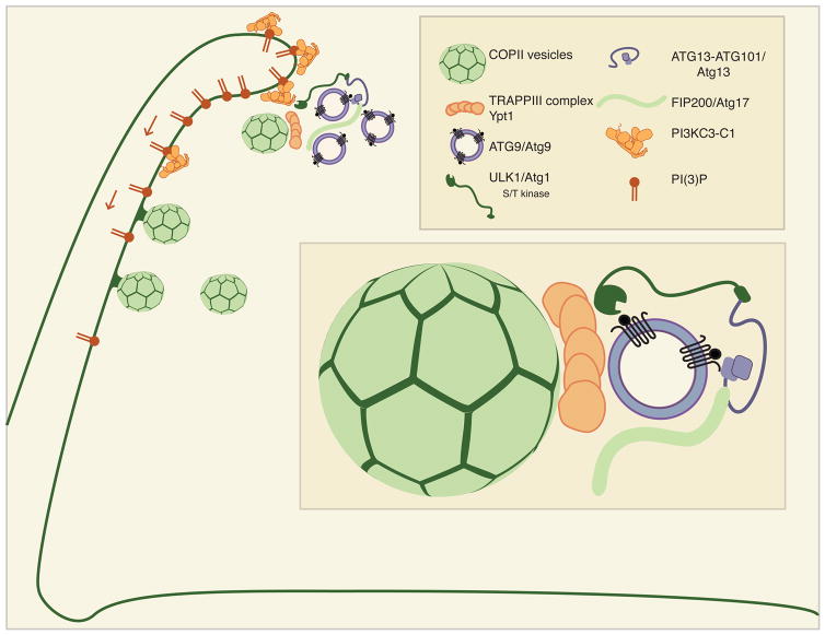
Autophagy is the process of cellular self-eating by a double-membrane organelle, the autophagosome. A range of signaling processes converge on two protein complexes to initiate autophagy: the ULK1 (unc51-like autophagy activating kinase 1) protein kinase complex and the PI3KC3-C1 (class III phosphatidylinositol 3-kinase complex I) lipid kinase complex. Some 90% of the mass of these large protein complexes consists of noncatalytic domains and subunits, and the ULK1 complex has essential noncatalytic activities. Structural studies of these complexes have shed increasing light on the regulation of their catalytic and noncatalytic activities in autophagy initiation. The autophagosome is thought to nucleate from vesicles containing the integral membrane protein Atg9 (autophagy-related 9), COPII (coat protein complex II) vesicles, and possibly other sources. In the wake of reconstitution and super-resolution imaging studies, we are beginning to understand how the ULK1 and PI3KC3-C1 complexes might coordinate the nucleation and fusion of Atg9 and COPII vesicles at the start of autophagosome biogenesis.

Keywords: Atg1; COPII; ULK1; Vps34; membrane fusion; membrane remodeling; nanoscale biology; phosphatidylinositol 3-kinase; tethering complex; vesicle.


Figures

Figure 1
Figure 1. The autophagy program
(A) Preautophagosome assembly site (PAS). (B) PAS elongates into phagophore to surround bulk cytosolic materials. (C) The geometry of phagophore closure. (D) The closed autophagosome. (E) The autophagosome merges with the lysosome; the new organelle is the autolysosome.
Figure 2
Figure 2
The autophagy initiation complex in yeast and mammals. (A) In budding yeast, vesicles containing Atg9 are phosphorylated by the S/T kinase Atg1, additionally, the N-terminus of Atg9 makes a direct contact with the HORMA domain Atg13. Atg13 in turn make contacts with the Atg17-29-31 scaffold. (B) Based on yeast observations, it is proposed that small vesicles containing ATG9 interact with the autophagy initiation complex- the S/T kinase ULK1, the HORMA domain containing ATG13 forms a heterodimer with the HORMA domain of ATG101, FIP200 is modeled on the structure of Atg17. However, the yeast interaction site in Atg9 is not conserved in humans. (C) ULK1 is inhibited by mTORC1. When mTORC1 is inactivated, ULK1 becomes activated, thereby leading to autophosphorylation within ULK1, ATG13, ATG101 and FIP200- multiple phosphorylation sites occur within ATG13, one is shown for clarity. ULK1 then phosphorylates ATG9, and PI3KC3-C1- at the N-terminus of BECN1 and within the C2 domain of VPS34. Inset: mTORC1 directly phosphorylates ATG14L of PI3KC3-C1. (D) From research in the budding yeast model system, the Intrinsically Disordered Regions (IDRs) within Atg1 and Atg13 as well as the position of Atg13-Atg17 contacts lead a proposed mechanism in which ATG proteins form a “meshwork.”
Figure 3
Figure 3. Autophagy specific PI3KC3-C1
(A) Yeast PI3KC3-C1 is composed of the lipid kinase Vps34, the scaffold and potential protein kinase Vps15, the regulatory subunit Atg6, the phagophore targeting subunit Atg14. A dimer of Atg38 binds to one heterotetramer. (B) Mammalian PI3KC3-C1 is composed of the lipid kinase VPS34, the scaffold and potential protein kinase VPS15, the regulatory subunit BECN1, the phagophore targeting subunit ATG14L. The binding of dimer of NRBF2 to the heterotetramer creates leads to the dimerization and the formation of a homopentamer.
Figure 4
Figure 4. Model for Autophagy initiation at ER Exit Sites, the Omegasome
The omegasome provides for the initiation of autophagy. The omegasome is enriched with the lipid PI3P that is generated by PI3KC3-C1. The ULK1 complex activates PI3KC3-C1 through phosphorylation. Arrows denote that PI3P can diffuse through the membrane. PI3P is also enriched at ER Exit Sites (ERES), where COPII vesicles emerge. Inset: COPII vesicles interact with the TRAPPIII complex, which binds to ATG9/Atg9 and ULK1/Atg1.

References

    1. Wen X, Klionsky DJ. An overview of macroautophagy in yeast. J Mol Biol. 2016;428:1681–99. - PMC - PubMed
    1. Bento CF, Renna M, Ghislat G, Puri C, Ashkenazi A, et al. In: Annual Review of Biochemistry. Kornberg RD, editor. Vol. 85. Palo Alto: Annual Reviews; 2016. pp. 685–713. - PubMed
    1. Shaid S, Brandts CH, Serve H, Dikic I. Ubiquitination and selective autophagy. Cell Death Differ. 2013;20:21–30. - PMC - PubMed
    1. Zaffagnini G, Martens S. Mechanisms of selective autophagy. J Mol Biol. 2016;428:1714–24. - PMC - PubMed
    1. Menzies FM, Fleming A, Rubinsztein DC. Compromised autophagy and neurodegenerative diseases. Nature Reviews Neuroscience. 2015;16:345–57. - PubMed

Publication types

MeSH terms

Substances